HESI A2: Nurse Hub A&P Review (Parts & Identification)
 Call Kai
 Learn
Learn Practice Test
 Spaced Repetition
 Match
Match Flashcards
 Knowt Play
1/80

Earn XP
Description and Tags
Name | Mastery | Learn | Test | Matching | Spaced | Call with Kai | Chat |
|---|
No analytics yet
Send a link to your students to track their progress
81 Terms

Identify this bone feature and define its anatomical purpose.
[SKELETAL]
Tubercle
A round bump. Most of these bumps are sites for muscle and ligament attachments

Identify this bone feature that is circled and define its anatomical purpose.a
[SKELETAL]
Tuberosity
A rounded bump that has a more gradual slope.

Identify this bone feature labeled by a blue star and define its anatomical purpose.
[SKELETAL]
Styloid process
A pointy process that attaches to ligaments that support the hyoid bone.
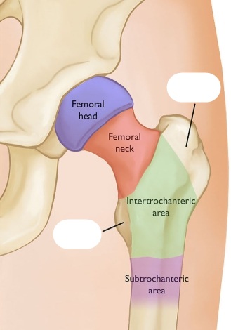
Identify this bone features covered by the white marks and define their anatomical purpose.
[SKELETAL]
Trochanter
A very large bump. These are found on the femur bones.

Identify this bone feature labeled by the blue stars and define its anatomical purpose.
[SKELETAL]
Condyle
A large, rounded process. In the skull it forms the lateral part of the temporomandibular joint (TMJ). They articulate with the temporal bone.
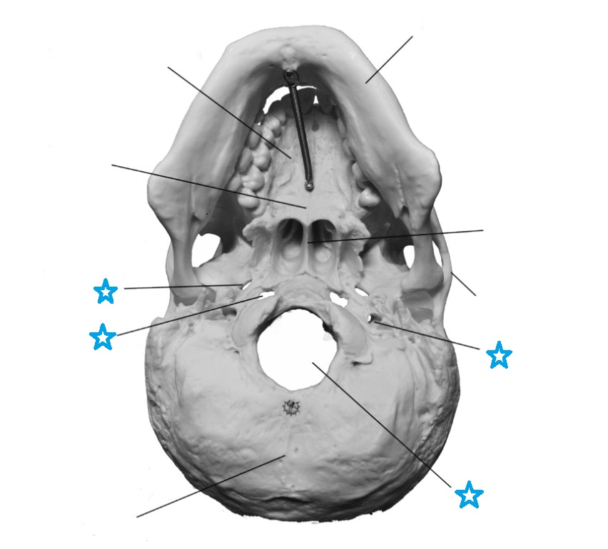
Identify this bone feature labeled by the blue stars and define its anatomical purpose.
[SKELETAL]
Foramen
A hole for arteries, veins and nerves.

Identify this bone feature labeled by the blue square and define its anatomical purpose.
[SKELETAL]
Sinus
A hollow cavity within a bone.
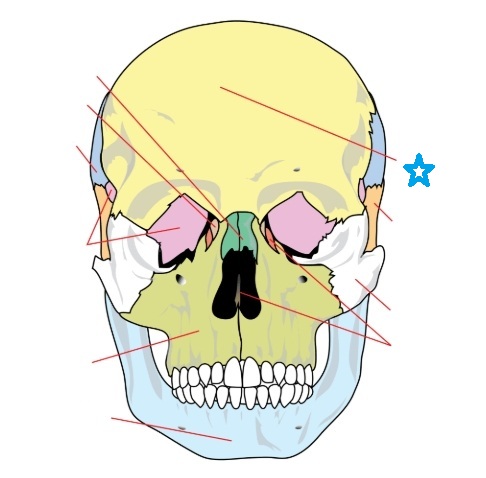
Identify this bone feature labeled by the blue star and define its anatomical purpose.
[SKELETAL]
Frontal bone
Is located on the anterior-superior aspect of the skull. If forms the anterior portion of the cranium and the superior portion of the orbits. It also contains sinuses (frontal sinuses) that secrete mucous to help flush the nasal cavity.
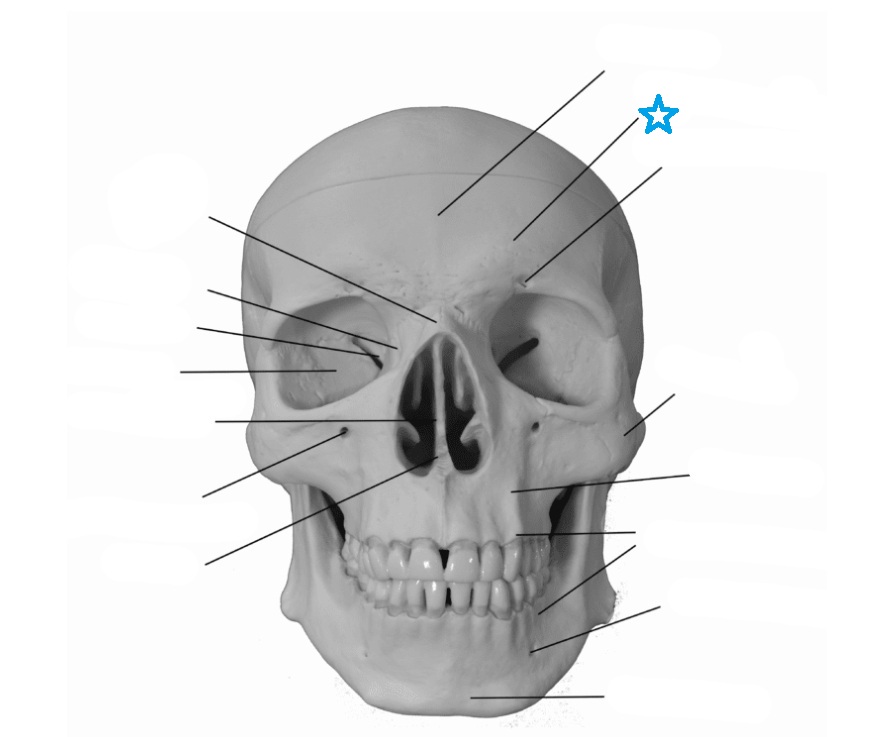
Identify this bone feature labeled by the blue star and define its anatomical purpose.
[SKELETAL]
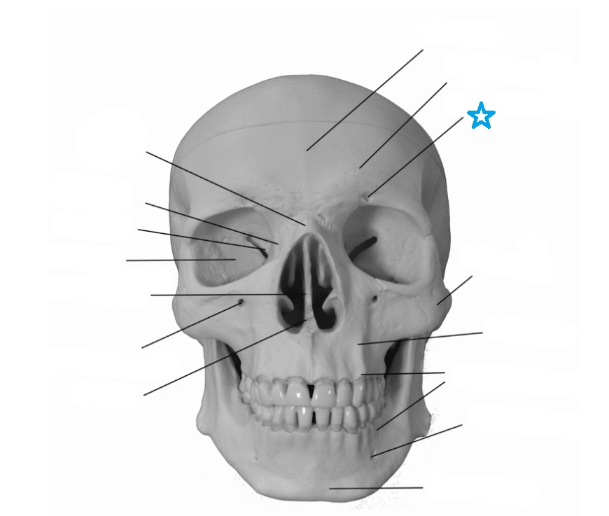
Supraorbital margin
A landmark on the frontal bone that is a thickened process above the orbits that helps to protect the eye.
Identify this bone feature labeled by the blue star and define its anatomical purpose.
[SKELETAL]
Suprorbital foramen
A passageway for blood vessels supplying the frontal sinus, eyebrow, and eyelid.

Identify this bone feature labeled by the blue star and define its anatomical purpose.
[SKELETAL]
Parietal bones
Paired bones that form the lateral margins of the cranium.

Identify this bone feature labeled by the blue star and define its anatomical purpose.
[SKELETAL]
Occipital bone
Located in the posterior region of the skull.

Identify this bone feature labeled by the blue star and define its anatomical purpose.
[SKELETAL]
Foramen magnum
Enclosed within the occipital bone and is a passageway for the spinal cord.
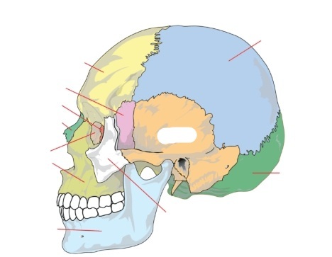
Identify this bone feature covered by the white mark and define its anatomical purpose.
[SKELETAL]
Temporal bones
On the lateral aspect of the skull.

Identify this bone feature labeled by the blue line in the box and define its anatomical purpose.
[SKELETAL]
Zygomatic process
Forms the posterior portion of the zygomatic arch. It articulates with the temporal process of the zygomatic bone.

Identify this bone feature labeled by the blue star and define its anatomical purpose.
[SKELETAL]
Mastoid process
A site of muscle attachments for some of the neck muscles. It also contains small air cavities called air cells that connect with the middle ear.

Identify this bone feature labeled by the blue star and define its anatomical purpose.
[SKELETAL]
External auditory canal
A tubelike structure that houses structures for the external and middle ear.

Identify this bone feature labeled by the blue star and define its anatomical purpose.
[SKELETAL]
Sphenoid bone
Forms part of the inferior portion of the cranium.
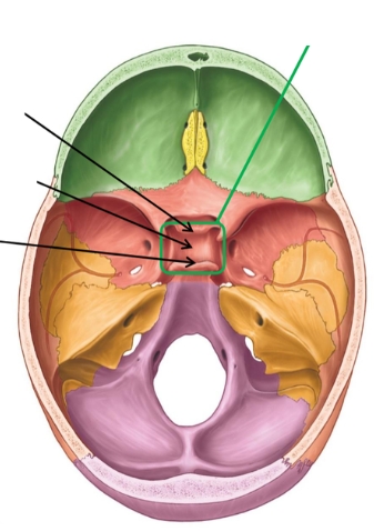
Identify this bone feature labeled by green box and arrows and define its anatomical purpose.
[SKELETAL]
Sella turcica
(Also known as the Turkish saddle) Is a grove in the central region of the sphenoid. The pituitary gland also resides in the sella turcica.
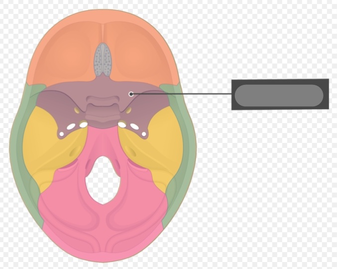
Identify this bone feature labeled by the grey box and define its anatomical purpose.
[SKELETAL]
Lesser wings
Extend laterally and are anterior to the sella turcica.

Identify this bone feature labeled by the grey box and define its anatomical purpose.
[SKELETAL]
Greater wings
Lateral to the sella turcica and form part of the floor of the cranium.

Identify this bone feature labeled by the blue star and define its anatomical purpose.
[SKELETAL]
Ethmoid bone
Located in the anterior and medial cranium. It forms the roof of the nasal cavity and the superior portion of the nasal septum and contains sinuses that secrete mucous to help flush the nasal cavity.

Identify this bone feature labeled by the grey box and define its anatomical purpose.
[SKELETAL]
Crista galli
A ridge of bone that extends superiorly. A portion of the membrane that surrounds the brain called the dura mater attaches to this ridge.

Identify this bone feature labeled by the blue star and define its anatomical purpose.
[SKELETAL]
Perpendicular plate
A ridge of bone extending inferiorly and forming the superior portion of the nasal septum.

Identify this bone feature labeled by the blue star and define its anatomical purpose.
[SKELETAL]
Maxilla
Located and the anterior aspect of the skull. It is superior to the mandible and inferior to the frontal bone. It forms the upper jaw.

Identify this bone feature labeled by the blue star and define its anatomical purpose.
[SKELETAL]
Mandible
Forms the lower jaw.

Identify this bone features labeled by the blue star and define its anatomical purpose.
[SKELETAL]
Zygomatic Bones
Located in the anterior portion of the skull. They connect with the maxilla, frontal and temporal bones and form the cheeks.

Identify this bone feature labeled by the white strip and define its anatomical purpose.
[SKELETAL]
Temporal Process
An extension of bone that connects with the zygomatic process of the temporal bone to form the zygomatic arch.

Identify this bone feature labeled by the blue star and define its anatomical purpose.
[SKELETAL]
Palatine Bone
Forms the hard palate. It connects with the palatine process of the maxilla to form the posterior portion of the hard palate. It is located between the maxilla and sphenoid bones.

Identify this bone feature labeled by the blue star and define its anatomical purpose.
[SKELETAL]
Vomer Bone
Forms the inferior aspect of the nasal septum. It articulates with the ethmoid, sphenoid, palatines and maxillary bones.

In the attached image, curve 1 represents ______ and curve 2 represents ______.
[SKELETAL]
1. Lordosis: Spinal curve with the convexity of the curve anterior.
2. Kyphosis: Spinal curve with the concavity of the curve anterior.
Hyperlordosis: An increased lordotic curve.
Hyperkyphosis: An increased kyphotic curve.

Based on the image of the spine identify the 4 main regions, the vertebrae numbers included in each, and whether their curve is lordosis or kyphosis.
[SKELETAL]
1. Cervical curve: C1-C7 // Curve: Lordosis
2. Thoracic Curve: T1-T12 // Curve: Kyphosis
3. Lumbar Curve: L1-L5 // Curve: Lordosis
4. Pelvic/Sacral Curve: S1-S5 // Curve: Kyphosis
![<p>[SKELETAL]<br><br>1. Cervical curve: C1-C7 // Curve: Lordosis <br><br>2. Thoracic Curve: T1-T12 // Curve: Kyphosis <br><br>3. Lumbar Curve: L1-L5 // Curve: Lordosis<br> <br>4. Pelvic/Sacral Curve: S1-S5 // Curve: Kyphosis </p>](https://assets.knowt.com/user-attachments/4252121a-96e8-4ab7-83ac-6e794b44b50d.jpg)

Identify the numbered structures pointed to by the lines.
[SKELETAL]
Ribs
True rib(s): 1-7
False rib(s): 8-10
Floating rib(s): 11-12
SternumManubrium
Body
Xiphoid
![<p>[SKELETAL]<br><br><strong>Ribs</strong></p><ol><li><p>True rib(s): 1-7</p></li><li><p>False rib(s): 8-10</p></li><li><p>Floating rib(s): 11-12<br><br><strong>Sternum</strong></p></li><li><p>Manubrium</p></li><li><p>Body</p></li><li><p>Xiphoid</p></li></ol><p></p>](https://assets.knowt.com/user-attachments/a1f24acb-65d5-45b7-9169-3cbbdb9fc2f7.webp)

Identify this bone feature labeled by the blue star and define its anatomical purpose.
[SKELETAL]
Glenoid cavity
AKA Glenoid fossa is a concave surface on the lateral aspect of the scapula. It forms the “socket” of the ball and socket joint of the shoulder.

Identify this bone feature labeled by the blue star and define its anatomical purpose.
[SKELETAL]
Spine
Located on the posterior surface of the scapula. It is a ridge of bone extending superiorly from medial to lateral.
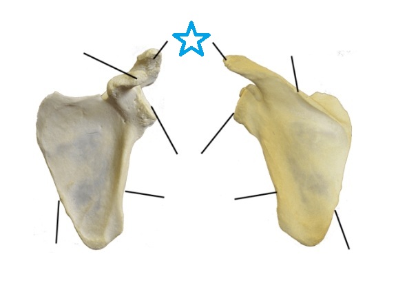
Identify this bone feature labeled by the blue star and define its anatomical purpose.
[SKELETAL]
Acromion process
The terminal end of the spine of the scapula. It is a large process and articulates with the clavicle. The acromion process marks the highest point of the shoulder.

Identify this bone feature labeled by the blue star and define its anatomical purpose.
[SKELETAL]
Coracoid process
Located on the anterior aspect of the scapula. This process is smaller than the acromion process and is located anterior and inferior to it.

Identify this bone labled by the blue star.
[SKELETAL]
Humerus
The proximal bone of the arm. It is a long tubular bone that articulates proximally with the scapula and distally with the radius and ulna.
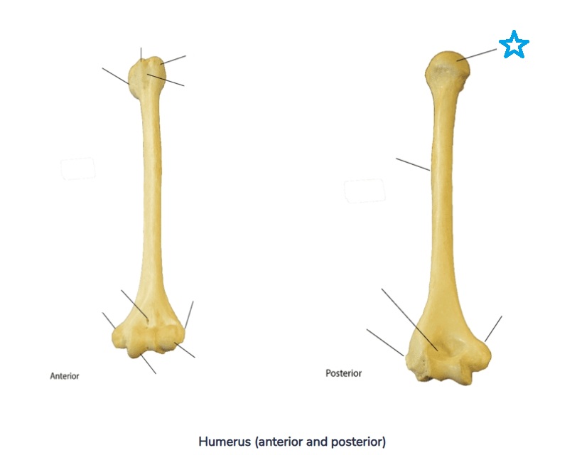
Identify this bone feature labeled by the blue star and define its anatomical purpose.
[SKELETAL]
Head of the humerus
The proximal rounded end of the bone.

Identify this bone feature labeled by the green strips and define its anatomical purpose.
[SKELETAL]
Anatomical neck
Is a small region that marks the end of the joint capsule between the humerus and the scapula.

Identify this bone feature labeled by the green strips and define its anatomical purpose.
[SKELETAL]
Surgical neck
Marks the beginning of the diaphysis.

Identify this bone feature labeled by the blue star and define its anatomical purpose.
[SKELETAL]
Greater tubercle
A rounded process on the lateral aspect of the proximal humerus.

Identify this bone feature labeled by the blue star and define its anatomical purpose.
[Skeletal]
Lesser tubercle
A smaller rounded process on the medial aspect of the proximal humerus.
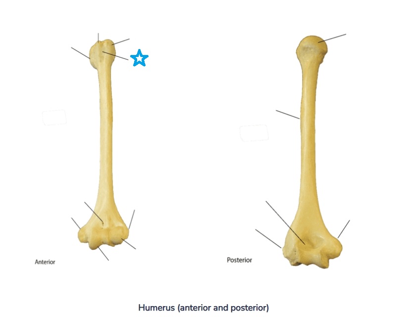
Identify this bone feature labeled by the blue star and define its anatomical purpose.
[SKELETAL]
Intertubercular groove (sulcus)
A groove between the greater and lesser tubercles.

Identify this bone feature labeled by the blue star and define its anatomical purpose.
[SKELETAL]
Deltoid tuberosity
A bump with a gradual slope on the lateral aspect of the humerus and is the site of attachment of the deltoid muscle.

Identify this bone feature labeled by the blue stars and define its anatomical purpose.
[SKELETAL]
Lateral epicondyle
A widened area on the lateral aspect of the distal humerus. It is an important site of muscle attachments for the wrist extensor muscles.
Identify this bone feature labeled by the blue star and define its anatomical purpose.
[SKELETAL]
Medial epicondyle
A widened area on the medial aspect of the distal humerus. It is a site of attachment for wrist flexor muscles.
Identify this bone feature labeled by the blue star and define its anatomical purpose.
[SKELETAL]
Capitulum
A small, rounded process at the distal end of the humerus on the lateral side. It articulates with the radius.
Identify this bone feature labeled by the blue star and define its anatomical purpose.
[SKELETAL]
Trochlea
A small spool shaped process at the distal medial end of the humerus. It articulates with the ulna.
Identify this bone feature labeled by the blue star and define its anatomical purpose.
[SKELETAL]
Olecranon fossa
A small indentation on the posterior distal aspect.
Identify this bone feature labeled by the blue star and define its anatomical purpose.
[SKELETAL]
Coronoid fossa
A small indentation on the anterior distal aspect.
Identify this bone feature labeled by the blue star and define its anatomical purpose.
[SKELETAL]
Olecranon process
A rounded process on the proximal end of the ulna.
Identify this bone feature labeled by the blue star and define its anatomical purpose.
[SKELETAL]
Trochlear notch
The ulna articulates with the trochlea of the humerus.
Identify this bone feature labeled by the blue star and define its anatomical purpose.
[SKELETAL]
Radial notch
A flat spot that articulates with the radius.
Identify this bone feature labeled by the blue star and define its anatomical purpose.
[SKELETAL]
Styloid process
Is of the ulna and is a needle-like process at the distal end.
Identify this bone feature labeled by the blue star and define its anatomical purpose.
[SKELETAL]
Radius
Is located in the forearm. It articulates with the ulna and carpal bones. The radius allows for rotation of the forearm.
Identify this bone feature labeled by the blue star and define its anatomical purpose.
[SKELETAL]
Head of the radius
Articulates with the capitulum of the humerus. This joint can rotate.
Identify this bone feature labeled by the blue star and define its anatomical purpose.
[SKELETAL]
Radial tuberosity
Is a bump on the proximal aspect of the radius. The biceps muscle attaches there.
Identify this bone feature labeled by the blue star and define its anatomical purpose.
[SKELETAL]
Styloid process
A needle-like process on the distal aspect of the radius.
Identify this bone feature labeled by the blue star and define its anatomical purpose.
[SKELETAL]
Carpal Bones
Carpal bones, a group of eight short bones in the human hand that form the wrist joint.
Identify this bone feature labeled by the blue star and define its anatomical purpose.
[SKELETAL]
Metacarpal and Phalanges
The metacarpals are the five long bones of the palm connecting the wrist to the fingers, while the phalanges are the bones of the fingers, providing structure and enabling movement.
Identify this bone feature labeled by the blue star and define its anatomical purpose.
[SKELETAL]
Femur bone
The upper leg bone and is the longest bone in the body.
Identify this bone feature labeled by the blue star and define its anatomical purpose.
[SKELETAL]
Head of the femur
A rounded process on the proximal end.
Identify this bone feature labeled by the blue star and define its anatomical purpose.
[SKELETAL]
Neck of the femur
The area that connects the head with the shaft.
Identify this bone feature labeled by the blue star and define its anatomical purpose.
[SKELETAL]
Greater trochanter
A large process located on the proximal lateral aspect of the femur.
Identify this bone feature labeled by the blue star and define its anatomical purpose.
[SKELETAL]
Lesser trochanter
A smaller process located on the proximal medial aspect.
Identify this bone feature labeled by the blue star and define its anatomical purpose.
[SKELETAL]
Medial and lateral condyles
Two large, rounded processes on the distal aspect of the femur
Identify this bone feature labeled by the blue star and define its anatomical purpose.
[SKELETAL]
Tibia
The larger of 2 bones of the lower leg. It articulates with the femur, fibula, patella and talus bones.
Identify this bone feature labeled by the blue star and define its anatomical purpose.
[SKELETAL]
Tibial condyles
Large, rounded processes on the proximal aspect of the tibia. The 2 condyles are named medial and lateral.
Identify this bone feature labeled by the blue star and define its anatomical purpose.
[SKELETAL]
Tibial tuberosity
A broad bump on the anterior aspect of the tibia.
Identify this bone feature labeled by the blue star and define its anatomical purpose.
[SKELETAL]
Medial malleolus
A rounded process on the distal medial aspect of the tibia. It is the bump on the inside of the ankle.
Identify this bone feature labeled by the blue star and define its anatomical purpose.
[SKELETAL]
Fibula
Lateral bone in the lower leg. It forms the lateral ankle and articulates with the tibia and talus bones.
Identify this bone feature labeled by the blue star and define its anatomical purpose.
[SKELETAL]
Fibular head
A rounded process on the proximal end of the bone.
Identify this bone feature labeled by the blue star and define its anatomical purpose.
[SKELETAL]
Lateral malleolus
A rounded process on the distal end of the bone. It forms the lateral ankle.
Identify this bone feature labeled by the blue star and define its anatomical purpose.
[SKELETAL]
Patella
Commonly known as the knee bone.
Identify this bone feature labeled by the blue star and define its anatomical purpose.
[SKELETAL]
Tarsals
A group of seven irregularly shaped bones in the foot that form the ankle and midfoot, providing support, stability, and enabling movement.
Identify this bone feature labeled by the blue star and define its anatomical purpose.
[SKELETAL]
Cartilagenous joints
Bones united by cartilage.
Examples include the intervertebral discs and the symphysis pubis.
Identify this bone feature labeled by the blue star and define its anatomical purpose.
[SKELETAL]
Synovial joints
Complex joints that consist of a number of parts. Synovial joints are freely movable and encapsulated by a synovial membrane.
Identify this bone feature labeled by the blue star and define its anatomical purpose.
[SKELETAL]
There are a number of types of synovial joints named for their shape:
Ball and socket joints consist of a rounded process and rounded socket. These include the hip and shoulder and allow for a variety of movements.
Hinge joints consist of a convex surface and concave socket. Examples include the joint between the humerus and ulna as well as in some of the phalanges. Hinge joints only move in one plane.
Condyloid joints consist of oval processes fitting into elliptical sockets. An example of this joint is the metacarpal phalangeal joint.
Gliding joints consist of flattened surfaces connected together. Examples include the carpal bones of the wrist.
Pivot joints consist of a cylinder fitting into a ring of bone. Examples include the joint between the atlas and axis of the spine and the joint between the radius and humerus.
Saddle joints consist of two bones having both concave and convex surfaces. An example is the carpal-metacarpal joint of the hand.
Identify this bone feature labeled by the blue star and define its anatomical purpose.
[SKELETAL]
Joints move according to their shapes.
The terms associated with movements include:
Flexion which consists of the humerus moving anterior in a sagittal plane.
Extension consists of the humerus moving posterior in a sagittal plane.
Abduction is the movement of the humerus away from the body in a coronal plane.
Adduction is moving the humerus toward the body in a coronal plane.
Internal rotation is moving the humerus along its long axis toward the body.
External rotation is moving the humerus along its long axis away from the body.
Inversion is the movement of the foot so the sole of the foot points medially.
Eversion is the movement of the foot so the sole of the foot points laterally.