RS 2 - Nose, Pharynx and Larynx
 Call Kai
 Learn
 Practice Test
 Spaced Repetition
 Match
 Flashcards
 Knowt Play
1/59

There's no tags or description
Looks like no tags are added yet.
60 Terms
External Landmarks of the nsoe
Root: The top bridge of the nose right between your eyebrows.
Dorsum: The length of the ridge running down the front of the nose.
Apex: The pointy tip of your nose.
Ala: The rounded, flaring outer "wings" on the sides of your nostrils.
Nares (nostrils): The two openings where air enters.
Nasal Septum: The dividing wall right down the middle that splits your nose into left and right sides.

Functions of the Nose
• Olfaction
• Respiration
• Filtration of particles
• Humidification of air
• Elimination of secretions from nasal mucosa, paranasal sinuses & nasolacrimal ducts
which bones in the neurocranium
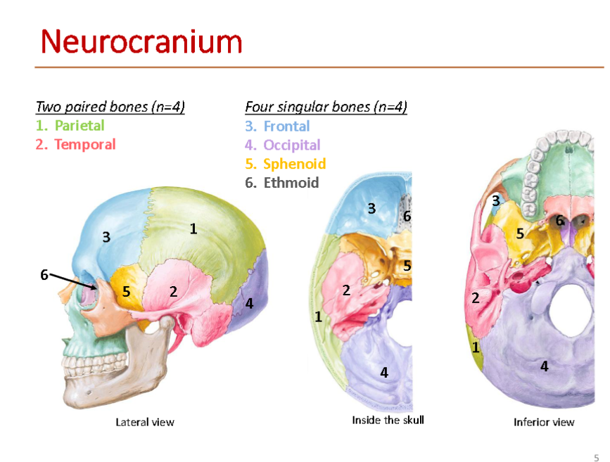
which bones in the Viscerocranium

cartilage that makes up the external nose
Lateral Cartilage (2)
Septal Cartilage
Alar Cartilage (2)
Bones that make up the external nose
Nasal Part of Frontal Bone
Nasal Bones
Frontal Process of Maxilla
function of Nasal Septum
which is the central dividing wall splitting your nasal cavity into left and right hallways.
The 3 Main Pieces of the Septum (The Dividing Wall
Septal cartilage (Grey): The flexible front part of the wall that lets the tip of your nose wiggle.
Ethmoid bone — perpendicular plate (Purple): The upper bony part of the wall coming down from the roof.
Vomer (Green): A thin, plow-shaped bone forming the lower-back part of the wall.
what bones make up the (Roof & Floor)
Roof: Framed from front to back by the nasal bone, frontal bone, ethmoid bone (cribriform plate) (which has tiny holes for smell nerves), and sphenoid bone.
Floor: Framed by the maxilla (front of your hard palate) and palatine bone (back of your hard palate).

Bones of the Nasal Cavity — Lateral Wall
Superior & Middle Nasal Conchae (Purple): Both of these top two shelves are actually parts of the ethmoid bone.
Inferior Nasal Concha (Tan): This bottom shelf is its own separate, independent facial bone (a favorite anatomy exam trick!).
Surrounding Wall Bones: The rest of the outer wall is pieced together by the nasal bone and frontal bone at the top-front, the maxilla in the middle-front, and the palatine bone and sphenoid bone at the back.

Nasal Cavity front entrance structures
Nasal Atrium
Nasal Vestibule
Nare
what conchae are part of nasal cavity
1. Superior Nasal Concha \(\rightarrow \) sits right above the Superior Nasal Meatus.
2. Middle Nasal Concha \(\rightarrow \) sits right above the Middle Nasal Meatus.
3. Inferior Nasal Concha \(\rightarrow \) sits right above the Inferior Nasal Meatus.
what are Areas of the Nasal Cavity
Olfactory Area - Superior 1/3 of nasal mucosa, function is for smell
Respiratory Area - Inferior 2/3 of nasal mucosa, function are warms and moistens air
Arterial blood supply for the nasal cavity derives from the
i. External carotid artery – maxillary a.
ii. Internal carotid artery – ophthalmic a
what is the Kiesselbach's Area
The Nosebleed Spot)
Because so many arteries meet right under the thin lining here—right where dry air hits first or a finger can reach—this is the most common spot where nosebleeds start.
Location: Found on the anteroinferior (front-bottom) part of the nasal septum (circled in green).
what does Anastomosis mean
ust a fancy word for a junction where multiple blood vessels meet and connect together.
General Sensory Innervation
Ophthalmic Branch of Trigeminal Nerve (CN V1)
(anterosuperior part of lateral wall & nasal septum)
Maxillary Branch of Trigeminal Nerve (CN V2)
(posteroinferior lateral wall & nasal septum)
Special Sensory Innervation and locattion
Olfaction function is to mediate sense of smell, location is olfactory muscosa the rest is respiratory

How a Smell Signal Travels to Your Brain
Receptors in olfactory epithelium ➔ Olfactory Nerves (pass through cribriform plate)
➔ Synapse on olfactory bulb ➔ axons of olfactory bulb form olfactory tract (conveys impulses to brain)
what is Anosmia
loss of olfaction
Often associated with loss/change in taste
what are common causes of anosmia
• Increasing age
• Excessive smoking
• Cocaine use
• Inhalation of noxious chemical
• Fracture of cribriform plate
• Sinusitis/Upper respiratory infections
what are Paranasal Sinuses and their functions
Air-filled, mucosa-lined spaces in bones of the skull
extensions of the respiratory part of the nasal cavity
• Voice resonation
• Warm and humidify air
• Decrease skull weight
what bones make up the paranasal sinuses
frontal
ethmoid
sphenoid
maxillary

where does Sphenoid Sinus drain?
Behind Superior Concha (spheno-ethmoidal recess)
where does Posterior ethmoidal cells drain
Superior Meatus
where does Frontal Sinus drain
Middle Meatus
where does Nasolacrimal duct drain
Inferior Meatus
where does Maxillary Sinus drain
middle meatus
where does Anterior & Middle ethmoidal cells drain
middle meatus
where does Sphenoid Sinus drain
Behind Superior Concha
(spheno-ethmoidal recess)
what is Sinusitis
➢ Inflammation of paranasal sinuses typical due to infections
➢ Maxillary sinus is most commonly infected due to the superior location of the maxillary ostium
Symptom of sinusitis
• Nasal congestion
• Facial pain
• Headache
• Toothache
• Fever
Treatments of sinusitis
• NSAIDs
• Saline nasal spray
• Neti pot
what is the Pharynx
Muscular tube connects
nasal cavity
oral cavity
larynx
esophagus
conduit for both digestive and respiratory tract
what is the Larynx
• Cartilaginous box suspended by hyoid bone
• Continuous with trachea
• Conduit for only respiratory tract
what are parts of the Pharynx
nasopharynx
oropharynx
laryngopharynx

where does Pharynx connect to on the vertebra
Muscular tube extending from cranial base to C6 vertebra
nasopharynx location and function
Posterior to nose & superior to soft palate
• Respiratory function
Oropharynx location and function
• Posterior to mouth; soft palate to superior border of epiglottis
• Digestive function
laryngopharynx location and function
Posterior to the larynx; superior border of epiglottis to cricoid
cartilage & pharyngo- esophageal junction
what group of Pharyngeal Muscles are their
Constrictor muscles: 3 groups
Internal Longitudinal muscles
what muscles make up the Constrictor muscles
• Superior pharyngeal constrictor
• Middle pharyngeal constrictor
• Inferior pharyngeal constrictor
• Constrict wall of pharynx during swallowing

whats the function of the Internal Longitudinal muscles
• 3 muscles
• Elevate and shorten the pharynx
during swallowing and speaking
how does Swallowing (Deglutition) happen

what provides General sensory to the nasopharynx
Maxillary division of Trigeminal nerve (CN V2)
what provides general sensory to the Oropharynx
Glossopharyngeal nerve (CN IX)
what provides general sensory to he Laryngopharynx
Vagus nerve (CN X)
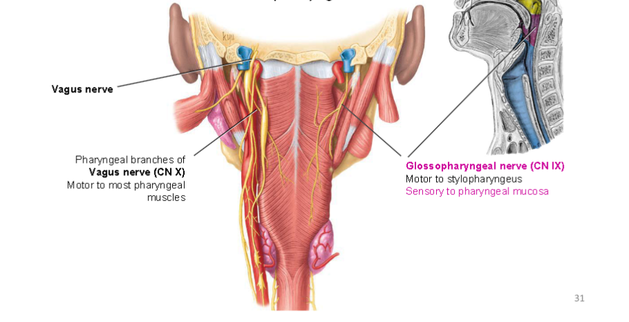
what provides Innervation to the Pharyngeal Muscles
Vagus n. (CN X)
• Motor to most pharyngeal muscles
via Pharyngeal branchesGlossopharyngeal n. (CN IX).
• Motor to the Stylopharyngeus muscle
how does Gag Reflex happen
➢ CN IX → Sensory irritation of pharyngeal mucosa
➢ CNS: medulla
➢ CN X → Motor innervation of pharyngeal muscles
where is Larynx located
➢ Located in anterior neck at level of C3-C6 vertebral bodies
➢ Connects the oropharynx to trachea
Larynx function
➢ Organ of voice production “Voice Box”
➢ Maintains airway, guards air lower respiratory tract, especially during swallowing
how many Laryngeal Cartilages does laryncs have
Consists of 9 cartilages: 3 Unpaired & 3 Paired
which are paied and unpaired
Paired
Corniculate cartilage
Arytenoid cartilage
Cuneiform cartilage*
Unpaired
Epiglottis
Thyroid cartilage
Cricoid cartilage
what are Laryngeal Folds
Inside your voice box (larynx), there are two pairs of horizontal tissue folds stacked on top of each other like double doors.
Vestibular fold
Vocal fold
what are vestibular folds
(false vocal cord)
• Does not contribute to sound production
• Protection of vocal fold
what are Vocal folds
(true vocal cord)
• Contains vocal ligament and vocalis muscle
• Intrinsic laryngeal muscles change position
and tension of vocal folds to produce sounds (phonation)
Laryngeal Fold Movement

Innervation of the Larynx
