Lower Limb Points for Palpation
 Call Kai
 Learn
 Practice Test
 Spaced Repetition
 Match
 Flashcards
 Knowt Play
1/29

Earn XP
Description and Tags
Points for palpation at the Hip, Knee and Toes
30 Terms
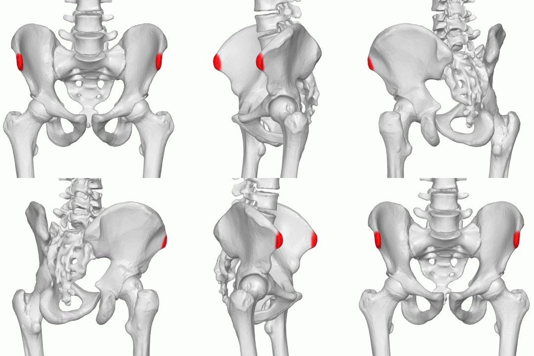
ASIS — Anterior superior iliac spine
Palpate the iliac crest and follow it anteriorly until you reach the prominent bony point at the front of the pelvis.

PSIS — Posterior superior iliac spine
Palpate the posterior iliac crest and follow it towards the midline to the bony prominence
Find the dimples in the lower back, just above the buttocks; the bony points are usually nearby.

Iliac crest
Trace the superior border of the ilium from the ASIS anteriorly to the PSIS posteriorly.

Greater trochanter
Palpate the lateral hip bony prominence on the outer hip while gently internally and externally rotating the femur.

Ischial tuberosity (Sitting Bone)
Position the patient appropriately and palpate the inferior buttock region to identify the ischial tuberosity.
The “sitting bone” — a bony point beneath the lower buttock.

Pubic tubercle
Locate the bony point just lateral to the pubic symphysis. Obtain explicit consent
A small bony point at the front of the pelvis, just beside the pubic symphysis.
Hip joint line
The hip joint line lies approximately 1 to 2 cm above the greater trochanter and can be palpated or estimated by identifying the anterior superior iliac spine (ASIS) and the pubic tubercle. The joint line approximately lies approximately 1.5cm inferior.

Femoral triangle
Borders:
Superior (Base): Inguinal ligament — stretches from the anterior superior iliac spine (ASIS) to the pubic tubercle.
Lateral: Sartorius muscle (medial border of the muscle).
Medial: Adductor longus muscle (medial border of the muscle).
Floor (from lateral to medial): Iliopsoas muscle -> Pectineus muscle
Note: highlight NAV. From lateral to medial -> nerve, artery, vein

Patella
Palpate the superior, inferior, medial and lateral borders of the patella with the knee relaxed.

Trochlear groove
Locate the groove between the femoral condyles, just proximal to the patella.
Feel the groove at the front of the femur, just above the kneecap. Make a V valley shape with each thumb the point of the V should be in groove

Medial femoral epicondyle
Palpate the bony prominence on the (Inner) medial distal femur (superior to the medial femoral condyle)
Remember
Condyles sit at or below the joint line: To feel a condyle, you must place your fingers directly on the seam where two bones meet (the joint space). •
Epicondyles sit above or outside the joint line: Epicondyles always sit further up the bone shaft, completely clear of the moving joint space.

Lateral femoral epicondyle
Palpate the bony prominence on the lateral distal femur)(superior to the lateral femoral condyle)

Adductor tubercle
Palpate the medial distal femur superior to the medial epicondyle to identify the adductor tubercle.
To palpate the adductor tubercle come down the fleshy part of the thigh on the medial aspect and the first bony point you come to will be the adductor tubercle or 2cm away from Medial epicondyle

Tibial plateau
Palpate the proximal tibial surface beneath the femoral condyles, on either side of the patellar tendon and joint line.
Feel the top of the shinbone, immediately below the knee joint line.

Tibial tuberosity
Trace the patellar tendon downwards from the inferior patella to its attachment on the anterior tibial tuberosity.
Feel the bony bump at the front of the shin, a few centimetres below the kneecap.

Head of fibula
Feel for the bony bump on the outer side of the upper shin, just below the knee
Palpate lateral side of leg / calf until hit first bony landmark

Medial condyle of the tibia
Palpate the broad medial prominence of the proximal tibia directly inferior to the medial femoral condyle.

Lateral condyle of the tibia
Palpate the lateral proximal tibia beneath the lateral femoral condyle, at the lateral tibial plateau.
Superior + medial to fibula head
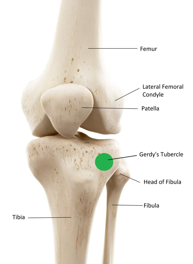
Gerdy’s tubercle
Palpate the anterolateral proximal tibia, just below the lateral joint line, where the iliotibial band inserts.


Collateral ligaments
MCL: Trace from the medial femoral epicondyle towards the medial proximal tibia.
LCL: Trace from the lateral femoral epicondyle towards the head of the fibula.

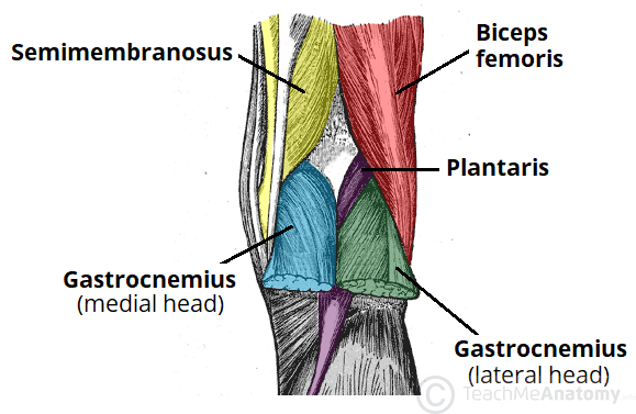
Popliteal fossa
Boundaries
Superior:
Medial - Semimembranosus and semitendinosus muscles
Lateral - Biceps femoris muscle
Inferior:
Medial - Medial head of gastrocnemius muscle
Lateral - Lateral head of gastrocnemius muscle

Ankle (Talocrural) joint line
Palpate horizontally across the anterior ankle joint, between the medial and lateral malleoli, following the joint line.
Feel across the front of the ankle, between the two ankle bones (malleoli).

Head of the Talus
Identify the tips of both malleoli.
Place thumb on the medial malleolus and index finger on the lateral malleolus.
Move anteriorly until you feel a dip behind the tendons.
Palpate the head of the talus.
Gently plantarflex and dorsiflex the foot to feel the talus move.
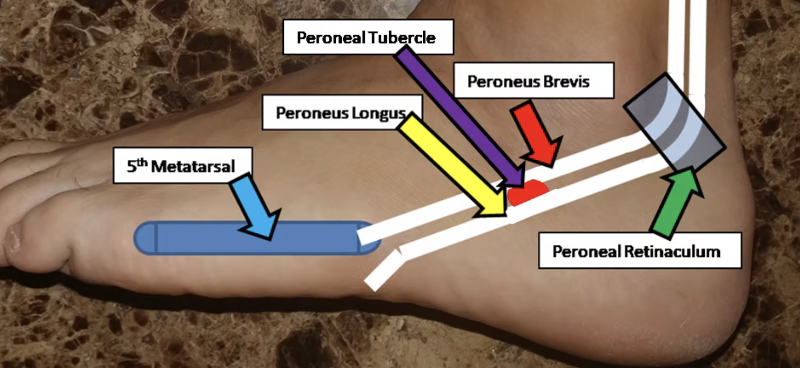
Peroneal Tubercle
Palpate the lateral calcaneus, anterior and inferior to the lateral malleolus, between the peroneal tendons.
Feel the outer side of the heel bone, just below and in front of the outer ankle bone.
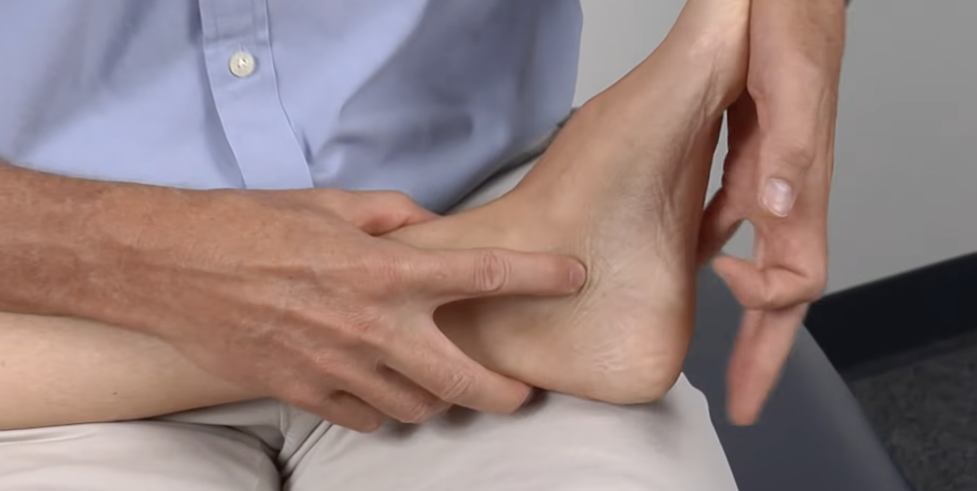
Sustentaculum Tali
Position the foot in neutral. (In image)
Palpate straight inferiorly from the medial malleolus.
Feel for the firm bony structure.
Gently invert and evert the foot to help confirm the landmark.
Continue slightly inferiorly to appreciate where the bone ends and the softer heel tissue begins.
Memory tip: Medial malleolus → straight down → last hard bony point = sustentaculum tali.
Spring Ligament
The spring ligament connects the Sustentaculum tali to the plantar surface of the navicular bone
and is located on the medial aspect of the foot, on the plantar (under) side


Tuberosity of the Navicular
Palpate the medial prominence of the navicular, anterior and inferior to the talar head.
From the sustentaculum tali, move anteriorly along the medial side of the foot until you feel the prominent navicular tuberosity.

First Metatarsal + Medial Cuneiform
First metatarsal: Trace backwards from the big toe to the first metatarsal shaft and base.
Medial cuneiform: Palpate just proximal to the base of the first metatarsal, between it and the navicular.

Cuboid Bone
Start at the base of the fifth metatarsal and move posteriorly towards the calcaneus until you feel a dip, then locate the firm bony prominence just beyond it — the cuboid.
On the outer side of the midfoot, between the heel bone and the bases of the fourth and fifth metatarsals.

Base of Fifth Metatarsal
The prominent bump on the outer edge of the foot, near the little toe side.
Trace backwards from the little toe along the fifth metatarsal until you feel its prominent base.

Metatarsal Heads (Plantar Surface)
Palpate the sole just behind the toes, then gently flex and extend the toes while keeping your fingers over the area; you should feel the metatarsal heads move beneath your fingers.
You can also trace each metatarsal forwards towards its head to confirm you are on the correct bone.