Mod 7 Skeletal and Smooth Muscle Histology
 Call Kai
 Learn
 Practice Test
 Spaced Repetition
 Match
 Flashcards
 Knowt Play
1/208

Earn XP
Description and Tags
Objective: 1. Identify and describe the cellular characteristics and the mechanism of contraction in the 3 muscle types. 2. Identify muscle microanatomy in microscopic images. 3. Describe the organization of contractile proteins in a sarcomere and the sliding filament theory. 4. Compare and contrast between the 3 types of muscles
209 Terms
What are the 4 types of tissue?
epithelial, connective, muscle, nerve
What are the 3 types of muscle tissues?
skeletal, cardiac, smooth

Each microfilament strand is assembled from bead-like subunits, called?
G-actin (G = globular)
What type of muscle is the tongue?
skeletal
What is the G-acting polymerized into?
F-actin (F = fibrous)
What is an F-actin?
two strands wrap around each other to form the microfilament
Is skeletal muscle voluntary or involuntary?
voluntary
What is the function of skeletal muscle?
locomotion/movement
Is cardiac muscle voluntary or involuntary?
involuntary
Is smooth muscle voluntary or involuntary?
involuntary
Where are smooth muscles?
tube like hollow organs (artery, GI tract, reproductive tract)
Why does the GI tract and fallopian tubes have partial contractions?
conduct peristalsis (smooth muscle push contents out)
What are microfilaments made up of?
actin
Are these all the same (microfilament = actin filament = F-actin)
yes

What does F-actin interact with to move on contractions?
myosin filaments
What do myosin and actin form?
the contractile ring
How do all kinds of muscle contractions occur?
sliding of actin filaments over myosin filaments

What does muscle have a lot of?
smooth ER

What do muscle cells not have?
any ribosomes on their surface
What is the function of smooth ER?
storage and release of calcium in skeletal and cardiac muscle
What does calcium do in the muscle?
activate contractile mechanisms
What is the smooth ER called in muscle?
sarcoplasmic reticulum
What is the cytoplasm of a muscle called?
sarcoplasm
What is the plasma membrane of a muscle called?
sarcolemma
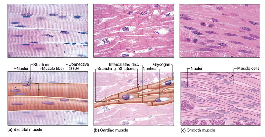
What is skeletal muscle that makes it show very evident striations?
multinuclei
What is the contraction like for skeletal muscle?
quick and strong
Which muscles has cells that are branched?
cardiac
What does it mean for cardiac cells to have branches?
one cell will have nucleus that is often surrounded by glycogen inclusion bodies
How does cardiac muscle use glycogen?
for energy

What does the connections between the cardiac muscle cells look like?
small bands
What are the small bands in cardiac muscle called?
intercalated discs
What are smooth muscles contractions like?
not very strong involuntary contrations
What is the shape of smooth muscle cells?
fusiform (football/spindle) and lack striations
How are smooth muscle cells arranged?
staggered
how is skeletal muscle cells arranged?
rows
Where does skeletal muscle come from?
embryonic mesenchymal cells
What do muscle fibers look like?
long multinucleated
What are the skeletal cells that differentiate (specialize) called?
myoblasts
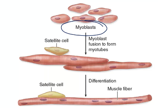
What is the process of the contractile proteins, actin and myosin forming?
embryonic mesenchymal cells → myoblasts → multinucleated tube-like structure → striated skeletal muscle fiber → forms contractile proteins myosin and actin
What makes skeletal muscle?
myoblast
What are the myoblasts that stay dormient (reserves) called?
satellite cells
Where can you find satellite cells?
attached to muscle fiber on the outside of the sarcolemma
What is the function of satellite cells?
muscle repair after damage by undergoing cell division and making new fibers when needed
What is the outer most layer (a dense connective tissue) that the muscle is covered in called?
epimysium

Where is epimysium?
under deep fascia
What does epimysium do?
connect bone to muscle
What is the epimysium layer made of?
fascicles

What is a fascicle covered by?
perimysium
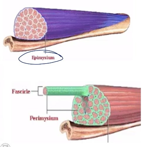
What is perimysium
thin dense connective tissue
Which is thinner, epimysium or perimysium?
perimysium

What is inside the fascicles?
numerous skeletal muscle fibers/skeletal muscle cells
What is each skeletal muscle fiber covered in?
endomysium

What are both mysium laters containing?
collagen 1 and 3 and fibroblasts
What supplies the epimysium?
large nerves, blood, and lymph vessels
What supplies the endomysium?
small blood vessels and nerve
What supplies the perimysium?
nerves, blood vessels, and lymph vessels

What is the order of layers from outside in?
Epimysium
Skeletal muscle
Perimysium
Fascicle
Endomysium
Muscle fiber/ cell
When shown a skeletal muscle transverse section, what should you see?
perimysium, endomysium, and epimysium

What is the “P” in this picture
perimysium

What is the En in the picture?
endomysium

How many muscle cells - muscle fibers?
3
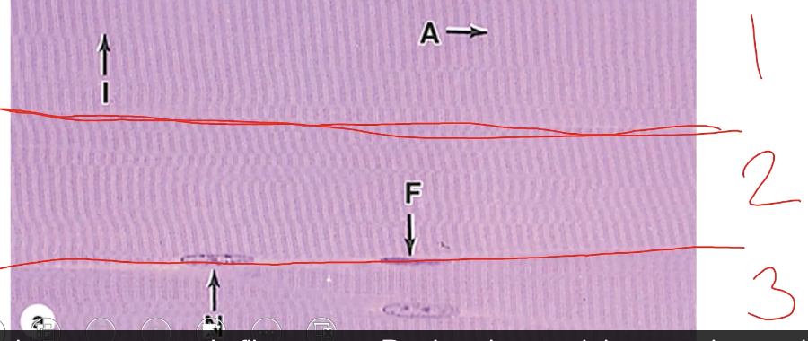
What do the light nucleus mean?
leptochromatic = highly active

What has become clear in this image?
striations (I and A bands)

What do you find when you cut through a muscle cell?
myofibrils

What is the I band consist of?
actin

What does the A band consist of?
mostly myosin (plus actin)

What is the middle lighter zone in the A band called?
H zone
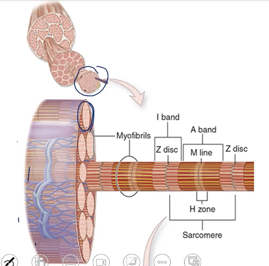
What is the H zone divided by?
M line
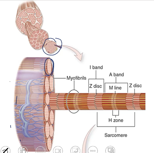
Draw out the structure of myofibril

explain the closer look at the lighter striation.
light bands are I bands and divided in the middle by a darker line called Z disc
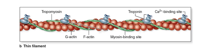
What are the two other proteins actin is always associated with?
tropomyosin and troponin
What is actin made of?
G-actin
What does G-actin form once polymerized
F-actin
What does every G-actin monomer have?
myosin binding site
What is the myosin binding site of the action monomer covered by
tropomyosin (when muscle is not activated)
What is the role of tropomyosin?
cover and expose binding site
What makes up the thin filament?
F-actin + tropomyosin + troponin
What does troponin bind to?
tropomyosin
How many subunits does troponin have?
3 (TnT, TnI, TnC)
What are the subunits of troponin?
TnT, TnC, and TnI
What does TnT do
bind to tropomyosin
What does TnC do
bind to Ca+
What does TnI do
mediates the interaction between actin and myosin

What is another name for myosin?
thick filament

What is myosin made of?
two tails and two globular heads
Where do the myosin heads face?
outward
What does it mean to say myosin is a motor protein?
uses ATP to do its function
What binding sites does myosin have?
actin binding site and ATP binding site

Sarcomere
z to z disc
What is thick filament called?
myosin filament
What is thin filament called?
actin filament
Where are the actin chains attached?
at Z disc
Where do the myosin bundles span?
entire A band
What forms the I band?
the part where actin doesn’t overlap myosin
What forms the H zone?
the part where myosin doesn’t overlap actin

sacromere in TEM images

muscle breakdown
How many subunits do tropinin have and what are they?
3 (TnT, TnI, TnC)
What is this process called: A series of binding and unbinding events of the myosin heads to the F-actin causes the sliding of Myosin bundles over the actin fibers, causing contraction of the sarcomere. It looks like the myosin heads are walking over the actin fibers.
sliding filament theory.
What is the t-tubule and terminal cisterna together called?
triad