medial epicondyl fx / elbow dx
 Call Kai
 Learn
 Practice Test
 Spaced Repetition
 Match
 Flashcards
 Knowt Play
1/28

There's no tags or description
Looks like no tags are added yet.
29 Terms
اطفال
2 مرکز استخوانیشدن مدیال اپیکندیل چه زمانی ظاهر و چه زمانی فیوز میشود؟
ظهور حدود ۴–۶ سالگی؛ فیوژن حدود ۱۵ سالگی.
چرا Fat-pad sign ممکن است منفی باشد؟
شکستگی مدیال اپیکندیل غالباً خارج مفصلی است؛ بنابراین حتی با وجود شکستگی، ممکن است افیوژن و جابهجایی fat pad دیده نشود.


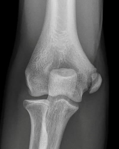
Direct fragmentation.
The fragmented appearance of the medial epicondyle (arrows) in a 13-year-old who sustained a direct blow to the medial aspect of the elbow.
Muscle avulsion

AP radiograph shows loss of normal smooth margins of the physis.
Displaced medial epicondylar fracture


Dislocation with incarcerati

Soft tissue attachments


الگوریتم درمانی مدیال اپی کندیل

Heterotopic calcification

Anteroposterior view of the elbow illustrates the bone and ligamentous structures which contribute to elbow stability.
(A, lateral collateral ligament; B, annular ligament; C, medial collateral ligament
The annular ligament and lateral collateral ligament complex provides stability to the proximal radioulnar joint and radial capitellar articulation.


Reduction by “pusher” techniques
Reduction by “puller” techniques in a supine position.


Median nerve entrapment.
A: Type 1. Entrapment within the elbow joint with the median nerve coursing posterior to the distal humerus. B: Type 2. Entrapment of the nerve between the fracture surfaces of the medial epicondyle and the medial condyle. C: Type 3. Simple kinking of the nerve into the anterior portion of the elbow joint.

Radioulnar synostosis.

Posterolateral rotary instability.
Posterolateral rotational instability is best demonstrated with the upper extremity over the head with the patient supine. The radial head can be subluxated or dislocated by applying a valgus and supination force to the forearm at the same time proximal axial compression is applied along the forearm.

Pathology associated with recurrent elbow dislocations.
The three components that allow the elbow to dislocate: a lax ulnar collateral ligament, a “pocket” in the radial collateral ligament, and a defect in the lateral condyle.

Mediolateral divergent dislocation.
۵. درمان غیرجراحی (Nonoperative Treatment)
اندیکاسیونها
شکستگی بدون جابهجایی یا با جابهجایی کمتر از ۲ میلیمتر.
بیمار با فعالیت کم تا متوسط.
نبودن incarceration، نقص عصبی قابلتوجه یا ناپایداری مهم.
روش درمان
اسپلینت یا گچ برای راحتی و محافظت؛ معمولاً ۲–۳ هفته در روشهای متداول.
در الگوریتم مورد ترجیح نویسندگان این فصل، بیحرکتی کوتاهتر، حدود ۷–۱۴ روز است.
سپس شروع زودهنگام ROM فعال.
در صورت نیاز، hinged brace برای محدودکردن فشار والگوس و اجازه فلکشن–اکستنشن کنترلشده.
در صورت محدودیت حرکت، فیزیوتراپی با تأکید بر کاهش درد و تورم و ب


افتراق درفتکی ارنج از شکستگی سوپرا کندیل
۱۶. جمعبندی یکدقیقهای
اگر در آزمون یک کودک یا نوجوان با posterior elbow dislocation دیدید، این ترتیب را به خاطر بسپارید:
قبل از reduction: معاینه عصبیعروقی، بهخصوص ulnar nerve و وضعیت شریان براکیال.
Reduction فوری: با sedation کافی؛ ترجیحاً بدون hyperextension و با اصلاح جابهجایی جانبی.
بعد از reduction: معاینه مجدد، رادیوگرافی و بررسی medial epicondyle، قطعات داخل مفصل و concentricity.
.
مفصل پایدار و هممرکز: درمان غیرجراحی و حرکت محافظتشده زودهنگام.
مفصل ناپایدار یا reduction غیرهممرکز: جستوجوی شکستگی یا بافت گیر افتاده و بررسی اندیکاسیون جراحی.
نقص جدید median nerve یا اختلال پایدار خونرسانی: احتمال entrapment یا آسیب جدی؛ ارزیابی فوری.
پیشگیری از stiffness: بیحرکتسازی کوتاه و متناسب با پایداری و آسیبهای همراه

Pulled elbow
در موارد عود مکرر که درد یا محدودیت فعالیت قابلتوجه ایجاد میکنند، گچ بالای آرنج با ساعد در سوپیناسیون یا وضعیت خنثی میتواند مؤثر باشد.
آیا جراحی لازم است؟
تقریباً هیچوقت.

Instability (PLRI)
یکی از مهمترین نکات مفهومی این قسمت است.
مکانیسم اصلی، نارسایی ساختارهای خارجی آرنج، بهویژه بخش اولنار Radial Collateral Ligament complex / Lateral Collateral Ligament Complex است. در کودکان و نوجوانان، عدم جوشخوردن غضروفی محل مبدأ RCL نیز میتواند عامل باشد.
تابلوی بالینی
احساس خالیکردن یا ترس از دررفتگی در بعضی فعالیتها
سابقه سابلوکساسیون یا دررفتگی گذرا
گاهی Contracture
معاینه معمولی ممکن است طبیعی باشد.