BIO230 2nd half
 Call Kai
 Learn
 Practice Test
 Spaced Repetition
 Match
 Flashcards
 Knowt Play
1/223

There's no tags or description
Looks like no tags are added yet.
224 Terms
Big questions — cancer

Cell polarization is essential for multicellular organisms
Polar = different at either end
apical domain (top)
basolateral domain (bottom and sides)
Polarized cells can:
have different functions at different cell regions
define inside vs outside
transmit signals from one end to the other

How does membrane trafficking send different proteins to different domains?
exocytosis directly to target domain
exocytosis to ANY domain then endocytosis followed by recycling to target domain

Membrane trafficking determines where proteins end up
some trafficking routes are polarized
protein are organized at sorting stations
different routes are balanced by retreival pathway

Constitutive secretion is the “default pathway”
most cargo move through constitutive secretion
no specific signal needed (ER still needed)
cell membrane gets more lipids
ALL eukaryotic cells

Regulated secretion can release material in response to a signal
regulated secretion vesicles are fully-formed but do not fuse with plasma membrane until signal is recieved
cell membrane also gets more lipids
in specialized eukaryotic cells
Regulated secretion can deliver extra membrane material
For:
cytokinesis
phagocytosis
plasma membrane repair

Secretion can release concentrated cargo
clathrin-coated vesicles can return membrane back to golgi
shrinks vesicle and makes cargo more concentrated

Endocytosed proteins have 3 options
recycling to same domain of plasma membrane
transcytosis to other domain (side) of plasma membrane
degradation in lysosome


Cholesterol uptake as an example of endocytosis
low density lipoprotein (LDL) with cholesterol attaches to LDL receptor on clathrin coat
uncoating of clathrin
fusion with early endosome, recycling of LDL receptors to plasma membrane
maturation into late endosome
arrival of hydrolytic enzymes
degradation of cholesterol
Three types of membrane changes during vesicle trafficking
forms from donor membrane into cytoplasm (extracellular or lumen)
fusion: vesicle merges with target membrane (extraceullar or lumen)
forms from donor membrane away from cytoplasm (extraceullar ONLY)
viruses or RNA/proteins
1) Vesicles coats are selective of cargo and specifically targeted

2) SNARE proteins help mediate vesicle fusion
both t-SNARE and v-SNAREs required
t-SNARE and v-SNARES must be on opposite membranes

3) ESCRT proteins can form vesicles away from cytsoplasm
form away from cytoplasm into lumen or extracellular space
vesicle formation machinery in cytoplasm

ESCRT proteins can form vesicles away from the cytoplasm
ESCRT-0 binds to signalling receptor with ligand attached (has multi-ubiquiation)
passes to ESCRT-1, passes to ESCRT-2
ESCRT-3 builds up, helps with budding
vesicle buds off with receptors into lumen or extracellular

Phosphoinositides “label” different membrane domains
Different membrane domains and compartments contain different lipids
Different types of phosphoinositides (PIPs) are found at different subcellular locations
All phosphoinositides (PIPs) share the same basic structure
Head group:
inositol sugar
phosphate group
Glycerophospholipid
glycerol
lipids

Phosphoinositides can be phosphorylated on the inositol sugar
Carbon attached to phosphate = carbon 1
Structural phosphate always part of PIPs
Count counterclockwise — carbons can be additionally phosphorylated

Phosphoinositides are named by their phosphorylated groups
PI(phosphorylation site positions)P(total number of phosphorylation sites
PI(3,4)P2
Phosphoinositides are interconverted by kinases & phosphatases
specific kinases and phosphotases for specific positions adding/removing
not every kinase and phosphotase exsists — have to reconvert

Different proteins bind to different PIPs

PI(4,5)P2 targets clathrin coat assembly

Adaptor proteins bind PI(4,5)P2, cargo, and clathrin
helps the bnding of clathrin inner coat and cargo receptors

Rab GTPases are molecular switches that can direct vesicles
Rab GTPases can act like molecular switches
GEF — exchanges GDP for GTP
GAP — activates GTPase

Rabs and PIPs combine to give membranes different identities
Rab5-GDP —→ Rab5-GTP by Rab5GEF
Inserts into membrane
Recruits PI 3-kinase
Phosphorylates PI —→ PI(3)P
PI(3)P recruits Rab5GEF
Positive feedback loop
Activates tethering proteins
Rabs and SNAREs work together in vesicle targeting & fusion


Polarized cells often have polar cytoskeleton organization
polar microtubules can transport vesicles and proteins to different ends of the cell
polarized actin can define cell shape and behaviours
intermediate filaments also contribute to cell polarity
The cytoskeleton undergoes dynamic rearrangements
Interphase crawling/migrating cell
microtubules radiate from cell centre
actin enriched at cell cortex
.
Mitosis
microtubule form mitotic spindle
actin at cell cortex disassembles
.
Cytokinesis
microtubules keep cell components separate
actin forms the contractile ring

Polar tubulin dimers form polar microtubules
monomeric protein a-tubulin and b-tubulin form dimers
tubulins can bind and hydrolyze GTP
tubulin heterodimers assembles head-to-tail to polarized protofilaments

Polar tubulin dimers form polar microtubules
a-tubulin is found at the minus-end
b-tubulin defines the plus-end
13 protofilaments associate to form a hollow microtubule

Microtubules exist in a state of dynamic instability
Tubulin dimers hydrolyze GTP after a while
T-form at end = growing (GTP cap)
D-form at end = shrinking
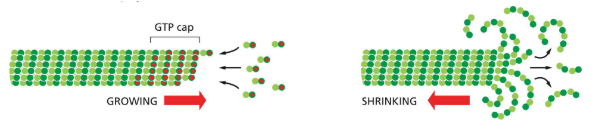
Microtubules exist in a state of dynamic instability
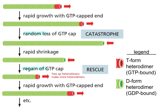
Depolymerization is ~100X faster at an exposed D-from heterodimer
growth with GTP-cap
loss of GTP-cap (catastrophe)
rapid shrinkage
regain of GTP-cap (rescue)
rapid growth with GTP-capped end
𝛾-tubulin helps to nucleate microtubules
y-tubulin (gamma) interacts with a-tubulin at minus-end
y-tubulin nucleates or stablizes the minus-end
y-tubulin protects microtubules from depolymerization at the minus end
plus-ends grow away from the nucleation site

Different patterns of nucleation can create microtubule patterns
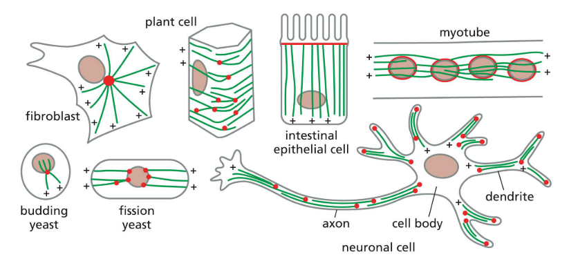
In many animal cells, 𝛾-tubulin is found near centrioles
Microtubules growing from y-tubulin ring complexes of centrosome
protein complex around the centrioles — pericentriolar meterial
has nucleating sites (y-tubulin ring complexes)

In many plant cells 𝛾-tubulin is found on other microtubules
Branch off of other microtubules

Vesicles and organelles can be transported along microtubules
MAPs = microtubule-associated proteins
Kinesins: walk towards plus-end
Dyneins: move towards minus-end
both can hold onto vesiles or organelles with (tail domain)
walk with head domain
both use ATP

Vesicles and organelles can be transported along microtubules — Tilapia example
fish such as tilapia can change colour using microtubule motors
dark: kinesins & dyneins compete for pigment-containing vesicles (melanosomes)
light: kinesins are inhibited, vesicles moved to minus ends near centrioles

Polar actin monomers form polar actin filaments
actin monomers are asymmetric (polar)
actin monomers can bind and hydrolyze ATP — cuts after a while
monomers assemble into polarized actin filaments, usually 2 strands twisted together

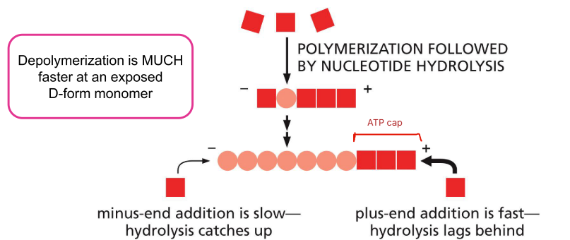
Actin filaments undergo treadmilling
Soluble subunits in T-form
Polymers mixture of T-form and D-form
Depolymerization is MUCH faster at an exposed D-form monomer
Rate of addition on plus end can equal depolymerization on the minus end = treadmilling
The ARP2/3 complex helps nucleate actin filaments
ARP2 & ARP3 have very simila structure to actin monomers
ARP2/3 complex nucleates the minus end of actin filaments and protects them from depolymerization
Plus-ends grow away from ARP2/3 complex
NPF activates ARP2/3


The ARP2/3 complex helps nucleate actin filaments
ARP2/3 can nucleate actin filaments on pre-existing filaments
Whole network can undergo treadmilling
Proteins (e.g. cofilin) sever minus of early actin treadmills for growing end of network to release them from ARP2/3 (more to use)
Proteins cap the plus ends to shape the network
POLAR network — for cell crawling
Actin treadmilling and adhesions help cells crawl
growing actin network pushes the cell leading edge forward
actin and myosin contract to bring lagging edge forward
has to grip on lamellipodium to move

Integrins anchor actin filaments to the extracellular matrix
Integrin heterodimers directly bind extracellular matrix proteins
Integrins indirectly interact with actin filaments
Actin-integrin-extracellular matrix interactions can provide the adhesion necessary for cell migration

Actin & myosins can generate contractile forces
There are many types of myosins
Myosins motor domains use ATP hydrolysis for energy
Myosins can hold onto vesicles or organelles with their other domain or myosins can help cells contract

Actin & myosins can generate contractile forces
Some myosins can “walk” towards the plus end of actin filaments
Actin and myosin work together to generate force (e.g. cell migration or muscle contraction)

Rho family small GTPases can influence actin organization
Rho family GTPases can act like molecular switches
Rho family GTPases (Rho, Rac, and Cdc42) can influence actin organization
Actin organization affects cell shape, cell polarity, and cell behaviour
Activation of Rho family GTPases can dramatically change actin
Healthy cells have a balance of active Rho family members: Rho, Rac, and Cdc42
Over-activation of different Rho family members leads to different actin organization patterns


Actin organization can influence cell shape & behaviour
Rac-GTP activation dominates at the leading tip to explore and to push the cell forward
Rho-GTP activation dominates at the back to pull the back of the cell
Local cytoskeletal organization can define cell movement
Bacterium releases chemoattractant
Binds to receptor
Most receptor binding = most of amount RAC
Least receptor binding = most of amount of RHO
Mutual inhibition

Anterior vs Posterior vs Dorsal vs Ventral
Front vs back vs back vs belly
Cytoskeletal organization can define cell polarity — c.elegans
Symmetry breaking defines anterior vs posterior
In C. elegans, sperm entry defines the posterior
Triggers cytoskeleton polarization in the fertilized egg
actin filaments are organized by a gradient of Rho-GTP activity
microtubules organized by centrosomes near where the sperm entered

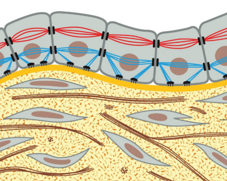
Cell cohesion is essential for multicellular organisms — epithelial tissue
Lots of cells, minimal ECM
mechanical stress transmitted cell to cell by cytoskeletal filaments anchored to cell-matrix and cell-cell adhesion sites
anchored basally to basal lamina

Cell cohesion is essential for multicellular organisms — Connective tissue
Lots of extracellular matrix
mechanical stress of tension and compression beared by extracellular matrix
Many animal cells are either connective or epithelial tissue
Epithelial tissue: cells directly connected to each other with minimal extracellular matrix
Connective tissue: cells dispersed through extracellular matrix

Epithelial cells line surfaces, cavities, and organs
protective layer on surface of organism (skin)
Absorptive lining on cavity of organism (digestive tract)
Surrounding and defining organs

Epithelial cells are polarized
Each cell surface must be different to perform different functions
These cell surfaces define the inside vs outside of the organism or tissue
ALL mature epithelial cells have these junctions in this order
tight junction
adherens junction
desmosome
gap junction
actin-linked cell-matrix junction
hemidesmosome
Tight junction
Tight junction — forms sealing strands, seals gap between cells, no diffusion
most apical
Cell-cell anchoring junctions
(adherens, desmosomes) sticks 2 cells together
adherens forms adhesion belts, connects actin filament bundle in one cell with next cell (all around)
desmosome connects intermediate filaments in one cell with next cell (just spots)
Channel-forming junction
Gap junction — allows passage of small water-soluble molecules from cell to cell
Cell-matrix anchoring junctions
Actin-linked cell-matrix junction, hemidesmosomes — anchors to basal lamina
anchors actin filaments in cell to ECM
anchors intermediate filaments in cell to ECM
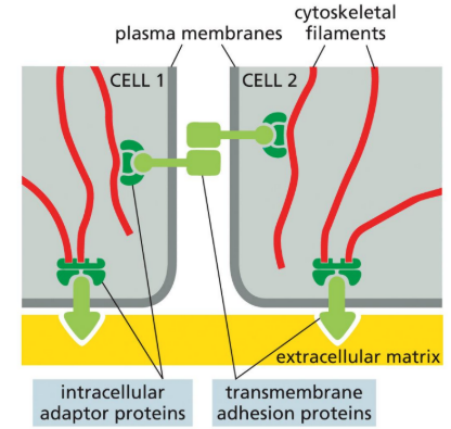
Junctional complexes can link to the cytoskeleton
Cell-cell junctions: usually mediated by cadherin family members
Cell-extracellular matrix junctions: usually mediated by integrins
Actin or intermediate filaments
Cadherins mediate cell-cell connections at adherens junctions
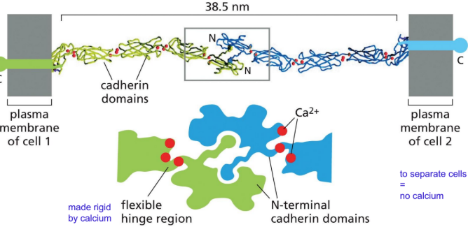
Cadherins are transmembrane proteins expressed by both cells
cadherins ONLY for adherens (family members for others)
Cadherin extracellular domains directly interact with each other
Cadherins interact homophilic interactions of their extracellular domains
interactions require Ca2+ ions
C-term in cytosol
N-term link together
Flexible hinge region made rigid by calcium (to separate cells = remove calcium)
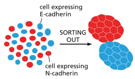
Cells can sort into groups based on homophilic cadherin interactions
No heterophilic interactions between different cadherins
Result: cells expressing different cadherins sort into two separate groups that do not interact
Can be used for differentiation

Cadherin intracellular domains indirectly interact with actin
Cadherins form homophilic interactions with their extracellular domains to directly link adjacent cells
Cadherins indirectly interact with actin filaments (other proteins needed)
indirectly link actin cytoskeleton between adjacent cells
Many cadherins interact in patches to form a strong adhesion belt
All along cell

Adhesion belts can mediate morphogenesis (tissue shapes)
Sheet of epithelial cells can migrate — cells stay intact due to adhesion belts
some can stick

Adhesion belts & sorting out can mediate morphogenesis
Adhesion belt contraction pulls cells to form a tube
Cells express different cadherins to establish new interactions and ensure neural tube closure
early embryonic tissue
cadherin type changes = different binding
forms brain/spinal cord

Cell adhesion is essential for embryo development & morphogenesis
Normal — cells move together to correct place
Mutant — uncoordinated, random, movement

Tight junctions define domains of the cell
Apical domain faces the surface, cavity, or organ
Basal domain faces the inside of the body
Basal + lateral = basolateral
Domains are defined and maintained by tight junctions
Occludins & claudins form tight junctions
All along cell = form sealing strands

Transmembrane occludins & claudins form homophilic interactions
Occludins & claudins — homophilic interactions in extracellular domains to directly link adjacent cells
4 TM proteins
many rows form functional tight junction
Tight junctions limit diffusion in the extracellular space — tracer molecule

Tight junctions limit diffusion of membrane proteins
Tight junctions prevent membrane proteins from moving between the apical and basolateral domains
can diffuse in own region

Tight junctions regulate what enters an organism — glucose from gut to blood
Tight junctions stop anything from directly diffusing from the gut lumen into connective tissue & blood
HIGH glucose conc in cell, LOW in gut and blood
sodium driven glucose transporter moves glucose into cell from gut (must apical domain)
passive carriers allow glucose to diffuse into connective tissue/blood (must basolateral)
Tight junctions keep the correct transporters in their correct domains of the cell
Integrin heterodimers mediate cell-to-cell-matrix junctions

Integrins anchor actin filaments to the extracellular matrix
Integrin heterodimers directly bind extracellular matrix proteins
Integrins have a transmembrane domain
Indirectly interact with actin filaments
Actin-integrin-extracellular matrix interactions can provide the adhesion necessary for cell migration

Cell-matrix junctions play roles in both epithelial & connective tissue
Epithelial tissue: adhesion to the basal lamina extracellular matrix
Connective tissue: cell interaction with the extracellular matrix and cell movement

Polarity cues can establish cell cohesion
Adherens junctions form first
Adherens junctions provide polarity cues to define apical from basolateral domains
activate PAR and Crumbs (signal for apical, inhibit basolateral)
activate Scribble (signal for basolateral, inhibit apical)
Tight junctions form apical to the adherens junctions after

An initial landmark can generate subsequent patterns
Primary landmark — structure, protein, signal, or process
Leads to…
Subsequent Elaboration (elaboration of polarity)
Can be…
molecules in a cell
cells in a tissue
E.g. cell crawling (Rac and Rho)
Cell polarity can be established by external or internal signals
Initial polarity signal was external (sperm entry)
Asymmetric cell division can unevenly distribute internal polarity cues between child cells

3 key components of cell polarity define a functional epithelium
1) intracellular trafficking
2) cytoskeleton organization
3) cell cohesion
=
functional epithelium
functional epithelium
a functional epithelium defines organs and organisms
Cell proliferation
increase in cell numbers via cell division
Cell differentiation
change in cell fate via cell signaling & differential genome expression
Cell morphogenesis
change in cell shape, interactions and/or location
Model organisms are used to study multicellular development
C. elegans
Drosophila
Zebrafish
Humans
Mice
Arabidopsis
Multicellular development(growth and change) is an ongoing process, even in adults
occurs in adults from stem cells
completely new developmental processes can also be initiated (pregnancy)
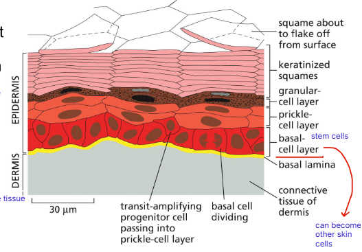
Skin in adults
Epidermis — epithelial tissue
Dermis — connective tissue
Basal cell layer — stem cells, can become other skin cells
Embryogenesis as a model for multicellular development
starts with egg fertilized with sperm
ending just before birth
egg — cleavage (hollow middle) = blastula — gastrulation = gastrula
forming the gut tube (mouth to anus)
from round ball to elongated structure

Key concepts in multicellular development during embryogenesis
blastula = cell proliferation
gastrula = cell differentiation and morphogenesis (and proliferation)

Morphogenesis = generation of shape
cell internalization
elongation
fine repositioning of cells
1. Cell internalization
Ingression/delamination
Invagination/involution
Morphogenesis during gastrulation forms 3 key tissue layers
Gastrulation = change from ball of cells to embryo with a gut & 3 germ layers:
ectoderm differentiates into — epidermis & nervous system
mesoderm differentiates into — muscles, connective tissue, bones, blood, kidneys
endoderm differentiates into — gut, lungs, pancreas, liver
These 3 types needed for every other type of cell

Cell internalization(moving inwards) by ingression/delamination forms the mesoderm
Ingression = (moving inwards) individual cells detach from the outer cell layer and migrate in (crawling in)
blastula cells are epithelial cells
epithelial-to-mesenchymal transition (EMT)
mesenchymal = loose,undifferentiated connective tissue
Delamination = (splitting into layers) cells form a new cell layer, the mesoderm
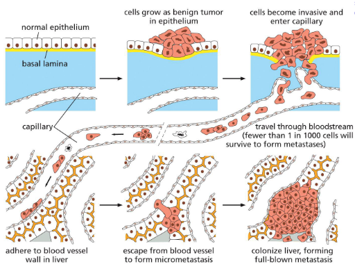
Epithelial-to-mesenchymal transitions must be carefully controlled
switching from type to type is rare
mess up can cause epithelial cancer
cancer cells look like mesenchymal cells

Cell internalization by invagination/involution forms the endoderm
Invagination = (making cavity or pouch) attached cells in epithelial cell sheet are pulled to middle of embryo while remaining attached
involution = (curling inwards) these cells curl in and grow to form endoderm