Anatomy and Physiology - Lymphatic System
 Call Kai
 Learn
 Practice Test
 Spaced Repetition
 Match
 Flashcards
 Knowt Play
1/41

There's no tags or description
Looks like no tags are added yet.
42 Terms
Lymphatic system - functions
Transport and house lymphocytes and other immune cells
Return excess fluid in body tissues to blood to maintain blood volume
Lymphatic system - components
Lymph vessels, lymphoid tissues and organs
Lymph is the fluid transported within lymph vessels
Excess interstitial fluid moves into lymphatic capillaries where it is then called lymph (basically recycled blood plasma, but MOST RESEMBLES ISF)
Interstitial fluid
Comes from substances that leak out of blood capillaries; originates from blood plasma
Lymph
Water, dissolved solutes, and small amount of protein (like the components of plasma)
On top of immune cells, it may also contain cell debris, microorganisms, cancer cells
NO RBCS
MOST RESEMBLES ISF

Movement of lymph
Lymph moves through vessels of larger and larger size: lymphatic capillaries → lymphatic vessels (have valves to prevent backflow) → lymphatic trunks → lymphatic ducts
Ultimately, fluid is returned to blood circulation after passing through jugular and subclavian veins

3 ways lymphatic system moves lymph
Contraction of nearby skeletal muscles and respiratory pumps, as seen in venous circulation
Pulsatile movement of blood in nearby arteries
Rhythmic contraction of smooth muscle in larger lymph vessel walls

Lymphatic capillaries
Closed-end vessels within connective tissue among most blood capillary networks; absorb excess interstitial fluid left during capillary exchange
Hydrostatic pressure of excess interstitial fluid pushes it into capillary (interstitial fluid HP > lymph HP)
Anchoring filaments link endothelial cells to surrounding structures prevent vessel collapse
Pressure of lymph inside vessel closes intercellular openings (one-way “flaps”) of capillary wall


Lymphatic flaps
Allows fluid, macromolecules, pathogens, etc. to enter the capillary but prevents their release

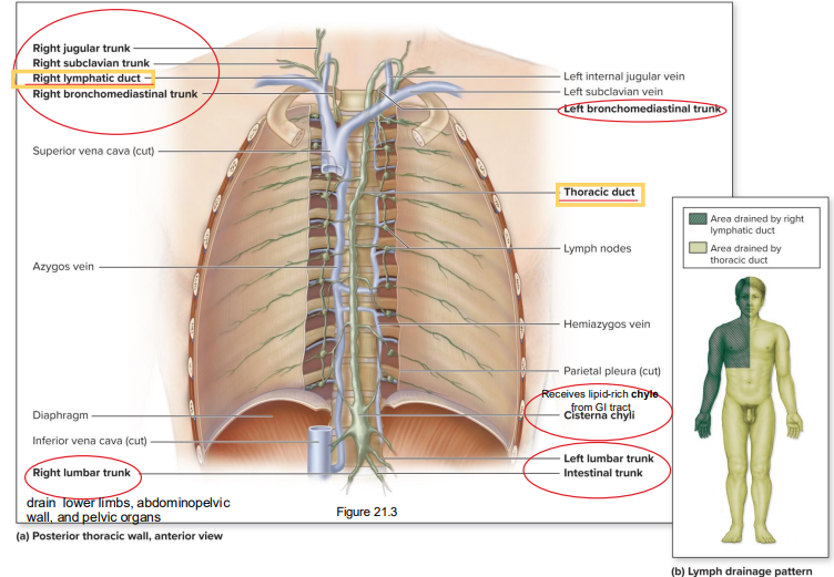
Lymphatic trunks
Jugular trunks: head and neck
Subclavian trunks: upper limbs, breasts, and superficial thoracic wall
Bronchomediastinal trunks: deep thoracic structures
Intestinal trunk (singular): most abdominal structures
Lumbar trunks: lower limbs, abdominopelvic wall, and pelvic organs


Lymphatic ducts
Lymph drains from lymphatic trunks into two ducts
Right upper quadrant of body is drained by the right lymphatic duct → empties into the right jugular and right subclavian veins
Left upper quadrant and lower quadrants (including all of the abdomen) are drained by the thoracic duct → empties into the left jugular and left subclavian veins
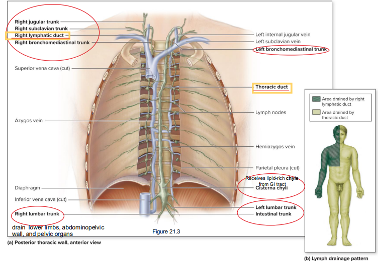

Cisterna chyli
Sac-like structure at the base of the thoracic duct
Both the intestinal trunk and left and right lumbar trunks drain into it
Receives lipid-rich chyle from GI tract

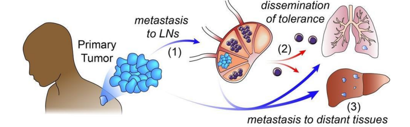
Lymphatic system’s role in metastasis
Wandering cancerous cells in the lymph establish secondary tumors (metastasis)
Ex: breast cancer may metastasize to the lung when cancerous cells break free from primary tumor and are transported in the lymph
In mice, cancer cells that spread to and reside in the lymph nodes appear to convince immune cells to help cancer spread to other organs

Lymphedema
Interstitial fluid accumulation due to interference with lymph drainage
Causes swelling and pain in affected area; may interfere with wound healing and contribute to infection
Most caused by blockage of lymph vessels (ex: trauma, parasitic infection of lymph vessels, tumors, radiation therapy scarring, removal of lymph nodes)
Controlled with compression garments, exercise, massage
Extreme cases known as elephantiasis: caused by infection with parasitic worms
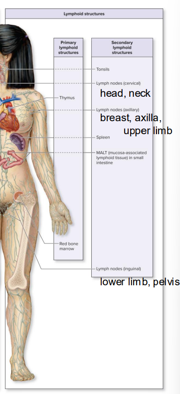
Primary lymphoid structures
Red bone marrow and thymus
Involved in formation and maturation of lymphocytes

Red bone marrow (primary lymphoid structure)
Located between trabeculae (meshwork/support beams) of spongy bone
Found in flat bones of skull, ribs, sternum, vertebrae, ossa coxae, heads of humerus and femur
Site of hematopoiesis: production of blood’s formed elements
Thymocytes
Type of stem cell
Mature formed elements (and thymocytes) leave the red bone marrow to enter the blood
Thymocytes enter the thymus, where they mature and differentiate into T-lymphocytes and then circulate in the blood

Thymus (primary lymphoid structure)
Involved in T-lymphocyte maturation
Lymphoid organ in the mediastinum
Larger in children than adults: grows until puberty, then regresses and is gradually replaced by adipose tissue
Consists of two thymic lobes, each surrounded by connective tissue capsule


Micrographs of child and adult thymus glands
Each lobule has outer cortex and inner medulla regions
Both regions are lymphoepithelial (have lymphatic cells + epithelial tissue)
Cortex: immature T-lymphocytes; darker purple
Medulla: mature T-lymphocytes; lighter purple
Trabeculae are red; capsules are pink and white

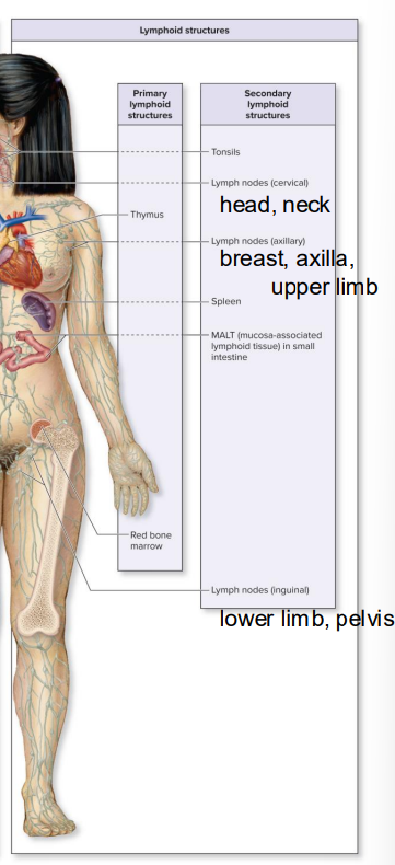
Secondary lymphoid structures
Lymphoid organs (lymph nodes, spleen) + aggregates of lymphoid nodules (tonsils, diffuse lymphoid nodules, MALT/mucosa-associated lymphoid tissue)
Do not form lymphocytes, but house them and other immune cells in a reticular connective tissue matrix
Sites of immune response initiation
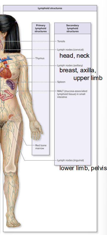
Lymphoid organs (secondary structures)
Organs have complete capsule of dense irregular connective tissue (examples: spleen and lymph nodes)
Aggregates of lymphoid nodules (secondary structures)
Lymphoid structures that either have an incomplete capsule or lack one (examples: tonsils, MALT, diffuse lymphoid nodules)
Node ≠ nodule

Lymph nodes (secondary lymphoid organ)
Small, bean-shaped structures about 10 mm long
Afferent lymphatic vessels bring lymph to node
An efferent vessel drains a lymph node; located at hilum (involuted portion of node)
Has a cortex and medulla


Lymph node cluster locations
Estimated 500-700 throughout the body
Cervical: head and neck
Axillary: breasts, axilla, upper limbs
Inguinal: lower limbs, pelvis


Cortex of lymph node
Contains lymphoid nodules
Reticular fibers support a germinal center: houses proliferating B-lymphocytes and some macrophages
Surrounding mantle zone contains T-lymphocytes, macrophages, and dendritic cells
Cortical sinuses are tiny open channels lined by macrophages
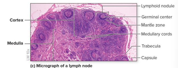
Immune cells found in parts of the lymph node’s cortex
Lymphoid nodule
Germinal center: proliferating B-cells, macrophages
Mantle zone: T-cells, macrophages, dendritic cells
Cortical sinuses: macrophages

Medulla of lymph node
Contains medullary cords: connective tissue fibers that support B-cells, T-cells, and macrophages
Medullary sinuses are tiny open channels lined with macrophages

Lymph flow through lymph nodes
Enter through several afferent vessels → flow through sinuses → exit through one efferent vessel
Lymph is monitored for presence of foreign material
Macrophages remove foreign debris from lymph
Lymphocytes may initiate immune response
Lymphoma
Malignant neoplasm from lymphoid structures
Nontender, enlarged lymph node
Possible night sweats, fever, and weight loss
Includes Hodgkin and non-Hodgkin lymphomas
Hodgkin lymphoma
Affects young adults and people over 60
If caught early, cured by excision, radiation, and chemotherapy
Non-Hodgkin lymphoma
More common
Some kinds aggressive, others slow-growing
Palpation of lymph nodes as a diagnostic tool
Lymph nodes house T- and B-lymphocytes of adaptive immune system → node enlargement can occur in response to infection
Nodes can be palpated in head and neck, but in other areas, can only be palpated when enlarged (axillary, inguinal regions)
What can be diagnosed from palpation of lymph nodes?
Palpation evaluates size and other characteristics; can point to:
Mononucleosis: sore throat, fever, swelling of all nodes
Cold: swelling limited to cervical nodes
More common with cancer: enlarged, hard, nontender nodes

Spleen (secondary lymphoid organ)
Largest lymphoid organ, filtering blood, not lymph
Located lateral to left kidney, inferior to the diaphragm, and posterolateral to stomach
Posterolateral aspect (outer) is convex and rounded
Anteromedial border (inner) is concave; contains the hilum: indentation where blood vessels and nerves enter (splenic artery supplies blood; splenic vein drains it)
Surrounded by connective tissue capsule
Trabeculae from capsule extend into spleen dividing red and white pulp
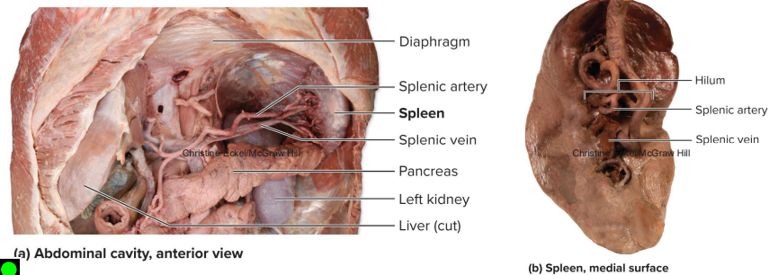
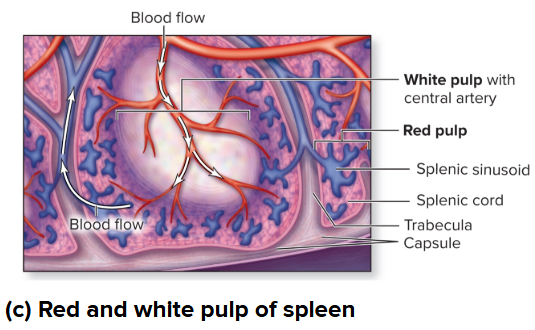
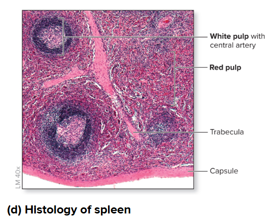
White pulp of spleen
White color from WBCs
Clusters of T- and B-lymphocytes and macrophages around central artery
White pulp monitors blood for foreign materials and bacteria
Initiate immune response when microorganisms detected in blood
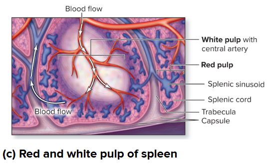
Red pulp of spleen
Red color from RBCs
Contains erythrocytes, platelets, macrophages, and B-lymphocytes
Splenic cords: cells and reticular connective tissue in red pulp
Splenic sinusoids: very permeable capillaries; macrophages in sinusoids of red pulp phagocytize bacteria, debris, and defective erythrocytes and platelets
Storage site for erythrocytes and platelets

Splenectomy
Surgical removal of the spleen; may be more prone to life-threatening infection
May be performed due to:
Ruptured spleen from abdominal injury (most common)
Infection, cyst, or tumor
Lymphoma or other cancer
Blood disorders (for example, sickle cell anemia)
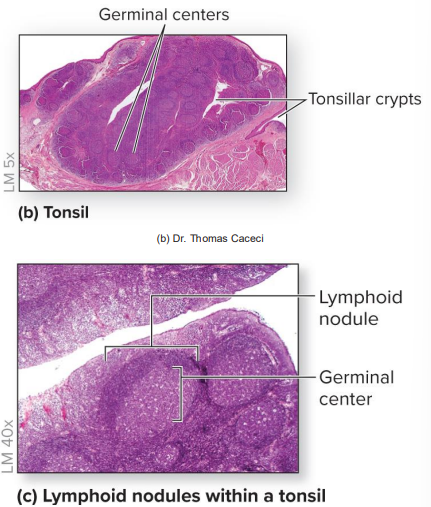
Tonsils (secondary lymphoid structure)
Immune surveillance of inhaled and ingested substances
Incomplete connective tissue capsule
Tonsillar crypts: invaginations (insertions, pockets) that trap material
Contain groups of lymphoid nodules, some with germinal centers


Tonsil locations
Pharyngeal tonsil in nasopharynx (pharynx connecting nasal cavity to larynx and esophagus); called adenoids when enlarged
Palatine tonsils (2x) in posterolateral oral cavity
Lingual tonsils along posterior one-third of tongue

Lymphoid nodules (secondary lymphoid structure)
Clusters of lymphoid cells with some extracellular matrix; not completely surrounded by connective tissue capsule
Cells in the nodule filter unwanted substances like bacteria
Pale center called the germinal center: site of proliferating B-lymphocytes and some macrophages
T-lymphocytes outside the center
In some areas, lymphoid nodules group together to form larger structures; see MALT
MALT (lymphoid nodule - secondary lymphoid structure)
Aggregations within the lamina propria (thin layer) of mucous membranes, where much absorption takes place
Located in GI, respiratory, reproductive, and urinary tracts
Help defend against foreign substances
Prominent in small intestines, especially ileum, as peyer patches
Peyer patches
Type of MALT
Gut-associated lymphoid tissue (GALT); large aggregations of lymphoid nodules within the wall of the GI tract
Tonsillitis and tonsillectomy
Acute tonsillitis
Inflammation and infection of tonsils (most commonly palatine tonsils)
Redden and enlarge
May partially obstruct pharynx
Fever, chills, sore throat, and difficulty swallowing
May be infected by viruses or bacteria
Chronic tonsillitis: persistent recurrent infections; may require tonsillectomy, surgical removal of tonsils