Lab 6- skeletal system

0.0(0)
Studied by 0 people
call kaiCall Kai
learnLearn
examPractice Test
spaced repetitionSpaced Repetition
heart puzzleMatch
flashcardsFlashcards
GameKnowt Play
Card Sorting

1/164

flashcard set

Earn XP

Description and Tags

part 1

Last updated 9:59 PM on 10/19/26
Name
Mastery
Learn
Test
Matching
Spaced
Call with Kai
Chat

No analytics yet

Send a link to your students to track their progress

165 Terms

1
New cards
Condyle
A round bone projection covered by cartilage that articulates with another bone. Where to find it: medial and lateral condyles on the distal femur and on the proximal tibia.
2
New cards
Crest
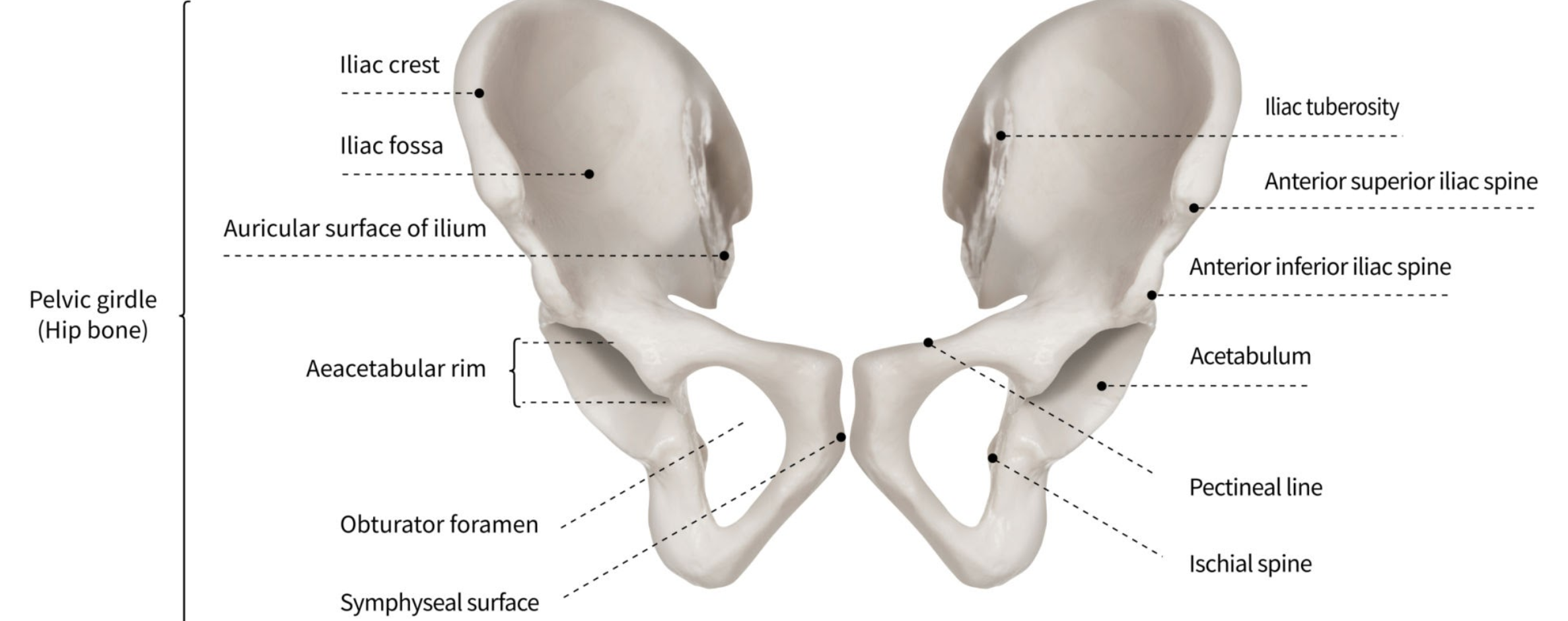
A ridge-like projection at the edge of a bone. Where to find it: iliac crest of the ilium; anterior crest of the tibia; median sacral crest.
3
New cards
Facet
A smooth, flat surface. Where to find it: superior articular facet of the sacrum.
4
New cards
Foramen
Foramen
An opening in a bone for the passage of blood vessels and/or nerves. Where to find it: obturator foramen of the os coxa; vertebral foramen; anterior and posterior sacral foramina.
5
New cards
Fossa

A depression at the surface of a bone. Where to find it: olecranon fossa on the posterior distal humerus -The olecranon fossa is the large, deep depression located on the posterior (back) side of the humerus, just above the elbow joint.

; iliac fossa of the ilium; coronoid and radial fossae of the humerus.

<p>A depression at the surface of a bone. Where to find it: olecranon fossa on the posterior distal humerus -<strong>The olecranon fossa is the large, deep depression located on the posterior (back) side of the humerus, just above the elbow joint.</strong></p><p>; iliac fossa of the ilium; coronoid and radial fossae of the humerus.</p>
6
New cards
Fovea
A small cone-shaped depression. Where to find it: on the head of the femur.
7
New cards
Head
An enlargement at the end of a long bone. Where to find it: head of the femur (fits the acetabulum); head of the humerus (fits the glenoid cavity); head of the radius, ulna, and fibula.
8
New cards
Process
Process
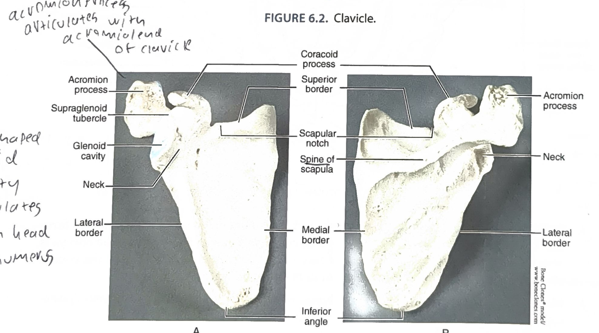
An obvious projection of bone. Where to find it: acromion and coracoid processes of the scapula; xiphoid process of the sternum; olecranon process of the ulna.
9
New cards
Sinus
An air-filled cavity within a bone. Where to find it: inside skull bones (e.g., frontal and maxillary sinuses; skull is covered in Lab 7).
10
New cards
Spine
Like a crest, but sharp. Where to find it: spine of the scapula; ischial spine; anterior superior iliac spine.
11
New cards
Styloid
A small projection at the end of a bone. Where to find it: styloid process of the radius (lateral) and of the ulna (medial) at the distal forearm/wrist.
12
New cards
Suture
The joint where two skull bones are fused together. Where to find it: between the cranial bones of the skull (a fibrous joint).
13
New cards

Trochanter

A large process at the end of the femur. Where to find it: greater trochanter (proximal, lateral) and lesser trochanter (distal to the head, medial) of the femur.
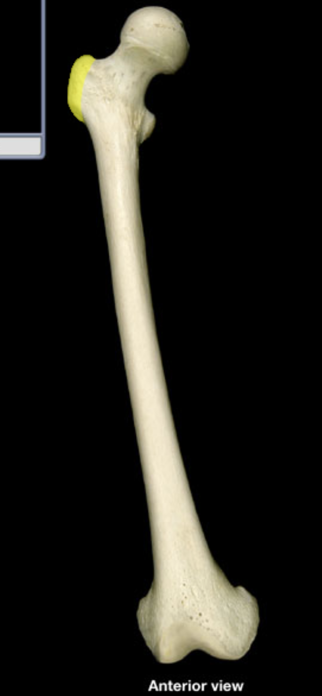
<p>A large process at the end of the femur. Where to find it: greater trochanter (proximal, lateral) and lesser trochanter (distal to the head, medial) of the femur.</p>
14
New cards

Tubercle

A small, rounded process, usually attached to tendons or ligaments. Where to find it: greater and lesser tubercles of the humerus; conoid and deltoid tubercles of the clavicle.

<p>A small, rounded process, usually attached to tendons or ligaments. Where to find it: greater and lesser tubercles of the humerus; conoid and deltoid tubercles of the clavicle.</p>
15
New cards
Tuberosity
A rough patch on the surface of a bone for muscle attachment. Where to find it: deltoid tuberosity of the humerus; tibial tuberosity; radial tuberosity; ischial tuberosity.
16
New cards
Fossa vs. fovea
Fossa = general depression on a bone (e.g., olecranon fossa on the posterior distal humerus). Fovea = small cone-shaped depression (on the head of the femur).
17
New cards
Which features are holes or openings in a bone?
Foramen (for passage of blood vessels/nerves) and sinus (an air cavity).
18
New cards

Which three features are rounded processes?

Tubercle (small round process attached to tendon/ligament), condyle (covered in cartilage, articulates with another bone), and trochanter (large process on the femur).

19
New cards

P68 Q1: Name the bone features that are a hole or opening in a bone.

Foramen (passage of blood vessels/nerves) and sinus (air cavity).

20
New cards
P68 Q2: Name the three rounded processes.

Tubercle (small rounded process attached to tendon/ligament); condyle (covered in cartilage, articulates with another bone); trochanter (large process, on the femur).

21
New cards
P68 Q3: What is the difference between a fossa and a fovea?
Fossa = general depression on a bone (e.g., olecranon fossa, posterior side of the distal humerus). Fovea = small cone-shaped depression (on the head of the femur).
22
New cards
P72: How many vertebrae fuse to form the sacrum?
5
23
New cards
P72: How many vertebrae fused in the coccyx (your specimen)?
4 (the number is variable)
24
New cards

P73 Q3: Humerus: two landmarks to tell superior from inferior end?

Superior end: large, smooth, spherical bone end (the head). Inferior end: more triangular, with a large triangular depression (olecranon fossa) for the ulna.

<p>Superior end: large, smooth, spherical bone end (the head). Inferior end: more triangular, with a large triangular depression (olecranon fossa) for the ulna.</p>
25
New cards
P73 Q4: Humerus: two landmarks to tell right vs. left side?
First hold the humerus with the head pointing medially. Then make the olecranon fossa face posteriorly (to the back). Once established, compare with an anatomical figure: the greater tubercle faces laterally and the lesser tubercle faces medially.
26
New cards
P76 Q2d: Femur: two landmarks to tell proximal from distal end?
Proximal end: large, smooth, spherical head. Distal end: two side-by-side rounded surfaces (medial and lateral condyles).
27
New cards
P76 Q2e: Femur: two landmarks to tell right vs. left side?
First the head of the proximal end faces medially. Then the larger, more sweeping medial condyle faces medially.
28
New cards
Part B Q1: Length of the LHS (large hanging skeleton) femur?
43 cm
29
New cards
Part B Q2: Predicted height of the LHS using (2.27 x femur length) + 67.99?
165.6 cm (2.27 x 43 + 67.99)
30
New cards
Part B Q3: Measured height of the LHS skeleton?
142 cm
31
New cards
Part B Q4: How does predicted height compare with actual height (LHS)?
Actual height is shorter than the predicted height (about 23.6 cm shorter).
32
New cards
Part B Q5: Why might the LHS prediction be off?
A long femur does not always equate to a tall height (anatomical variation). Also, the LHS has knee joint stiffening that prevents a fully tall posture.
33
New cards
Part B Q6: Length of the SSM (small skeleton model) femur?
20.2 cm
34
New cards
Part B Q7: Predicted height of the SSM?
113.84 cm (2.27 x 20.2 + 67.99)
35
New cards
Part B Q8: Measured height of the SSM?
81 cm
36
New cards
Part B Q9: How does predicted height compare with actual height (SSM)?
The actual height is way shorter than the predicted height.
37
New cards
Part B Q10: Why might the SSM prediction be off?
Anatomical variation: femur length does not always predict actual height. Also the height formula may not be accurate for a model.
38
New cards
Part B Q11: Why was the LHS measurement so much closer to the actual height than the SSM?
The formula is for male skeletons and the LHS might be male. The formula is formulated for the correct sex.
39
New cards
Part C Q2: What does "articulation" mean?
A joint: any point in the body where two or more bones meet.
40
New cards

The three ways bones can articulate?

Synovial (fluid-filled, freely moving: shoulder, knee, hip); cartilaginous (slightly moving; bones joined by cartilage with no joint cavity, e.g., intervertebral discs); fibrous (locked by dense connective tissue, e.g., cranial sutures).

41
New cards

Part C Q3: What bones make up the pelvic girdle?

Eight bones: two ilia, two ischia, two pubic bones (pubis), a fused sacrum, and a fused coccyx. (The femur is NOT part of it.)

<p>Eight bones: two ilia, two ischia, two pubic bones (pubis), a fused sacrum, and a fused coccyx. (The femur is NOT part of it.)</p>
42
New cards
Part C Q4: Three anatomical similarities between the upper and lower limb?
1) Both go from big to small as we move proximal to distal: single bone, then two parallel middle bones, then small extremity bones. 2) Both attach to the axial skeleton via a girdle and have ball-and-socket synovial joints. 3) Both hands and feet have 5 digits.
43
New cards
Part C Q5: Three anatomical differences between the upper and lower limb?
1) Pectoral girdle: sternoclavicular joint allows the scapula a wide range of motion; pelvic girdle: sacroiliac joint is more rigid, maximizing force transfer from the ground up. 2) Glenoid cavity enables max range of motion; the hip's acetabulum is deep to emphasize security and weight bearing of the femoral head. 3) The forearm can rotate fully (pronate or supinate); the tibia/fibula cannot access this range of motion.
44
New cards
Part C Q6: Compared with the other toes, which bone is missing in the big toe?
The middle phalanx (the big toe, hallux, has only proximal and distal phalanges).
45
New cards
Part C Q7: Why is the anterior connection of the two os coxae fibrocartilage and not bone?
It allows the pelvic girdle slight mobility during locomotion; it absorbs vibrations; if it were osseous the pelvic girdle would more easily fracture; it also widens during child birth.
46
New cards
Part C Q8: What is the common name of the pollex?
The thumb
47
New cards
Part C Q9: Name the three bones that make up the ankle.
Tibia (medial malleolus), fibula (lateral malleolus), and talus.
48
New cards
Part C Q10: What is the common name of the patella?
Kneecap
49
New cards
Part C Q11: In the pectoral girdle, what structure is similar to the acetabulum of the pelvic girdle?
The glenoid cavity (glenoid fossa) of the scapula.
50
New cards
Part C Q12: What does the word tubercle mean?
A rounded prominence; a bony bump on the bone's surface (usually attached to bone/ligament, per lab: tendons/ligaments).
51
New cards
Part D Q1a: Thalidomide / phocomelia: bones missing if a malformed HAND attached to the trunk?
Humerus, radius, ulna (the glenoid cavity may be affected too).
52
New cards
Part D Q1b: Bones missing if a malformed FOOT attached to the trunk?
Femur, patella, tibia, fibula.
53
New cards
Part D Q1c: Which bones would show normal development (hand/upper extremity)?
Carpals (8), metacarpals (5), phalanges (14 total, including the thumb); the clavicle and scapula would also be normal.
54
New cards
Part D Q1c: Which bones would show normal development (foot/lower extremity)?
Tarsals (7), metatarsals (5), phalanges (14); the os coxae would also be normal.
55
New cards
Part D Q2a: Elderly X-ray: which bones are broken?
A lateral section shows fractures in the tibia and fibula.
56
New cards
Part D Q2b: How long should it take the broken bone to heal?
6-12 months to restore full pre-injury bone matrix density.
57
New cards
Part D Q3: Torso model: sternum?
Articulates with the clavicle at its clavicular notch.
58
New cards
Margin Q: Femur vs. humerus: what is the difference?
Femur (upper leg): head fits the acetabulum, has greater and lesser TROCHANTERS, distal condyles articulate with the tibia, patellar surface. Humerus (arm): head fits the glenoid cavity, has greater and lesser TUBERCLES, deltoid tuberosity, distal trochlea (ulna) and capitulum (radius), olecranon fossa.
59
New cards
Margin Q: Which forearm bone interfaces with the thumb?
The radius (lateral, thumb side).
60
New cards
Margin Q: How do you tell the tibia from the fibula?
The tibia is the thicker, weight-bearing, medial bone (tibial tuberosity, anterior crest, articulates with the femur). The fibula is skinnier, lateral, and does not articulate with the femur.
61
New cards
Sternum: 3 key facts
1) Proximal/superior: clavicular notch articulates with the clavicle. 2) Distal/inferior: ends in the pointed xiphoid process. 3) Key feature: three parts, manubrium (superior), body (main portion), xiphoid process (inferior); jugular notch at the top. Breastbone, axial skeleton.
62
New cards
Clavicle: 3 key facts
1) Lateral (acromial) end articulates with the acromion process of the scapula. 2) Medial (sternal) end articulates with the sternum. 3) Key feature: conoid tubercle and deltoid tubercle; part of the pectoral girdle with the scapula.
63
New cards
Scapula: 3 key facts
1) Acromion process articulates with the acromial end of the clavicle. 2) Glenoid cavity (pear-shaped) articulates with the head of the humerus. 3) Key features: coracoid process, spine of the scapula, scapular notch, medial/lateral/superior borders, inferior angle. Shoulder blade.
64
New cards

how to find orientation of scapula?

Find the posterior side: Locate the spine of the scapula (the prominent, bony ridge that juts out). This side must face you (posterior). The relatively smooth, slightly concave side (the subscapular fossa) rests against the ribcage (anterior).

Find the inferior side: Point the sharpest, most tapered point of the bone (the inferior angle) straight down toward the ground. The spine and the large bony hooks (acromion and coracoid process) should now be at the top (superior).

  • Check the lateral socket: Look for the glenoid cavity (the shallow, pear-shaped socket where the humerus attaches). Because your arm is on the outside of your body, this socket must always face outward (laterally


<p><strong>Find the posterior side:</strong> Locate the <strong>spine</strong> of the scapula (the prominent, bony ridge that juts out). This side must face you (posterior). The relatively smooth, slightly concave side (the subscapular fossa) rests against the ribcage (anterior).</p><p><strong>Find the inferior side:</strong> Point the sharpest, most tapered point of the bone (the <strong>inferior angle</strong>) straight down toward the ground. The spine and the large bony hooks (acromion and coracoid process) should now be at the top (superior).</p><ul><li><p><strong>Check the lateral socket:</strong> Look for the <strong>glenoid cavity</strong> (the shallow, pear-shaped socket where the humerus attaches). Because your arm is on the outside of your body, this socket must always face outward (<strong>laterally</strong></p></li></ul><p></p>
65
New cards

Os coxa (hip bone): 3 key facts

1) Posterior: ilium joins the sacrum at the sacroiliac joint. 2) Anterior: the two os coxae join at the symphysis pubis (fibrocartilage); lateral: acetabulum receives the head of the femur. 3) Key feature: fusion of three bones (ilium = proximal, pubis = medial, ischium = distal); iliac crest, ASIS/AIIS, ischial tuberosity, obturator foramen.
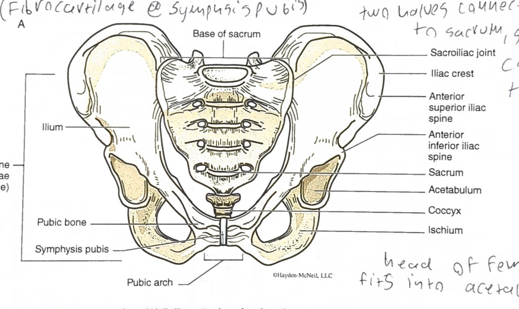
<p>1) Posterior: ilium joins the sacrum at the sacroiliac joint. 2) Anterior: the two os coxae join at the symphysis pubis (fibrocartilage); lateral: acetabulum receives the head of the femur. 3) Key feature: fusion of three bones (ilium = proximal, pubis = medial, ischium = distal); iliac crest, ASIS/AIIS, ischial tuberosity, obturator foramen.</p>
66
New cards

Sacrum: 3 key facts

1) Proximal: base of the sacrum connects up to the spine. 2) Lateral: joins the two os coxae posteriorly (between the ilia) at the sacroiliac joints; distal: apex articulates with the coccyx. 3) Key feature: 5 fused vertebrae; promontory, anterior/posterior sacral foramina, median sacral crest, sacral hiatus. Axial skeleton.

<p>1) Proximal: base of the sacrum connects up to the spine. 2) Lateral: joins the two os coxae posteriorly (between the ilia) at the sacroiliac joints; distal: apex articulates with the coccyx. 3) Key feature: 5 fused vertebrae; promontory, anterior/posterior sacral foramina, median sacral crest, sacral hiatus. Axial skeleton.</p>
67
New cards
Coccyx: 3 key facts
1) Proximal: attached to the apex of the sacrum. 2) Distal: free end (tailbone); no bone articulates below it. 3) Key feature: formed from a variable number of fused vertebrae (4 in the lab specimen); part of the axial skeleton.
68
New cards
<p>Humerus: 3 key facts</p>

Humerus: 3 key facts

1) Proximal: head articulates with the glenoid cavity of the scapula. 2) Distal: trochlea articulates with the ulna; capitulum articulates with the radius. 3) Key features: greater tubercle (wider) and lesser tubercle (narrower), deltoid tuberosity, olecranon fossa (posterior, receives the olecranon of the ulna). Only bone of the arm.

<p>1) Proximal: head articulates with the glenoid cavity of the scapula. 2) Distal: trochlea articulates with the ulna; capitulum articulates with the radius. 3) Key features: greater tubercle (wider) and lesser tubercle (narrower), deltoid tuberosity, olecranon fossa (posterior, receives the olecranon of the ulna). Only bone of the arm.</p>
69
New cards

humerus oritentation

  • Hold the bone vertically with the rounded head at the top and facing inward (medially). Look at the bottom end: if the massive, deep hole (olecranon fossa) is facing you, you are looking at the back (posterior) of the bone.

  • Superior (Top / Proximal): Look for the large, smooth, rounded head of the humerus. This fits into the shoulder socket.

  • Inferior (Bottom / Distal): This end is flattened, broad, and flares out into the elbow joint.

  • Medial (Inner): The rounded head of the humerus always faces inward toward your body (to meet the scapula). At the bottom end, the medial epicondyle is a very prominent, large bony bump that sticks out significantly further than the bump on the other side (this is your "funny bone").

  • Lateral (Outer): The top end has the greater tubercle sticking out to the side. The bottom end has the much smaller lateral epicondyle.

  • Posterior (Back): Look at the bottom (distal) end of the bone. You will see a single, massive, deep triangular depression called the olecranon fossa. This must face the back—it's where the tip of your elbow locks in when you straighten your arm.

  • Anterior (Front): The bottom end has two smaller, shallower indentations side-by-side (the coronoid and radial fossae). At the top end, you will see a distinct vertical groove (the intertubercular groove) separating the two bony bumps (tubercles) near the head.


<ul><li><p>Hold the bone vertically with the rounded head at the top and facing inward (medially). Look at the bottom end: if the massive, deep hole (olecranon fossa) is facing you, you are looking at the <em>back</em> (posterior) of the bone.</p></li><li><p><strong>Superior (Top / Proximal):</strong> Look for the large, smooth, rounded <strong>head</strong> of the humerus. This fits into the shoulder socket.</p></li><li><p><strong>Inferior (Bottom / Distal):</strong> This end is flattened, broad, and flares out into the elbow joint.</p></li><li><p><strong>Medial (Inner):</strong> The rounded <strong>head</strong> of the humerus <em>always</em> faces inward toward your body (to meet the scapula). At the bottom end, the <strong>medial epicondyle</strong> is a very prominent, large bony bump that sticks out significantly further than the bump on the other side (this is your "funny bone").</p></li><li><p><strong>Lateral (Outer):</strong> The top end has the <strong>greater tubercle</strong> sticking out to the side. The bottom end has the much smaller <strong>lateral epicondyle</strong>.</p></li><li><p><strong>Posterior (Back):</strong> Look at the bottom (distal) end of the bone. You will see a single, massive, deep triangular depression called the <strong>olecranon fossa</strong>. This <em>must</em> face the back—it's where the tip of your elbow locks in when you straighten your arm.</p></li><li><p><strong>Anterior (Front):</strong> The bottom end has two smaller, shallower indentations side-by-side (the coronoid and radial fossae). At the top end, you will see a distinct vertical groove (the <strong>intertubercular groove</strong>) separating the two bony bumps (tubercles) near the head.</p></li><li><p></p></li></ul><p></p>
70
New cards

Ulna: 3 key facts

1) Proximal: trochlear notch articulates with the trochlea of the humerus; radial notch articulates with the radius. 2) Distal: head of the ulna articulates with the ulnar notch of the radius. 3) Key features: olecranon process (elbow, posterior hook), styloid process (distal, medial). Medial forearm bone.

<p>1) Proximal: trochlear notch articulates with the trochlea of the humerus; radial notch articulates with the radius. 2) Distal: head of the ulna articulates with the ulnar notch of the radius. 3) Key features: olecranon process (elbow, posterior hook), styloid process (distal, medial). Medial forearm bone.</p>
71
New cards

Radius: 3 key facts

1) Proximal: head of the radius articulates with the capitulum of the humerus. 2) Distal: ulnar notch articulates with the ulna; carries the wrist and thumb side. 3) Key features: styloid process (distal, lateral), radial tuberosity. Lateral forearm bone; the radius is the bone that interfaces with the thumb.

<p>1) Proximal: head of the radius articulates with the capitulum of the humerus. 2) Distal: ulnar notch articulates with the ulna; carries the wrist and thumb side. 3) Key features: styloid process (distal, lateral), radial tuberosity. Lateral forearm bone; the radius is the bone that interfaces with the thumb.</p>
72
New cards

Carpals: 3 key facts

1) Proximal row articulates with the forearm bones (wrist); distal row articulates with the metacarpals. 2) Eight bones in two rows. Proximal row, lateral to medial: scaphoid, lunate, triquetrum, pisiform. Distal row, lateral to medial: trapezium, trapezoid, capitate, hamate. 3) Key feature: trapezium is the most lateral bone of the distal row (under the thumb); pisiform may be fused to the triquetrum.
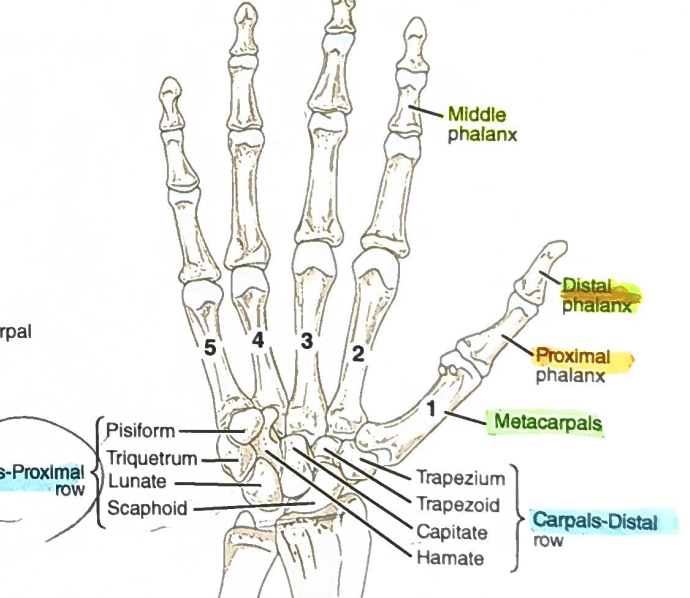
<p>1) Proximal row articulates with the forearm bones (wrist); distal row articulates with the metacarpals. 2) Eight bones in two rows. Proximal row, lateral to medial: scaphoid, lunate, triquetrum, pisiform. Distal row, lateral to medial: trapezium, trapezoid, capitate, hamate. 3) Key feature: trapezium is the most lateral bone of the distal row (under the thumb); pisiform may be fused to the triquetrum.</p>
73
New cards
Carpal mnemonic (medial to lateral, proximal then distal row)
Pisiform, Triquetrum, Lunate, Scaphoid, Hamate, Capitate, Trapezoid, Trapezium = "Peter Told Lisa Skeleton Hands Can Talk Today"
74
New cards

Metacarpals: 3 key facts

1) Proximal: articulate with the distal row of carpals. 2) Distal: articulate with the proximal phalanges. 3) Key feature: five bones of the palm named by position, 1st (thumb) to 5th (pinky).

<p>1) Proximal: articulate with the distal row of carpals. 2) Distal: articulate with the proximal phalanges. 3) Key feature: five bones of the palm named by position, 1st (thumb) to 5th (pinky).</p>
75
New cards

Phalanges of the hand: 3 key facts

1) Proximal: proximal phalanges articulate with the metacarpals. 2) Distal: distal phalanges are the fingertips. 3) Key feature: 14 total; each finger has proximal, middle, distal; the thumb (pollex) has only proximal and distal.
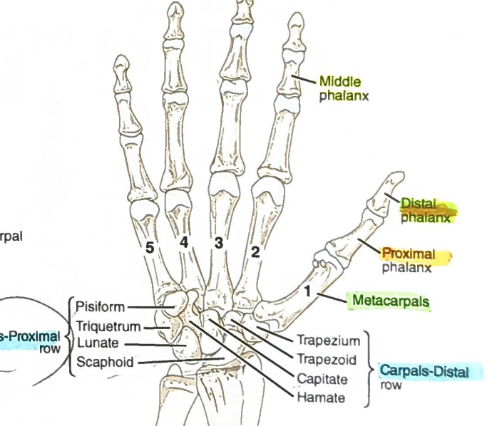
<p>1) Proximal: proximal phalanges articulate with the metacarpals. 2) Distal: distal phalanges are the fingertips. 3) Key feature: 14 total; each finger has proximal, middle, distal; the thumb (pollex) has only proximal and distal.</p>
76
New cards
<p>Femur: 3 key facts</p>

Femur: 3 key facts

1) Proximal: head articulates with the acetabulum of the os coxa. 2) Distal: medial and lateral condyles articulate with the tibia (patellar surface sits at the kneecap). 3) Key features: greater trochanter (lateral, opposite the head), lesser trochanter (distal to the head, medial), neck, fovea on the head. Only bone of the upper leg.

<p>1) Proximal: head articulates with the acetabulum of the os coxa. 2) Distal: medial and lateral condyles articulate with the tibia (patellar surface sits at the kneecap). 3) Key features: greater trochanter (lateral, opposite the head), lesser trochanter (distal to the head, medial), neck, fovea on the head. Only bone of the upper leg.</p>
77
New cards

femur orientation?

  • Hold the bone vertically with the head at the top and the prominent linea aspera ridge facing you (so you are looking at the posterior side).

    • If the spherical head points to your right, it is a right femur.

    • If the spherical head points to your left, it is a left femur.


  • Superior (Proximal): Look for the large, smooth, spherical head of the femur. This end fits into the acetabulum (the hip socket) of the pelvis.

  • Inferior (Distal): The bottom end is broad, flat, and flares out into two massive knobs called the condyles, which rest on the tibia to form the knee joint.

  • Medial (Inner): The spherical head of the femur must always point medially (inward toward the midline of the body) to connect with the pelvis. If you look closely at the head, there is a small pit called the fovea capitis for ligament attachment.

  • Lateral (Outer): Opposite the head at the superior end is a massive, prominent bony block called the greater trochanter. This always points laterally (outward).

  • Posterior (Back): Run your finger down the shaft (diaphysis) of the bone. On the posterior side, you will feel a distinct, rough, raised ridge running vertically down the middle—this is the linea aspera, a major muscle attachment site. Additionally, at the distal end (the bottom), there is a very deep, U-shaped notch separating the two condyles called the intercondylar fossa.

  • Anterior (Front): The anterior shaft is relatively smooth and slightly convex (it bows slightly forward). At the distal end, the two condyles merge together into a smooth, shallow depression called the patellar surface, where the kneecap glides.


<ul><li><p>Hold the bone vertically with the head at the top and the prominent linea aspera ridge facing you (so you are looking at the <em>posterior</em> side).</p><ul><li><p>If the spherical head points to your <strong>right</strong>, it is a <strong>right</strong> femur.</p></li><li><p>If the spherical head points to your <strong>left</strong>, it is a <strong>left</strong> femur.</p></li></ul></li><li><p></p></li><li><p><strong>Superior (Proximal):</strong> Look for the large, smooth, spherical <strong>head</strong> of the femur. This end fits into the acetabulum (the hip socket) of the pelvis.</p></li><li><p><strong>Inferior (Distal):</strong> The bottom end is broad, flat, and flares out into two massive knobs called the <strong>condyles</strong>, which rest on the tibia to form the knee joint.</p></li><li><p><strong>Medial (Inner):</strong> The spherical <strong>head</strong> of the femur <em>must</em> always point medially (inward toward the midline of the body) to connect with the pelvis. If you look closely at the head, there is a small pit called the <strong>fovea capitis</strong> for ligament attachment.</p></li><li><p><strong>Lateral (Outer):</strong> Opposite the head at the superior end is a massive, prominent bony block called the <strong>greater trochanter</strong>. This always points laterally (outward).</p></li><li><p><strong>Posterior (Back):</strong> Run your finger down the shaft (diaphysis) of the bone. On the posterior side, you will feel a distinct, rough, raised ridge running vertically down the middle—this is the <strong>linea aspera</strong>, a major muscle attachment site. Additionally, at the distal end (the bottom), there is a very deep, U-shaped notch separating the two condyles called the <strong>intercondylar fossa</strong>.</p></li><li><p><strong>Anterior (Front):</strong> The anterior shaft is relatively smooth and slightly convex (it bows slightly forward). At the distal end, the two condyles merge together into a smooth, shallow depression called the <strong>patellar surface</strong>, where the kneecap glides.</p></li></ul><p></p>
78
New cards
Patella: 3 key facts
1) Kneecap; sits at the anterior knee on the distal femur (patellar surface). 2) The tibial tuberosity is just inferior to it on the tibia. 3) Placed in the skeleton after the femur and tibia; one per leg.
79
New cards

Tibia: 3 key facts

1) Proximal: medial and lateral condyles articulate with the condyles of the femur. 2) Distal: medial malleolus forms part of the ankle with the talus and the lateral malleolus of the fibula. 3) Key features: tibial tuberosity (below the kneecap), anterior crest (bony line down the midline). Thicker, medial lower-leg bone.
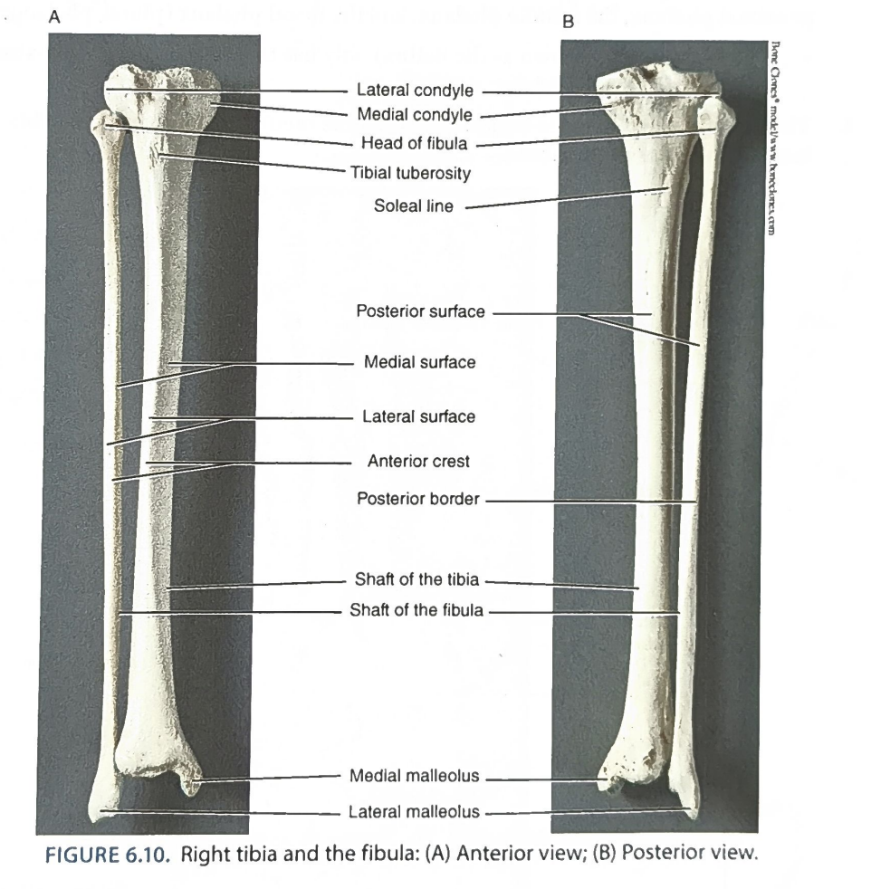
<p>1) Proximal: medial and lateral condyles articulate with the condyles of the femur. 2) Distal: medial malleolus forms part of the ankle with the talus and the lateral malleolus of the fibula. 3) Key features: tibial tuberosity (below the kneecap), anterior crest (bony line down the midline). Thicker, medial lower-leg bone.</p>
80
New cards

Fibula: 3 key facts

1) Proximal: head (round knob) articulates with the tibia; it does NOT articulate with the femur. 2) Distal: lateral malleolus articulates with the talus to form the lateral ankle. 3) Key feature: skinnier than the tibia; lateral bone of the lower leg.

<p>1) Proximal: head (round knob) articulates with the tibia; it does NOT articulate with the femur. 2) Distal: lateral malleolus articulates with the talus to form the lateral ankle. 3) Key feature: skinnier than the tibia; lateral bone of the lower leg.</p>
81
New cards

Tarsals: 3 key facts

1) Proximal: talus fits between the tibia and fibula to form the ankle. 2) Distal: articulate with the metatarsals. 3) Key features: seven bones: talus (superior), calcaneus (heel; large distal, posterior bone), navicular, cuboid, medial, intermediate, and lateral cuneiforms.

<p>1) Proximal: talus fits between the tibia and fibula to form the ankle. 2) Distal: articulate with the metatarsals. 3) Key features: seven bones: talus (superior), calcaneus (heel; large distal, posterior bone), navicular, cuboid, medial, intermediate, and lateral cuneiforms.</p>
82
New cards

Metatarsals: 3 key facts

1) Proximal: articulate with the distal tarsals (cuneiforms/cuboid). 2) Distal: articulate with the proximal phalanges. 3) Key feature: five bones named by position, 1st (proximal to the big toe) to 5th (proximal to the pinky toe).

<p>1) Proximal: articulate with the distal tarsals (cuneiforms/cuboid). 2) Distal: articulate with the proximal phalanges. 3) Key feature: five bones named by position, 1st (proximal to the big toe) to 5th (proximal to the pinky toe).</p>
83
New cards

Phalanges of the foot: 3 key facts

1) Proximal: proximal phalanges articulate with the metatarsals. 2) Distal: distal phalanges are the tips of the toes. 3) Key feature: 14 total; each toe has proximal, middle, distal; the big toe (hallux) has only proximal and distal.

<p>1) Proximal: proximal phalanges articulate with the metatarsals. 2) Distal: distal phalanges are the tips of the toes. 3) Key feature: 14 total; each toe has proximal, middle, distal; the big toe (hallux) has only proximal and distal.</p>
84
New cards
Which bone? The breastbone with manubrium, body, and xiphoid process
Sternum
85
New cards
Which bone? Shoulder blade with glenoid cavity and acromion process
Scapula
86
New cards
Which bone? Articulates with the sternum medially and the acromion process laterally
Clavicle
87
New cards
Which bone? Fusion of ilium, ischium, and pubis
Os coxa (hip bone)
88
New cards
Which bone? Five fused vertebrae with sacral foramina and a promontory
Sacrum
89
New cards
Which bone? Only bone of the arm; head fits the glenoid cavity
Humerus
90
New cards
Which bone? Forearm bone with the olecranon process (elbow)
Ulna
91
New cards
Which bone? Lateral forearm bone on the thumb side with a head that meets the capitulum
Radius
92
New cards
Which bone? Only bone of the upper leg; head fits the acetabulum
Femur
93
New cards
Which bone? Thicker lower-leg bone with the tibial tuberosity and anterior crest
Tibia
94
New cards
Which bone? Skinnier lower-leg bone with a lateral malleolus; does not touch the femur
Fibula
95
New cards
Which bone? Heel bone
Calcaneus
96
New cards
Which bone? Tarsal that sits between the tibia and fibula to form the ankle
Talus
97
New cards
Which bone? Most lateral carpal of the distal row, under the thumb
Trapezium
98
New cards
Articulate
When two bones come together to form a joint, they are said to articulate with each other.
99
New cards
Axial skeleton
Skull, vertebral (spinal) column, and rib cage (sternum is included).
100
New cards
Appendicular skeleton
The pectoral and pelvic girdles and the arms and legs.