BCEM3201 - Globins & Cooperative Binding
 Call Kai
 Learn
 Practice Test
 Spaced Repetition
 Match
 Flashcards
 Knowt Play
1/43

There's no tags or description
Looks like no tags are added yet.
44 Terms
Globins
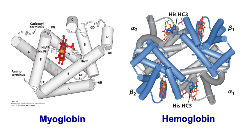
family of proteins, involved in oxygen transport and storage
Why does oxygen need to be carried?
It’s poorly soluble in aqueous solutions
Prosthetic group
Cofactors that binds tightly to proteins
Heme
Porphyrin ring with Fe2+
Proteins bind oxygen through Fe embedded in heme
Myoblobin (Mb) or Hemoglobin (Hb) without heme
apoprotein
Myoblobin (Mb) or Hemoglobin (Hb) with heme
holoprotein
Myoblobin (Mb) or Hemoglobin (Hb) without O2 bound
deoxymyoglobin or deoxyhemoglobin
Myoblobin (Mb) or Hemoglobin (Hb) with O2 bound
oxymyoglobin or oxyhemoglobin
Porphyria
“Vampire Disease”: Blood disorder that causes body to produce less heme
Why is porphyria the “vampire disease”?
Sensitivity to sunlight
“Fangs” (facial disfigurement and receding gums)
“Blood drinking” (urine is dark red)
Aversion to garlic (sulfur content of garlic attacks porphyria, leads to acute pain)
Which residues in globin are essential for function?
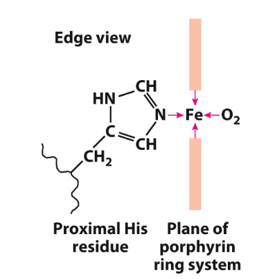
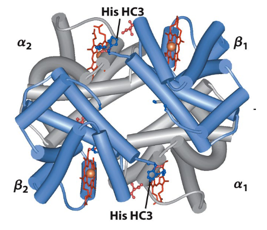
Proximal and Distal His residues

Proximal His
binds to Fe to stabilize it's position
Distal His
Increases binding affinity to O2 by forming H-bond with bound O2 (helps transition to R form in Hb)
Myoglobin (Mb)
Binds 1 oxygen
Single subunit (monomer)
153 AA
16700 g/mol
8 helices (A-H)

Oxygen binds to myoglobin of a Kd of..?
0.26 kPa
Myoglobin is found in _____ only
muscle tissues
During oxygen deprivation, myoglobin..?
releases bound O2 during oxygen deprivation
pO2 in lungs is 13 kPa, meaning..?
myoglobin binds oxygen well
pO2 in tissues is 4 kPa, meaning..?
myoglobin will not release oxygen
Hemoglobin (Hb)
O2 transport (64,500 MW, 4 times Mb)
4 subunits (seperate polypeptide chains)
2α and 2β subunits – quaternary structure
transports O 2 from lungs to all parts of the body
In Hb, binding of one ligant affects affinities of..?
any remaining unfilled sites
Cooperativitiy in Hb
Positive and negativecooperativity
Positive cooperativity
First binding event increases affinity at remaining sites (recognized by sigmoidal binding curves)
Negative cooperativity
First binding event reduces affinity at remaining sites
Sigmoidal curve
Shows transition from low-affinity to high-affinity state
More sensitive to small differences in O2 concentration

Sigmoidal curve: when pO2 is high..?
Hb binds oxygen
Sigmoidal curve: when pO2 is low..?
Hb releases oxygen
Multisubunit proteins and cooperativity

Two states of Hb
T-state “Tense” (weaker O2 binding, Kd high)
R-state - “Relaxed” (Stronger oxygen binding, Kd low)
How does Hb switch states?
O2 binding results in small changes at binding site, which propagates to other sites via protein-protein interactions
O2 binding causes heme to assume planar conformation, shifting position of proximal His and attached F hexlix

Allosteric Protein
binding of ligand (modulator) to one site (other than active site) affects binding properties of different site on same protein
has other shapes or conformations induced by moduators
Can be positve (activators) or negative (inhibitors)
Homotropic regulation
Normal ligand of protein is allosteric regulator
Heterotropic regulation
Different ligand affects binding of normal ligant
BPG
Negative heterotropic allosteric regulator of Hb
Reduces affinity for O2 binding by stabilizing T-state
Small negatively charged molecule, binds to positve charged central cavity of Hb

At higher altitudes, our bodies produce more..?
3-BPG
BPG concentration increases, decreasing the affinity of Hb to O2, releasing O2 to tissues (since higher altitudes have limited O2)
BPG concentration can be adjusted to compensate for..?
altitude
Carbon monoxide poisoning
CO binds Hb 250 times stronger than O2
CO binding pushes Hb to R-state, increasing O2 affinity, so bound O2 cannot be releasing into tissues

Sickle Cell anemia
Glu6 —> Val in β chain of Hb
New Val side chain can bind to different Hb molecule, forming large linear chain of Hbs
Red blood cells assume sickle shape and clog capillaries
Hill equation
n = actual # of ligands bound
h = phenomenological # of ligands bounds (ie in hemoglobin, h = 2.5-3.0 typically, not 4)

h values and cooperativity
h=1 - non-cooperativity
h>1 - positive cooperativity
h<1 - negative cooperativity

Hill’s model
gives the correct qualitative behavior of Hb

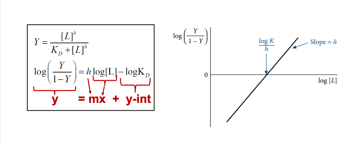
Hill plot
Linearization of hill equation to determine h and KD
Plot of log (Y/1-Y) vs log [L] gives straight line with slope of h and x-intercept of (log KD)/h
Cooperative vs Non-cooperative bind in Scatchard plot. How about a Hill plot?
Cooperative binding appears non-linear in a Scatchard plot
However, Hill plot of “simple” binding produces straight line
