Cancer therapies surgery in the management of the oncology patient
 Call Kai
 Learn
 Practice Test
 Spaced Repetition
 Match
 Flashcards
 Knowt Play
1/31

There's no tags or description
Looks like no tags are added yet.
32 Terms
What needle do you use for a FNA biopsy?
21G 1” needle + 2 or 5ml syringe
Why should cytology & FNA slides not be put in same packaging
Formalin from cytology damages cells of FNA
Describe the process of carrying out a tru-cut biopsy?
GA / Sedation + LA @ stab incision over mass
Aseptic Prep
1-3mm Stab incision
Multiple needle cores
Gently remove
Formalin +/- Impression smear
Consider Culture as well
fungal / mycobacterial infection?

Describe the process of an incisional biospy?
Wedge of tissue
Ideally composite
near edge —> more likely to have normal tissue
don’t extend beyond edge of tumour
compare normal to tumour tissue
Keep within future field of Tx
What are the different ways you can stage tumours?
Blood test
Urine test
FNA local LNs
Thoracic imaging for distant mets —> rads (2 views to confirm) & abdo U/S
liver, spleen, LNs mets of mast cell tumours
Bone marrow aspirates

Why should you check both lateral views when checking for lung mets?
Can only see the lesions when they are surrounded by air filled lung —> contrast more visible

What is the TNM staging for tumours?

Describe surgery as a tx for tumours
Complete excision / no detectable dx
primary closure
Cytoreduction + adjunct tx
Palliation
What borders would you use when removing a mast cell tumour?
Mod grade —> 2cm lateral margin & deep facial plane
High grade —> 3cm lateral margin & deep margin
do margins meet? if yes = primary closure

What can help you when you are carrying out a primary closure?
Towel clamp —> use this before placing suture —> shouldn’t be tearing through tissues
Take bite through fascia to subdermal fascia on top, take towel clamp off & should hold
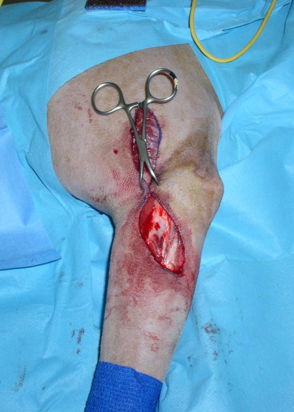
What should be done after primary closure of leg after tumour surgery?
Bandaged in extended position for 2 weeks (robert jones bandage) —> changed twice a week
Secures fibrous tissue → then restricted exercise
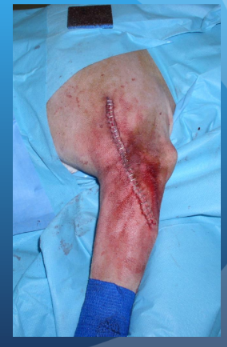

What is this showing?
Bright red granulation tissue + pale pink ep tissue migrating across —> heals but takes 8-12wks ish
What do you need to consider when healing by second intention?
Second intention healing is better than Incomplete excision
Work out the costs of sedation & rebandaging twice a week (poss w/ sedation)—> may be the same as referral
What type of tumour is this?
Mast cell tumour
Cannot get deep plane margins to excise → cytoreductive surgery


What is this showing?
Haemangiosarcoma on GI body wall
What is the role of surgical palliation?
Relieve clinical signs
Improve function
Prolong / improve quality of life
What are the different cytoreductive options?
Radiotherapy (1x per week for 4 weeks OR 3x per week for 4 weeks)
Chemotherapy
Electrochemotherapy
Immunotherapy - vaccine for malignant melanoma
What are some examples of paraneoplastic Syndromes?
Hypercalcaemia —> anal sac adenocarcinoma
Hypoglycaemia
Anaemia —> splenic haemoangiosarcoma

How should the patient be prepped for tumour surgery?
Wide clipping —> wider than you need just in case
Local n. blocks
Gentle scrub technique —> avoid metastasis / degranulation
metastatic cells can be released
mast cell tumour —> proteases interfere w/ healing, histamine release
Peri-op ABs
20 mins pre-op
q. 2hrs intra-op
up to 24hrs post-op
What are halstead’s principles of surgery?
Gentle tissue handling
Accurate haemostasis
Preserve local blood supply
Aseptic technique
Close tissue w/o tension
Careful tissue approximation
Avoid dead space
Surgical margins
What is the goal when removing a tumour?
Which Halstead principle is most important to follow when considering tissue margins?
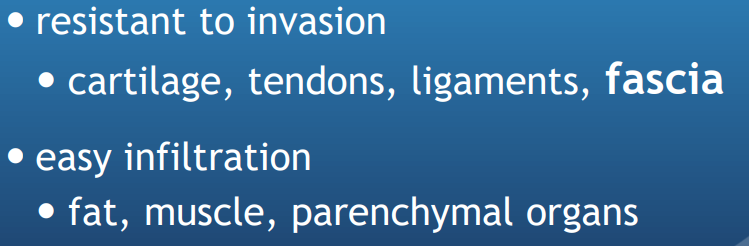
What tissues are resistant to invasion?
What tissues are easier for tumours to infiltrate?
Surgical margins
Goal= complete excision
& closure tissues without tension
Surgical margins
List the 4 different surgical margins that can be used for tumour removal
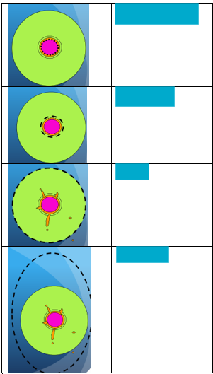
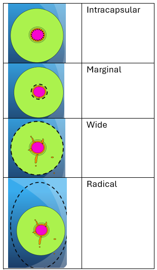
Intracapsular margin
What does this involve?
What types of tumours is it used for?
Why might tumour reoccur?
Intracapsular margin
Piecemeal removal
intracapsular dissection
Used for benign disease
Failure due to microscopic tissue

Marginal margins
What does this technique involve?
Why might failure occur?
In what situations would you do this?
Marginal margins
en bloc removal
both mass and pseudo capsule
Failure d/t satellite or skip metastases
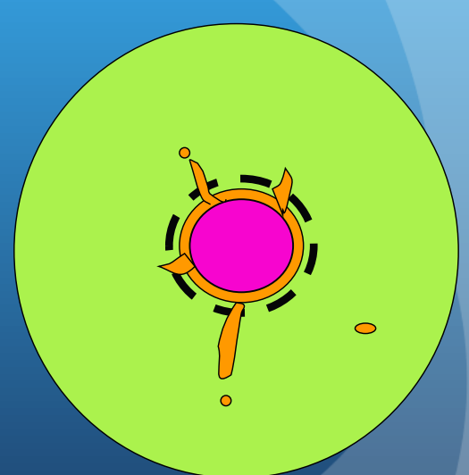
Used for benign tumours or cytoreduction
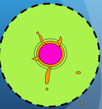
Wide margins
What does this involve?
Why might failure occur?
What types of tumours is it good for?
Wide margins
En bloc excision
mass + pseudocapsule + reactive zone + margin
Failure d/t skip metastasis
Good for skin & S/C tumours
Radical margins
What does this involve?
Why might failure occur?
What types of tumours is it good for?
Radical margins
en bloc removal
mass + entire tissue compartment
failure due to distant metastases
Used for poorly localised or high grade malignancies
When would surgery with wide margins fail?
Skip metastasis
Outgrowths of tumour (scattered cells & satellite mets —> branching down BV / lymphatic)
Generally good for skin & S/C tumours
How do you avoid tumour seeding?
Change gloves & instruments before closing so don’t spread tumour cells elsewhere
When would you remove LNs?
only remove if evidence mets (may be movement towards removal of tumours when staging)
enlarged, firm or fixed
microscopic confirmation (FNA)
What must you do once you have removed the mass?
Mark margins - will tell you if you have a clean edge once it gets to histopath
Bread slice (2cm) if it is large
What wound protection can you utilise?
Modified SPICA
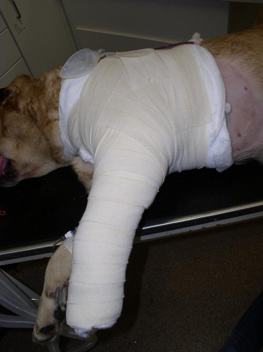
Robinson Sling (stops backwards movement of hindlimb for any wounds in the flank area)
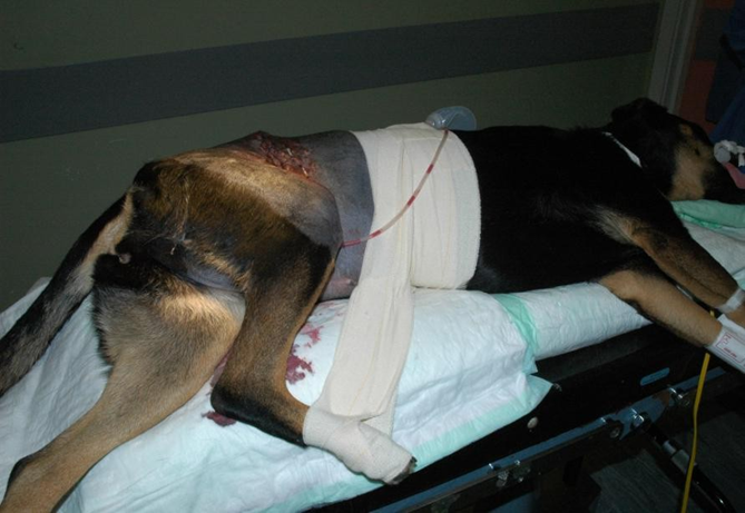
(for both don't use vet wrap which is too stretchy)
When would you re-examine the animal?
1, 2, 3 months
6, 9 & 12 months
then every 6 months
evaluate surgery site & regional LN