Urine examination, cells & casts
 Call Kai
 Learn
Learn Practice Test
 Spaced Repetition
 Match
Match Flashcards
 Knowt Play
1/36

There's no tags or description
Looks like no tags are added yet.
Name | Mastery | Learn | Test | Matching | Spaced | Call with Kai | Chat |
|---|
No analytics yet
Send a link to your students to track their progress
37 Terms
What are the 2 physical abnormalities of urine that would make you reflex to microscopy?
Abnormal color & clarity (hazy, cloudy)
What are the 4 (sometimes 5) chemical abnormalities (positive tests) of urine that would make you reflex to microscopy?
Blood
Protein
Nitrite → bacteria
Leukocyte esterase
lab-dependent: glucose
How do you prepare a urine sample for microscopy? What all may you need to identify in a urine sample?
Sample prep:
10mL of well-mixed urine → centrifuge 1500-2000rpm 5 min → remove 9 ml → resuspended 1 mL → load drop on KOVA slide → low power/low light or high power 40x w/ medium light
Identification:
Epi cells, WBCs, RBCs, Casts, Crystals, Organisms
If you do a screening test and see a dark/brown color, hazy/turbid clarity, have a positive blood, protein, nitrite, leukocyte esterase, and glucose test, what can the clinical significance be?
Dark/brown color: Blood
Hazy/Turbidity clarity: Hematuria vs. hemoglobinuria/myoglobinuria or pathogen
Positive tests:
Blood: RBCs or RBC casts
Protein: Casts, cells
Nitrite: Bacteria, WBCs
Leukocyte esterase: WBCs, WBC casts, bacteria
Glucose: Yeast
What is the difference between hematuria, hemoglobinuria and myoglobinuria?
Hematuria: Intact & whole RBCs, after centrifuge: red sediment & seen under microscope
Hemoglobinuria: free hemoglobin, after centrifuge: uniformly red liquid & no RBCs under microscope
Myoglobinuria: free myoglobin (muscle protein), after centrifuge: red/dark liquid & no RBCs under microscope
What are the 3 types of epithelial cells you may see in urine? Which cell has a subtype?
Squamous epithelial cells
Transitional epithelial cells
Renal Tubular epithelial cells
Oval Fat Bodies

What kind of cell is this? Describe how it looks. Where does it come from?
How do you report these cells (what power and how many)?
Usually these cells are considered normal findings in small numbers. If they are found in large amounts, what 2 diseases can they indicate?
Squamous epithelial cell: large w/ eccentric nucleus
Comes from distal part of urethra
LPF (10x) count in 10 fields: rare (0-5), few (5-10), moderate (10-25), many (25+)
Moderate-Many can indicate…
UTI
Possible cancer (if clumped/sheeted)

What kind of cell is this (compared to squamous)? Describe how it looks. Where does it come from?
How do you report these cells (what power and how many)?
Usually these cells are considered normal findings in small numbers. What is one case they may be found in many numbers but would still be considered normal in? If they are found in large amounts, what 2 diseases can they indicate?
Transitional epithelial cell: smaller than squamous. Spherical/oval or caudate shaped with large, round & central nucleus
Comes from renal pelvis, ureters, bladder, & upper portion of male urethra
High power (40x) & count 10 fields: rare, few, moderate, or many
Normal: high amount after catheterization
Moderate-Many caused by….
Malignancy (clumps & abnormal morphology)
Viral infection (clumps & abnormal morphology)
Why can shape vary so much for the transitional epithelial cells?
Due to their ability to absorb water

What kind of cell is this? Describe how it looks. Where does it come from?
How do you report these cells (what power and how many)?
These cells are only found in disease, even if there are only few amounts. What 2 diseases are they associated with? Include what can lead to tubular necorsis.
Renal tubular Epithelial cells (RTEC): smaller than transitional epi, very large nucleus & offset, cell can be round/oval/cuboidal & granular appearance
Comes from: PCT, DCT, & collecting duct
High power (40x) & 10 fields: rare, few, moderate & many
Disease:
Tubular necrosis
heavy metal/drug toxicity, viral infections, pyelonephritis, malignancy
Renal graft rejection
Renal Tubular epithelial cells are often mistaken for what other cell type? How do you determine if you are seeing a RTEC or the other cell?
Often mistaken for WBCs → refer to leukocyte esterase test (was it positive or negative?)
Positive esterase: likely WBC and not RTEC
Negative esterase: likely RTEC

What is the subtype of the renal tubular epithelial cells (pictured)? Describe their appearance & what they are.
What 3 diseases are these cells found in?
Oval Fat Bodies (RTEC): highly refractile, makes Maltese cross appearance w/ polarized light
RTECs that have lipids in them (hyperlipidemia)
Diseases:
Nephrotic syndrome
Lipid nephrosis
Terminal kidney disease

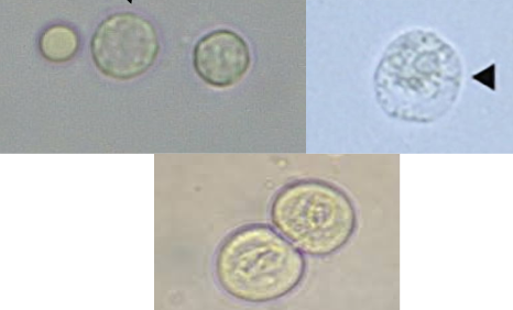
What kind of cell is this? Describe how it looks & what “normal” is.
How do you report these cells (what power and how many)?
If they are found to be >5 in urine, what is this called? What diseases are they associated with?
WBC (one is compared to RBC): normal amount is 0-5/HPF
Neutrophils: look glittery (bottom image) due to Brownian mvmnt. → may swell in hypotonic/alkaline urine
Lymphocytes: round nucleus, clear cyto, uncommon
Report: 40x (10 fields): 0-5, 5-10, 10-25, TNTC
Diseases:
Neutrophils: Infection → Pyelonephritis, cystitis, urethritis
Inflammation → Prostatitis, glomerulonephritis, acute interstitial nephritis
Lymphocytes → transplant rejection & malignancy
If WBCs are found in urine what are they correlated with? What test is usually positive? If it is positive but you don’t see WBCs why would that be? If that test is negative, why would that be?
Main correlation: bacterial infection
Test: Leukocyte esterase is usually positive
If positive but no WBCs seen: they have been lysed
If negative: WBCs are lymphocytes → don’t interact with reagent on test

What cell is this? Describe how it looks. How are they reported and what is the general reference for “normal”?
What two reasons may the finding still be “normal” if >5/HPF? What are the diseases associated with higher findings of this cell? What is the main disease you should think of first?
RBCs: usually small & slightly orange/green in color, no nucleus ; ref <5 normal
Can also be crenated (burst) due to hyperstenuric urine
Can also be enlarged in hypotonic urine
Can be very “faded” due to lysis in dilute/alkaline urine → ghost RBC
Report: by # seen in HPF, over 50 = TNTC
Normal >5:
Menstrual contamination
Strenuous exercise
Abnormal >5:
Glomerulonephritis
Renal vascular injury (stones, tumor)
Hypertension (high BP)
Acute infections (pyelonephritis, cystitis, urethritis)
What 3 things are sometimes confused with RBCs? How can you check if you are mis-identifying this for an RBC?
Oil droplets, crystals, yeast
Add glacial acetic acid → will lyse RBCs if present
If the stuff disappears → were RBCs
If stuff still there → No RBCs were present
What are RBCs correlated with? What will urine usually look like if it has RBCs?
What test is used to check for RBCs in urine?
What does it mean if the strip is positive but you don’t see any RBCs?
What if it’s negative but you see RBCs?
Correlation: Indicates damage to glomerular membrane or vascular injury in urinary tract
Urine will look red/brown and cloudy
Test: Hematuria
Positive but no RBC seen: RBCs lysed, hemoglobinuria or myoglobinuria
Negative but think RBCs seen: misidentification
What are casts mostly made of? What secrets this and where are the casts formed?
Made of: Tamm-Horsfall protein
Secreted by: RTEC of DCT
Formed in: DCT and collecting ducts
What 4 things can cause an increase in cast formation?
Dehydration (increased urine concentration)
Acidic urine
High protein concentration
Urinary stasis
What are the 2 normal casts that can be found in urine?
What are the 3 abnormal casts that can be found in urine? One abnormal cast has 3 subtypes, list them.
Normal:
Granular
Hyaline
Abnormal:
Waxy
Fatty
Cellular
RBC
WBC
Epithelial cells
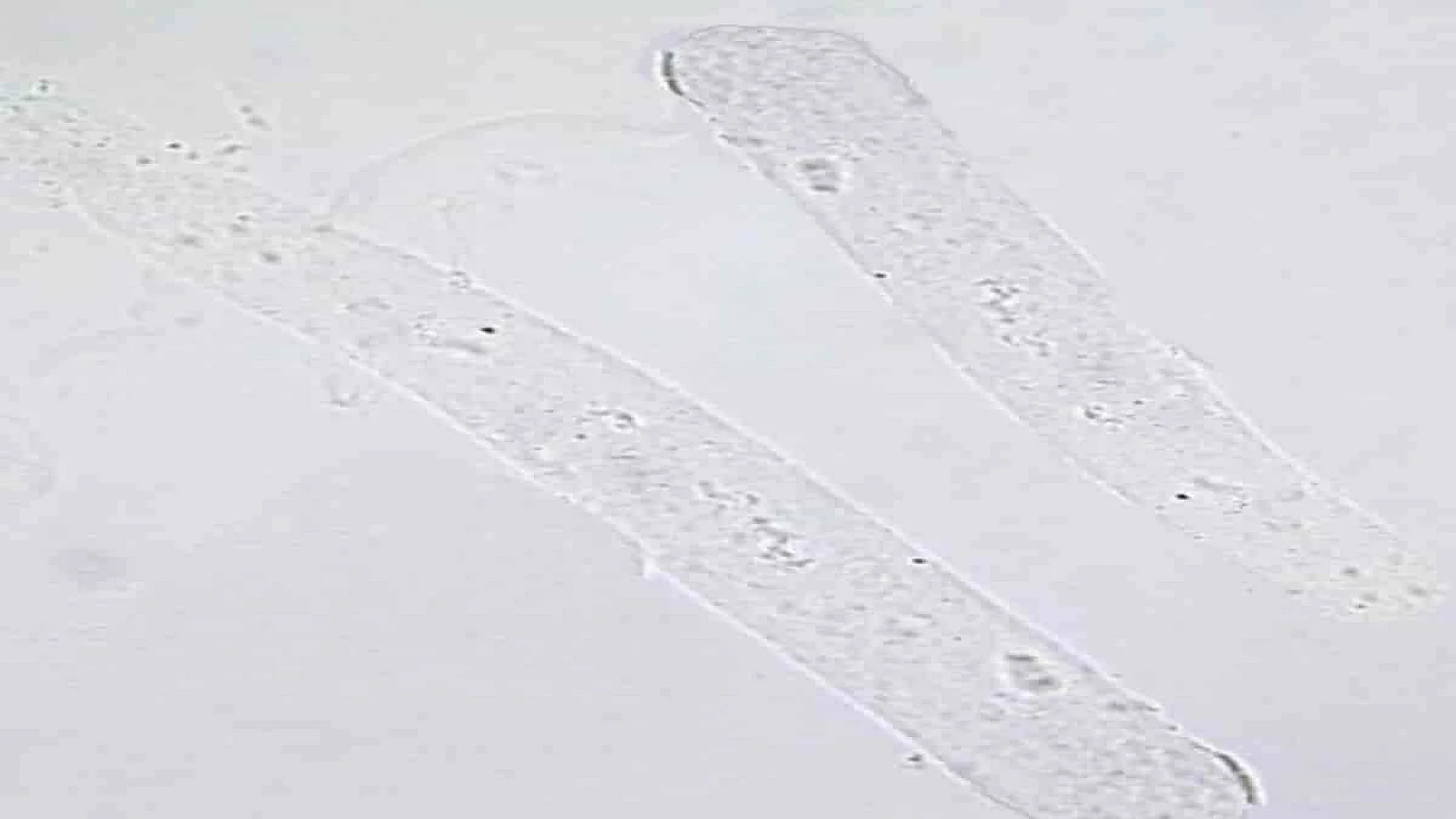
What kind of cast is this? Describe how it looks. What is the reference range?
Why may you see these in normal urine? If more than the normal reference range are seen, what diseases does it indicate?
Hyaline cast: colorless & smooth cylindrical shape
Reference: <2/LPF
Normal: strenuous exercise, heat exposure, dehydration, stress
Abnormal: >2
Glomerulonephritis
Pyelonephritis
Chronic renal disease

What kind of cast is this? Describe how it looks & what it’s made up in non-pathological conditions. What is it made up of in pathological conditions?
What is the reference range? Although this is usually a normal cast, what two diseases is it associated with?
Granular cast: may be coarse or fine granules
Non-pathological: lysosomes
Pathological: broken-down RBCs or WBCs
Reference: <2/LPF
Diseases:
Pyelonephritis
Prolonged renal disease

What kind of cellular cast is this? What disease is it associated with?
RBC cast
Disease:
Glomerulonephritis: damage to glomerular membrane

What kind of cellular cast is this? What diseases are they associated with?
WBC cast
Diseases:
Infection or Inflammation of nephron
Pyelonephritis (inflammatory renal disease)

What kind of cellular cast is this? What disease is it associated with, include the 2 subtypes.
Epithelial cell cast (RTEC in protein)
Diseases:
Advanced renal tubular damage
Acute nephritis
Tubular necrosis

What kind of cast is this? How is it formed and what disease is it associated with?
Waxy cast → “cracked” & “broken off” ends
Formed: extreme urinary stasis
Disease: Severe chronic renal failure

What kind of cast is this and what is it made of? What 2 diseases is it associate with?
Fatty cast → oval fat bodies (RTEC w/ lipids) or free fat droplets
Diseases:
Nephrotic syndrome
Tubular necrosis
How can casts be examined under a microscope?
Examine under low power & low light
Count casts & report avg. # → identify under high power (40x)
What are 5 things could potentially be contaminants from another part of the body (not originally in urine)? These contaminants would need further testing to determine origin. What is the most common contaminant?
Yeast (common) → further testing needed, cuz present in UTI
Trichomonas
Sperm
Parasites → further testing needed, cuz could be UTI
Bacteria → further testing needed, cuz could be UTI

What artifact is this?
Starch granules

What artifact is this?
Fiber strands

What artifact is this?
Hair

What artifact is this?
Talc (baby powder)

What artifact is this?
Pollen grain

What artifact is this?
Glass

What artifact is this?
Air bubbles

What artifact/contaminant is this?
Yeast