Topic 10: Protective Mechanism of Brain - Part 1
 Call Kai
 Learn
Learn Practice Test
 Spaced Repetition
 Match
Match Flashcards
 Knowt Play
1/16

There's no tags or description
Looks like no tags are added yet.
Name | Mastery | Learn | Test | Matching | Spaced | Call with Kai | Chat |
|---|
No analytics yet
Send a link to your students to track their progress
17 Terms
meninges
Protective layers of the brain that function to protect and stabilize the CNS
Includes: dura, arachnoid, and pia mater
dura mater
Meninges layer that is dense, thick, tough, and fibrous.
attatched the inner surface of teh skull, overlies the archnoid mater
Structures: falx cerebri, tentorium cerbelli, falx cerebelli

falx cerebri
Structure of the dura mater: longitudinal, separates cerebrum
acts as a protective layer around the cerebrum

tentorium cerebelli
Structure of the dura mater that is located above the cerebrum, arises from the temporal bone and from the tentorial notch
separates cerebrum from cerebellum and brainstem

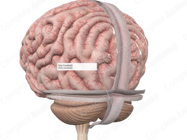
falx cerebelli
Structure of the dura mater that separates the cerebellar hemispheres
epidural space
Potential space around the dura mater that is in between the dura and the bone

subdural space
Potential space around the dura mater that is between the dura and the arachnoid

hematoma
A rupture of a blood vessel inside the head
arachnoid mater
The middle meningeal layer, thin spiderweb-like
Structure: arachnoid trabecula, arachnoid granulations
located above subarachnoid space

arachnoid trabecula
Structure of the arachnoid mater that is a fibrous, elastic connective tissue
Function: maintains subarachnoid space and allows for cerebrospinal fluid to occupy the space

arachnoid granulations
Structure of the arachnoid mater (called “villi”) function to drain cerebrospinal fluid from the subarachnoid space

subarachnoid space
Layer of space located between arachnoid and pia mater
contains arachnoid trabecula (to maintain space) and cerebrospinal fluid
part of the venous system (large blood vessels)
encloses the entire CNS

pia mater
Layer of the meninges that is thin, transparent, delicate, and vascular
Layer adheres to entire brain surface (like leggings)
Layer in which choroid plexus is formed

choroid plexus
Network of vascular capillaries (small, numerous blood vessels)
Located mostly in lateral ventricles and 4th ventricle
Formed in Pia Mater
Produces: cerebral spinal fluid, works to turn Blood —— CSF

middle meningeal artery
Largest artery in the CNS, most likely to hemorrhage - supply blood to meninges

meningeal veins
Veins within the superior sagittal sinus - supply blood to meninges

meningitis
Inflammation of the meninges - 2 types
Viral - mild, common, quick recovery
Bacterial - severe, possible brain damage, potentially life-threatening