Clinical Standards and Guidelines/Measurement Techniques and Quantification
 Call Kai
 Learn
 Practice Test
 Spaced Repetition
 Match
 Flashcards
 Knowt Play
1/67

There's no tags or description
Looks like no tags are added yet.
68 Terms
You are scanning a patient with mirror-image dextrocardia. Your transducer is on the chest, positioned for the PLAX view that you just obtained. From this position, how do you rotate the transducer to obtain a PSAX view, and where will the transducer marker be pointed?
Clockwise rotation (transducer marker pointing to the patient’s left hip)
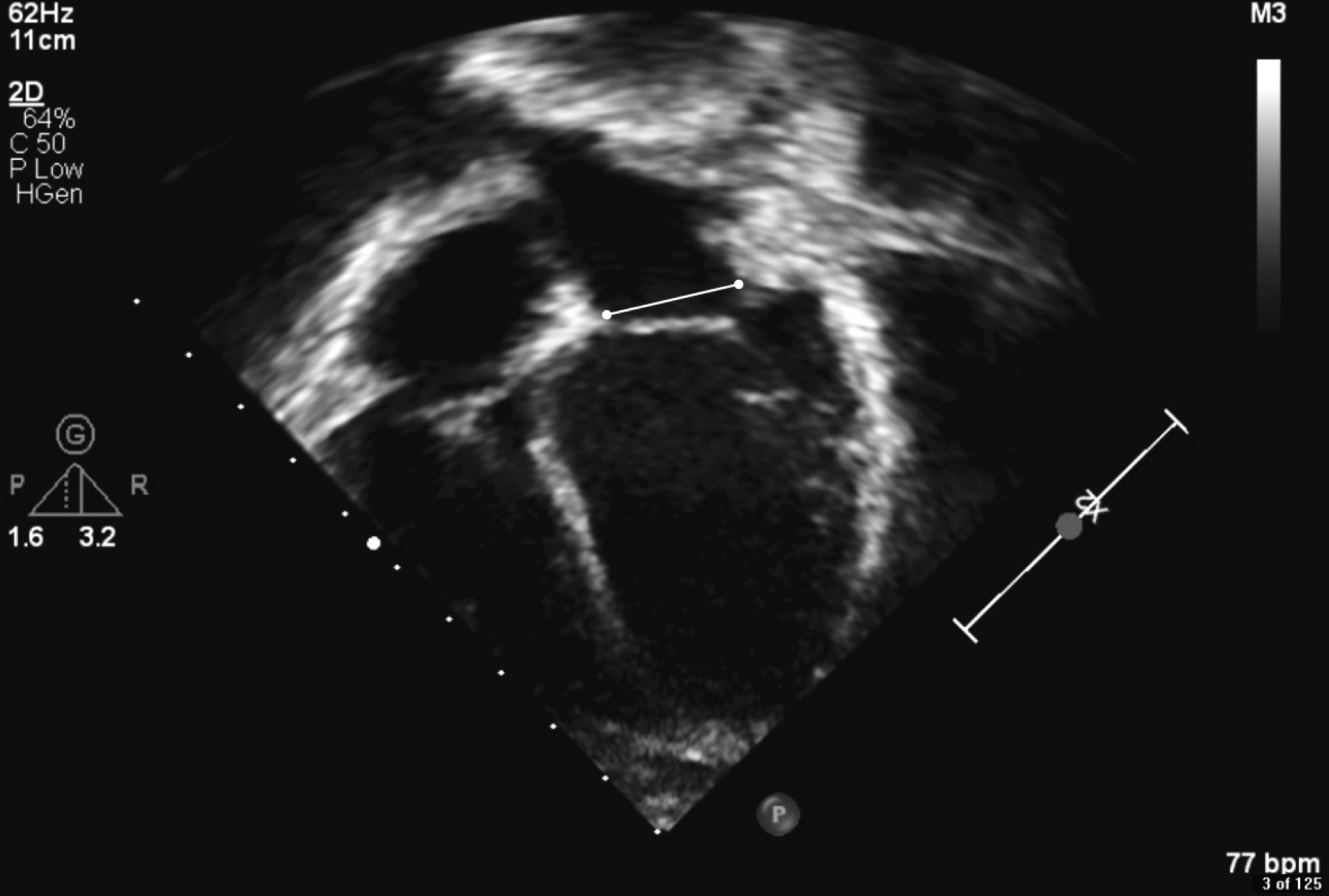
In this image of the mitral valve annulus, how would you best characterize the placement of the calipers?
Incorrect for measurement of the annulus because the mitral valve is closed
Under normal circumstances, a PDA is generally seen closest to which segment of the aortic arch?
Aortic isthmus

What is the problem with this spectral Doppler tracing?
Poor angle of isonation
You may use the simplified Bernoulli equation to calculate a pressure gradient that would be considered accurate in a patient who has?
Pulmonary valve stenosis
While undergoing chemotherapy two years ago, the patient you are about to image had a reduced left ventricular ejection fraction of 40$ and a global longitudinal strain (GLS) of -12%. Your echocardiogram now shows a normal LVEF and a normal GLS. Which value represents a normal GLS?
-20%
What does dP/dt measure?
Ventricular contractility

In this subcostal short-axis image, not all cardiac information is visible. What maneuver should the operator perform to improve the imaging?
Move the “tail” of the transducer toward the feet (more inferiorly)
You visualize an L wave in the spectral Doppler tracing from a patient with abnormal relaxation and a slower heart rate. Which option best describes the timing of the L wave?
Diastasis
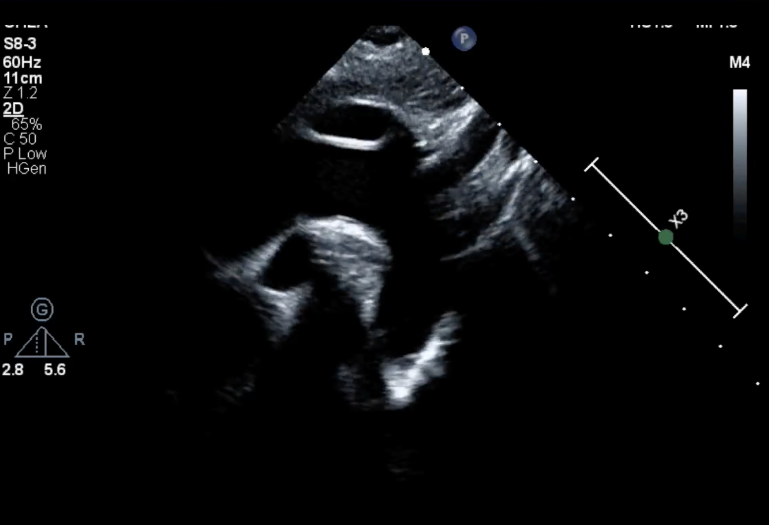
Where is the proximal transverse arch measured?
Between the innominate artery and the left common carotid artery
You obtain a peak velocity of 2.3 m/sec across the aortic valve from the apical five-chamber view. The spectral Doppler signal is clear and parallel to the blood flow. If this patient also has a large VSD, what do you think of the measurement of the aortic valve peak velocity?
It is underestimated
An infant boy has been diagnosed with a large VSD. His vital signs include a blood pressure of 70/50 mmHg. The tricuspid valve regurgitation (TR) jet is trivial, with an incomplete spectral Doppler signal and a peak velocity measured at 2.5 m/sec. What is your estimation of RVSP?
70 mmHg
Which statement about continuous-wave (CW) Doppler is true?
Continuous-wave Doppler can measure high velocities
A 5 MHz curvilinear array transducer is best suited for which of these tasks?
Obstetric/fetal imaging
In which scenario is contrast echocardiography using agitated saline not useful?
Blalock-Taussig shunt latency
For a person reading an echocardiogram, which echocardiographic view best displays the spatial relationship between the great arteries?
PSAX
Which type of transducer should be used for 3D/4D imaging?
Matrix array probe
A patient has a perimembranous VSD, and spectral Doppler shows a peak velocity across the defect of 4 m/sec. What is the peak gradient through the defect?
64 mmHg
What is the normal Qp/Qs value?
1

Which dimension of the tricuspid valve is being measured?
Anterior-posterior diameter
Increased LA volume is a marker of?
Chronic left ventricular diastolic dysfunction

What type of artifact is associated with the atrial septum seen in this image?
Image dropout artifact
You are about to perform an echocardiogram on a young adult with a parachute mitral valve and mitral stenosis. What will the mitral valve inflow pattern, recorded with pulsed-wave Doppler, tell you about diastolic function?
Findings are not reliable; diastolic function cannot be assessed in this setting
Which view is the best to demonstrate the azygos vein draining into the SVC?
Suprasternal sagittal view
The use of agitated saline during echocardiography is not helpful for diagnosing?
Aortopulmonary collaterals
You are asked to perform an echocardiogram on a patient whose heart, on chest x-ray, was noted to be located in the right chest (dextrocardia). There is a question as to whether the patient has congenital heart disease. Which of the following represents the best initial echocardiographic approach?
Subcostal long axis sweep
Your patient is a neonate who weighs about 6.5 pounds (3 kilograms) with mild hypoplasia of the left-sided structures. A biventricular repair is being contemplated. In the PLAX view, which of the following transducers would be most appropriate for obtaining measurement?
12 MHz phased array
If the ascending aortic diameter is 12 mm, what measurement for the proximal aortic arch is considered hypoplastic?
7 mm
You are calculating LVEF with the area-length method. Which of these views will you use to obtain the left ventricular length?
Subcostal long axis
A 5-year old patient with mitral stenosis due a single papillary muscle (parachute mitral valve) and no other form of congenital heart disease is being seen by a cardiologist. The mean pressure gradient for this mitral valve was recorded as 8 mmHg by echocardiography. The mean gradient tells you that the degree of mitral valve stenosis is?
Moderate

Which number represents systole?
1
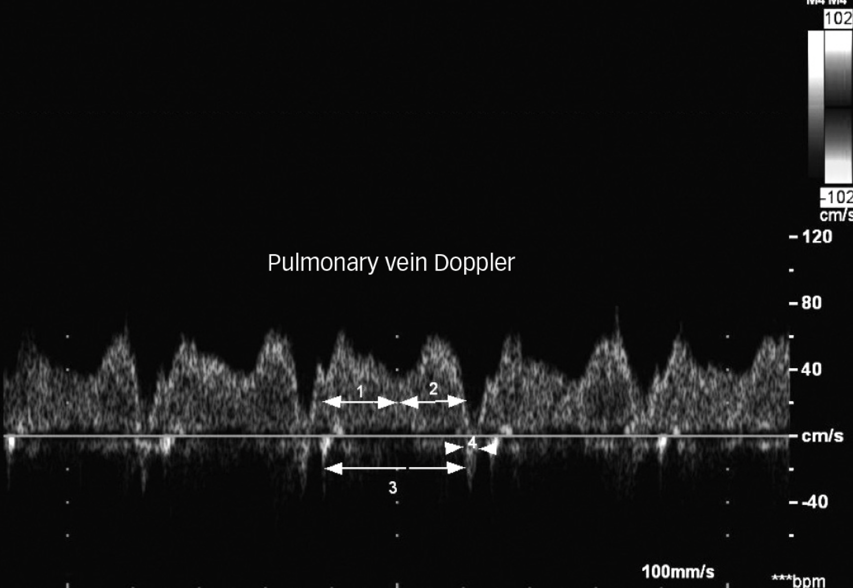
Which number represents early diastole (ventricular filling)?
2
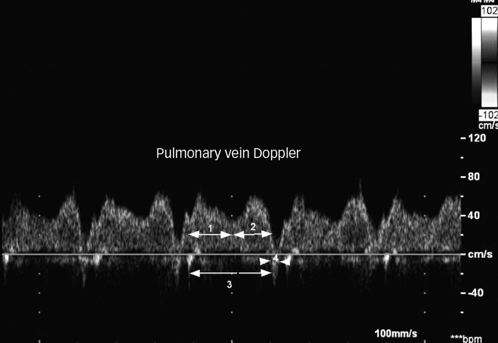
Which number represents the combined portions of 1 and 2?
3

Which number represents atrial contraction?
4
Global left ventricular longitudinal strain?
Is negative when normal
What is the purpose of adjusting TGCs?
Compensate for attenuation by boosting signal amplitudes at different depths
Which of the following echocardiographic views is best to demonstrate a left superior vena cava?
Suprasternal coronal view
What is the best transducer position and maneuver to optimize visualization of the innominate (brachiocephalic) vein?
High left parasternal view with the transducer notch/dot at 3 o’clock; transducer should be angled superiorly

This image is an apical four-chamber view in the setting of L-looped ventricles. What is being measured?
Right ventricular length
While using a parasternal long-axis view, you sweep toward the tricupisd valve and suspect a VSD because you see red color flow that might be crossing the ventricular septum. What will you do further to investigate the red color flow profile?
Use continuous-wave spectral Doppler (if the flow is systolic, a VSD is possible)

This image from a spectral Doppler tracing of the descending abdominal aorta most likely demonstrates which of the following?
Aortic valve regurgitation (diastolic flow reversal)
As mitral valve stenosis becomes worse, mitral valve pressure half-time?
Becomes longer
Which of the following values is not needed for the echocardiographic calculation for cardiac output?
Peak velocity across the aortic valve
In the presence of Ebstein anomaly, a Celemajer index greater than 1 indicates?
Poor prognosis in the setting of Ebstein anomaly of the tricuspid valve

What value would be most useful in diagnosing the anomaly demonstrated?
Displacement index
A patient has a perimembranous VSD, and spectral Doppler shows a peak velocity across the defect of 4 m/sec, with a blood pressure of 120/70. What is the estimated RVSP?
56 mmHg (120 mmHg - 64 mmHg)

Which of the following cardiac defects has the spectral Doppler profile shown in this image?
Coarctation
In HLHS, the anatomic variant with mitral stenosis and aortic atresia is most likely to be associated with?
Endocardial fibroelastosis
A patient has an ASD with left-to-right shunting. Which of the following is most likely the Qp/Qs ratio for this patient?
1.5:1

This spectral Doppler image was obtained from the pulmonary vein of an infant. Which statement best describes the state of the left ventricular diastolic function in this patient?
Triphasic pulmonary venous inflow waves are seen during atrial filling and do not indicate diastolic disease

Which letter marks the distal transverse aortic arch?
C

Which letter on the spectral tracing corresponds with isovolumic contraction time
C
Which of the following standard imaging views is best to evaluate the individual pulmonary veins?
Suprasternal coronal view
Tricuspid valve stenosis is least associated with?
Obligatory left-to-right shunting
Which of the following factors will not affect pressure half-time in the calculation of mitral valve area?
BSA
An L wave is most often seen in which of the following settings?
Abnormal relaxation and slower heart rates
What does a prolonged pulmonary A wave suggest in a pediatric patient?
Diastolic dysfunction
What is the normal value for right ventricular fractional area change (RVFAC)?
>35%

Identify the letter on the echocardiogram that corresponds with late ventricular diastolic filling
C
The mean gradient across your patient’s aortic valve is 45 mmHg, and there is preserved left ventricular systolic function. What is the most likely diagnosis?
Severe AS
In a 2-year old boy with coarctation of the aorta, you use an 8 MHz transducer for the entire study, but you cannot obtain an ideal Doppler signal. What is your next step?
Change to a 5 MHz transducer
What value, expressed as a percentage, is calculated by this formula: (LVEDV - LVESV)/LVEDV x 100?
Ejection fraction by biplane Simpsons method
Z-scores are often used to compare certain important cardiovascular structure measurements in the pediatric population. What is a Z-score for a specific cardiac structure?
A value based on the standard deviation around a mean value for that particular cardiac structure which varies by BSA
The Qp/Qs ratio represents the ratio of?
Pulmonary blood flow to systemic blood flow
Which of these measurements is not needed when you are calculating the myocardial performance index for a left ventricle?
R-R interval
Your patient has normal left ventricular systolic function. Continuous-wave Doppler measurement of the peak velocity across the aortic valve is 3.5 m/sec. Based on this measurement, what is the severity of aortic valve stenosis?
Moderate
What is the formula for calculating left ventricular volume when you are using the area-length method?
5/6 x short axis basal area x LV length
When does mitral regurgitation occur in the cardiac cycle?
Ventricular systole, closest to the QRS complex