10-11
 Call Kai
 Learn
Learn Practice Test
 Spaced Repetition
 Match
Match Flashcards
 Knowt Play
1/51

There's no tags or description
Looks like no tags are added yet.
Name | Mastery | Learn | Test | Matching | Spaced | Call with Kai | Chat |
|---|
No analytics yet
Send a link to your students to track their progress
52 Terms
Small Intestine
Principal site of digestion and absorption of amino acids, carbohydrates, lipids electrolytes and water; divided into duodenum, jejunum and ileum, bile and pancreatic enzymes enter the duodenum via the hepatopancreatic ampulla
Duodenum
Approximately 25 cm long; receives chyme, bile, and pancreatic secretions and is distinguished histologically by Brunner glands in the submucosa
Jejunum
Approximately 2.5 m long; has prominent plicae circulares, and villi but lacks Brunner glands and large aggregated Peyer patches
Ileum
Approximately 3.5 m long; has fewer/smaller plicae and villi and is characterized by prominent aggregated Peyer patches
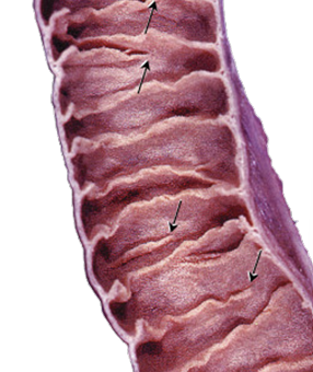
Plicae Circulares
Permanent folds of mucosa and submucosa that increase absorptive surface area; prominent in the distal duodenum and jejunum and decrease toward the ileum
Villi
Finger-like projections of mucosa that cover the small intestine and contain blood capillaries and lymphatic lacteals for nutrient transport
Crypts of Lieberkuhn
Small openings between villi that contain absorptive and goblet cells, paneth cells, enteroendocrine cells and stem cells

Microvilli
Tiny apical plasma-membrane projections on enterocytes that form the brush border and provide the greatest increase in intestinal surface area
Small Intestinal Surface Area
Plicae circulares increase area about 3-fold
Enterocytes
Most abundant intestinal epithelial cells; specialized for absorption and transport from the lumen to circulation and contain a brush border that greatly increases surface area, microvili contain contractile microfilaments, bound by tight junctions, used in lipid absorption and processing
Goblet Cells
Unicellular mucus-secreting glands that increase in number from the duodenum toward the ileum and protect/lubricate the intestinal epithelium, contain accumulation of mucinogen granules at the apex
Paneth Cells
Granule-rich cells at the base of intestinal crypts (in the mucosa) that secrete antimicrobial substances including lysozyme and defensins and help regulate normal intestinal flora
Defensins
Antimicrobial proteins from Paneth cells that form ion channels/increase membrane permeability of target organisms such as bacteria and parasites
Lysozyme
Antimicrobial enzyme secreted by Paneth cells that damages bacterial peptidoglycan and increases bacterial permeability
Enteroendocrine Cells
Diffuse neuroendocrine cells concentrated in crypts that release peptides with endocrine and paracrine effects to coordinate digestion secretion and motility, can reach the lumen but some will not
Distribution of GI Hormones

Gastrin
Produced primarily by G cells in the gastric antrum; stimulates gastric secretion and proliferation of gastric epithelium
CCK
Produced by enteroendocrine cells of the small intestine; stimulates pancreatic enzyme secretion and gallbladder contraction/movement
Secretin
Produced by s cells in the duodenum; stimulates secretion of water and bicarbonate
Motilin
Produced in the small intestine; stimulates motility of the stomach and small intestine and contributes to the migrating motor complex
Duodenum features
Brunner’s glands in the submucosa, alkaline-mucous and bicarbonate-secreting glands (pH of 8.1-9.3 to neutralize the acidic chyme entering the small intestine)
Jejunum features
NO Brunner’s glands, NO aggregate Peyer’s patches
Ileum features
Peyer’s patches (decide whether immune intervention is needed or not), M cells (endocytose proteins to allow lymphocytes to respond to antigens)
Large Intestine features
Teniae coli (thickened bands of longitudinal muscularis externa, forms →), haustra (saculations), epiploic/omental appendages (fat filled pouches of visceral peritoneum), crypts with no plicae/villi, increased number of goblet cells as you go further down
Appendix
Has numerous lymphatic nodules extending into the submucosa and contains Paneth cells; its prominent lymphoid tissue contributes to immune function
Rectum
Terminal portion of the large intestine beginning around S3; contains lymphatic nodules
Anal Canal
Terminal portion of the GI tract extending from the rectal ampulla to the anus and controlled by internal involuntary and external voluntary anal sphincters, keratinized near the opening, anal glands present in the submucosa
Liver
Exocrine (secretes bile into ducts) and endocrine (produce/release albumin, clotting factors, transport and lipoproteins into blood) organ, processes, stores (vitamin A, iron, thyroid hormone, vitamin D), synthesizes, detoxifies, and secretes
Liver blood flow
From portal triad to central vein, classic lobule= blood drainage unit
Classic hepatic lobule
Blood flow to the central vein from the hepatic artery and portal vein, moves from corners to the center (blood to the center)

Portal Lobule
Bile flow towards the bile duct (from the hepatocytes) (bile to the border)

Hepatic acinus
Supplies oxygenated blood to the hepatocytes
Zone 1 closest to portal triad, most oxygen, most active zone
Zone 3 closest to central vein, least oxygen, most likely to be injured in an ischemic event
Liver lymph flow
Opposite direction of blood, from hepatocytes through Space of Disse toward portal region, DO NOT MIX: bile and lymph, bile and blood (jaundice), lymph with blood
Sinusoid vs Space of Disse
Sinusoid= blood space, Space of Disse= exchange/lymph space
Portal Triad
Contains branches of the hepatic portal vein, hepatic artery, and bile duct; mnemonic VAD = Vein Artery Duct
Hepatic Sinusoids
Specialized vascular spaces where portal venous and arterial blood mix and flow past hepatocytes toward central veins (sinusoids= blood)
Blood Flow Through Liver
Portal vein + hepatic artery → sinusoids → central vein → sublobular vein → collecting vein → hepatic vein → inferior vena cava
Bile Flow Through Liver
Hepatocytes → bile canaliculi → canals of Hering → bile ductules → intrahepatic bile ducts → hepatic ducts → common hepatic duct → common bile duct → duodenum
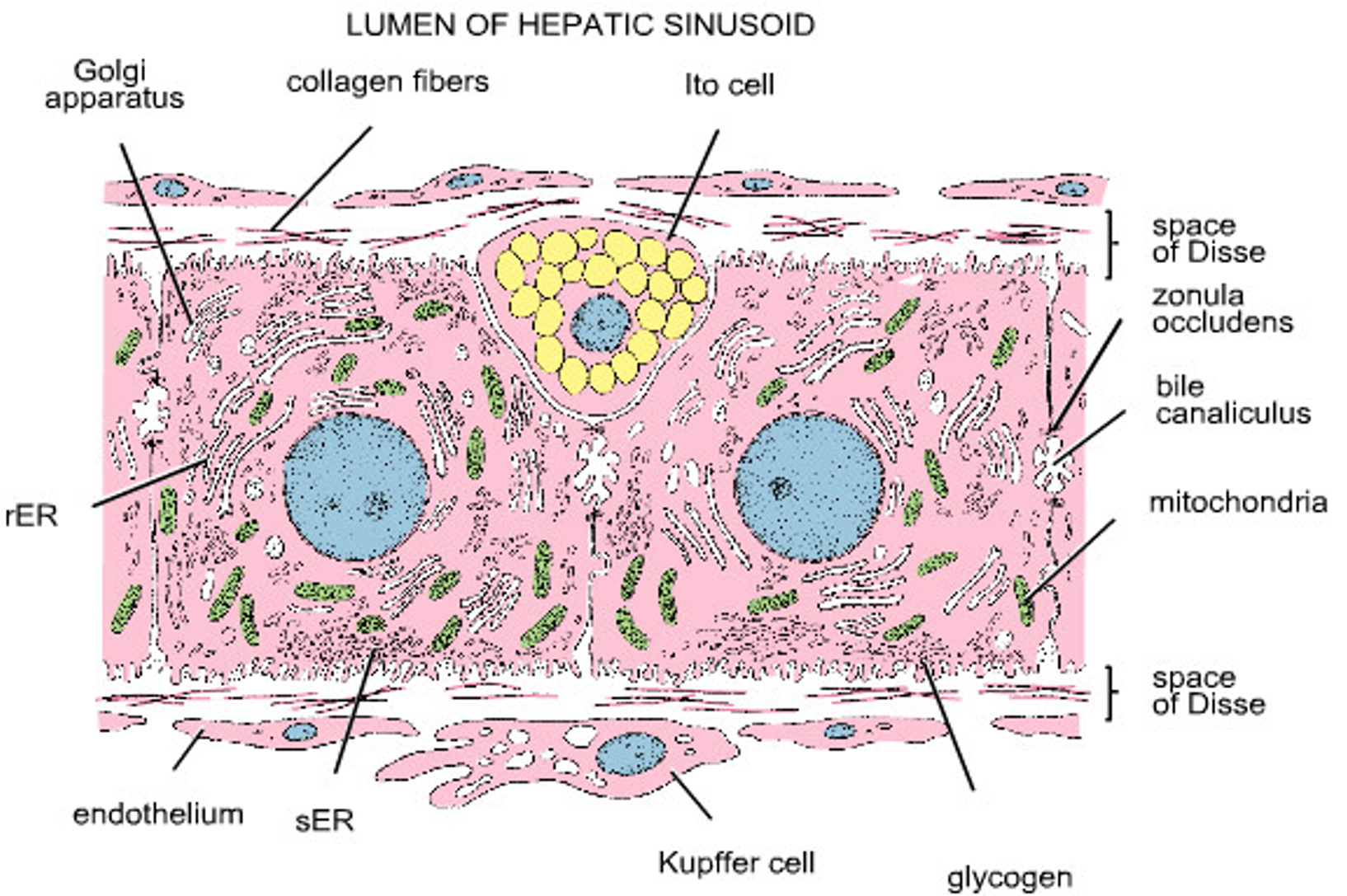
Hepatic Stellate/Ito Cells
Cells located in the Space of Disse that normally store vitamin A; when activated by chronic injury they produce collagen and contribute to hepatic fibrosis
Kupffer Cells
Resident macrophages within hepatic sinusoids that remove bacteria, debris and aging blood cells from portal blood, clean the blood before it leaves the liver

Bile Functions
Provides bile salts for lipid absorption and serves as a route for excretion of cholesterol phospholipids conjugated bilirubin electrolytes and metabolites of drugs/toxins
Bile Canaliculi
Tiny channels formed between adjacent hepatocytes that collect bile; they are sealed from the blood-facing surface by tight junctions and are not independently epithelial-lined ducts, between hepatocytes, not between hepatocytes and blood
Bile acid cycle
Most bile acids are secreted into the intestine, reabsorbed mainly in the ileum, and returned through portal blood to the liver for reuptake and resecretion into bile, small fraction newly synthesized from cholesterol and conjugated with glycine or taurine
Canals of Hering
Short transitional passages connecting hepatocyte bile canaliculi to cholangiocyte-lined bile ductules
Hepatic pathway
Sinusoids → central vein → subobular vein → collecting vein → hepatic vein → inferior vena cava
Gallbladder
Muscular sac that stores and concentrates bile between meals by absorbing water and electrolytes; does not produce bile, lipids entering the duodenum → CCK release from enteroendocrine cells→ gallbladder contraction
Gallbladder features
Simple columnar epithelium, microvilli, junctional complexes, large concentrations of mitochondria in apical and basal cytoplasm and lateral plications, recognition pattern= highly folded mucosa and simple columnar epithelium, no muscularis mucosae or submucosa
Gallbladder Bile Pathway
Liver → right/left hepatic ducts → common hepatic duct; bile can enter the gallbladder through the cystic duct between meals and leave through the cystic duct into the common bile duct during digestion (cystic duct= two way connection)
Pancreas
Mostly retroperitoneal organ with both exocrine (produces digestive enzymes and bicarbonate entering the duodenum through ducts) and endocrine functions (islets release hormones into the blood)
Pancreas features
Dark-staining exocrine acini surround scattered pale islets of Langerhans; unlike salivary glands the pancreas lacks striated ducts and myoepithelial cells
Pancreatic exocrine excretions
Acini= make enzymes (many stored as inactive zymogens to prevent digestion of pancreas), ducts= bicarbonate (helps neutralize chyme)
Centroacinar Cells
Pale cells located in the center of pancreatic acini that represent the beginning of the duct system; key histologic feature of the pancreas, unlike salivary glands with striated ducts, secretin stimulates release of bicarbonate (CCK= acinar cells)