Invasive Procedures
 Call Kai
 Learn
 Practice Test
 Spaced Repetition
 Match
 Flashcards
 Knowt Play
1/73

There's no tags or description
Looks like no tags are added yet.
74 Terms
femoral stick purpose
to obtain blood from the femoral artery or vein for analysis when there is no other peripheral site available
femoral stick contraindications
overlying cellulitis or hematoma
known venous or arterial thrombosis
known graft site
femoral stick anatomical landmarks
pt supine, legs extended
palpate artery 2 fingerbreaths below inguninal ligament
vein 1cm medial to artery
NAVY mnemonic
NAVY mnemonic
Nerve
Artery
Vein
“Y” shape of the groin

femoral stick supplies
20-30cc syringe
19G, 1.5” needle
heparinized syringe if ABG
chlorhexidine
alcohol swab
4×4
tubes for blood analysis
femoral stick preparation
informed consent if not an emergency
assemble needle and syringe
palpate artery w/ left hand, do not remove
swab w/ chloro-prep, allow to dry
swab w/ alcohol (skip if determining EtOH levels)
femoral stick procedure
90 angle to vessel
straight down w/ aspiration until blood withdrawn
withdraw desired amount
apply pressure to artery for at least 10 min
place blood in tubes for analysis
femoral stick complications
hematoma
infection
thromboembolus
septic arthritis of hip
femoral nerve damage
psoas abscess
penetration of viscous
intraosseous line
placement of a line into the marrow cavity of a bone to provide emergency access for medication and fluid resuscitation

intraosseous line placement purpose
emergency “venous” access
provides rapid access only when a peripheral line not available
can administer any fluids, bloods, or meds (except adenosine)
IO line CI
previous attempt in same bone
fracture
overlying infection
IO line anatomical landmarks
sandbag under knee for support
medical surface of tibia
1-3cm below tibial tuberosity
flat portion easily palpable
IO line placement supplies
IO or bone marrow needle
10cc non-leurlock syringe
chloro-prep
IV fluids, assembled w/ tubing
Is informed consent for an IO line necessary?
not necessary as this is a life saving, emergency procedure
IO line placement procedure
insert needle w/ stylet in place at 90 angle to bone using rotating motion
alternatively, 60 angle inferiorly
when resistance “gives way” stop advancing
remove stylet, withdraw marrow

How is IO line placement confirmed?
when marrow is withdrawn, or free flow of IV fluid w/ no evidence of soft tissue extravasation

IO line complications
accidental soft tissue placement
infection
fracture
IO line clinical pearls
No “2nd tries”
tibia, humerus, and sternum are only recommended sites, but iliac crest and distal femur
remove as soon as peripheral line is obtained
max 24 hrs
central venous lines
insertion of large bore catheter into a large proximal vein for administration of fluid or medication
central venous line indications
unable to obtain peripheral access
central venous monitoring - Swan-Ganz, CVP
frequent blood draws
chemotherapy
hyperalimentation
central venous lines anatomy
internal jugular vein
subclavian vein
femoral vein
tip of catheter should be in the SVC for jugular and subclavian placement
IVC below renal veins for femoral placement
central venous line - Internal Jugular
right side preferred
posterior approach just inferior to external jugular, aiming for midsternal notch
anterior approach at apex of triangle b/w 2 heads of SCM and clavicle, aiming for ipsilateral nipple

central venous line - subclavian vein
right side preferred
palpate tubercle of clavicle 1/3 to ½ across
1-2 cm below this spot
aim for sternal notch w/ minimal angle off skin
“Hit” clavicle and use the other hand to coax underneath - pops right into vein

central venous line - femoral vein
same landmarks as femoral stick
angle superiorly
tip of catheter should be in IVC, below renal vessels
central venous lines supplies
central venous kiit - multiple lumen catheters, percutaneous introducers
chlorhexidine, goggles, gown, drapes
flushes and IV fluid for line
dressing kit
central venous lines preparation
always obtain informwed consent unless emergenct
anxiolytics or sedation may be required
locate landmarks
positioning is key
assistant may be required
central venous lines procedure
clean and drape
reconfirm landmarks
local anesthetic
insert catheter using Seldinger technique
draw blood from each port
flush w/ saline, clamp off
suture line in place
Seldinger technique
J-type guide wire curls spontaneously minimizing damage to intima of vessel
should be pointed inferiorly in subclavian approach to prevent wire from going up jugular instead of down into SVC
insert large bore needle w/ syringe attached through skin, constantly withdrawing until venous flashback is obtained, stop advancing and hold needle in place
remove syringe, insert guidewire through needle until only 2-3cm remain protruding, NEVER LET GO OF GUIDE WIRE
remove needle, leaving guide wire in place
enlarge entry point using tip of a scalpel blade
pass dilator through catheter, advance to skin leaving 1-2cm of wire exposed
slowly remove guide wire from skin through catheter until end of wire appears out open end of catheter
advance catheter/dilator complex into vessel, use forward pressure and twisting motion advancing catheter throguh skin to hub od catheter
remove dilator and guide wire simultaneoulsy from catheter

central venous lines aftercare
always confirm venous placement w/ blood draw
if aterial placement suspected, run ABG
CXR to r/o PTX, and confirm placement in SVC
some institutions will not allow use of line until placement confirmed by CXR
central venous lines complications
bleeding - local, internal, and hematoma
infection
PTX, hemothorax, hydrothorax
air, catheter, and venous embolus
arrhythmia
knotting and kinking
Lumbar puncture (LP)
removal of fluid from dural space
LP purpose
to obtain CSF for analysis - meningitis or other neuro dz, subarachnoid hemorrhage
to remove excess fluid (in normal pressure hydrocephalus)
LP CI
cerebral edema - increasing HA
mass lesions - tumor, brain abscess
localizing sxs
rapid deterioration
papilledema
hx or physical suggesting sinusitis, otitis media, or endocarditis
LP anatomical landmarks
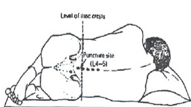
L4-L5 or L5-interveterbral space

LP positioning
horizontal line b/w posterior iliac crests localizes proper vertebral space
easiest to locate space when pt is maximally flexed
may also be done when pt is sitting
LP supplies
betadine, gloves, mask, gown, goggles
LP tray - 3cc syringe, 22/25G needles, 1% lidocaine, 25/22G spinal needle, 3-way stopcock, manometer, sterile field, tubes 1-4, 4×4, band-aid
LP preparation
always obtain informed consent
positioning is EVERYTHING
locate landmark and mark w/ fingernail or surgical marking pen
mask and gown
open tray using sterile technique
pour proviodine
gloves
get comdy
prep w/ betadine
drape
LP procedure
find fingernail mark and reconfirm
raise “wheal” w/ lidocaine
inject to bone w/ 3cc of lidocaine changing to larger needle when skin adequately anesthetized
insert spinal needle stylet in, bevel up aiming for umbilicus
when “pop”, remove stylet and check for fluid
if no fluid, reinsert stylet and continue to advance, checking every 1-2mm for penetrance
measure opening pressure
withdraw 4 tubes in succession 1-3cc each
replace stylet and remove needle slowly
hold pressure for several minutes, apply band-aid
LP aftercare
lay flat for 4 hrs, prone vs supine
IV or PO fluids encouraged
caffeine - coffee, coke, no-doze
LP complications
post LP HA
intraspinal epidermoid tumors
epidural, subdural, or SAH
infection
herniation
LP MC tests
tube 1 and tube 4 - CBC
tube 2 and tube 3 - protein, glucose, Gram stain, culture
LP other tests
VDRL
ANA
protein electrophoresis
crypto, toxo, viral cultures
other fungal cultures
cytology
thoracentesis
removal of fluid from thoracic cavity
thoracentesis indications
to withdraw fluid for analysis to aid in dx of pleural effusions (Dx tap)
to decrease pleural volume to adi in adequate ventilation and oxygenation (therapeutic tap)
thoracentesis absolute CI
overlying cellulitis
inability to determine proper anatomy
thoracentesis relative CI
COPd
loculated effusion
small effusion
thoracentesis anatomical landmarks
compare CXR to pt
percuss for dullness to find the fluid level
mid-scapular or posterior axillary line
above rib
thoracentesis supplies
prep sponges
sterile field
10cc syringe w/ 25 and 22G needles
14G angiocath w/ 20cc syringe
sterile IV tubing
vacuum bottle
thoracentesis preparation
informed consent
pt sitting up over bedside table, supplemental oxygen
locate site, mark w/ surgical pen or fingernail
clean skin, sterile drapes
thoracentesis procedure
anesthetize skin, constantly withdrawing
switch to larger needle, perpendicular, constantly withdrawing until pleural fluid return
insert angiocath w/ syringe same distance and withdraw fluid
remove needle from catheter and discard
attach IV tubing (turned off) to angiocath and then vacuum bottle
turn on IV tubing, collect as much fluid as required
remove angiocath, do not reinsert needle
pressure for several minutes, band-aid or occlusive dressing
thoracentesis fluid analysis
cell count
LDH
protein
culture
cytology
amylase
glucose
RF
thoracentesis complications
pneumothorax
draining fistula
bleeding
pulm edema
hypoxia d/t V/Q mismatch
laceration of liver or spleen
abdominal paracentesis
removal of fluid from abdominal cavity
abd paracentesis indications
dx - etiology, infection of pre-exisiting ascites
therapeutic
abd paracentesis CI
overlying cellulitis, surgical scar
full urinary bladder
abd paracentesis anatomical landmarks
avoid rectus sheath and vasculature
infraumbilical approach
L or R LQ
abd paracentesis supplies
prep sponges
sterile field
25 and 22G needle, 10 cc syringe
1% lidocaine
18G angiocath w/ 20 cc syringe
sterile IV tubing, vacuum bottle
abd paracentesis preparation
insert foley catheter
positioning pt on side being tapped encourages bowel away from needle
prep sponges
sterile field
abd paracentesis procedure
anesthesize area
insert 18G angiocath “Z” track if possible
constant drawback
withdraw and try again if no return
extract fluid similar to thoracentesis
abd paracentesis fluid analysis
cell count
protein
LDH
glucose protein cytology
culture
abd paracentesis complications
bleeding
infections
leak
vascular collapse and renal failure
hyponatremia
anterior nasal packing
stopping epistaxis
anterior nasal packing indications
to arrest hemorrhage in epistaxis - nose pickers, infections, dryness, vascular abnormalities, coagulation disorders, neoplasms
anterior nasal packing CI
sig facial fractures
posterior bleed should be handled by ENT
recurrent bleeds should be handled by ENT
anterior nasal packing anatomy
Kisselbach’s plexus
anterior ethmoidal artery
perforating branches of sphenopalatine artery

anterior nasal packing supplies
head lamp, goggles, gown
nasal speculum
appropraite size bayonet forceps
topical epinephrine or cocaine
2×2 gauze or cotten pledgets
vaseline gauze packing
antibiotic ointment
suction
anterior nasal packing preparation
pt sitting upright w/ head forward and nose pinched for 10 mins
attempt to control HTN if present
anxiolytics if required
IV access, blood work if required
anterior nasal packing procedure
ask pt to blow nose forcefully to remove any clots that may be acting as wicks
inspect both nares w/ speculum to attempt to localize bleeding
pumping arterioles may require cautery w/ sliver nitrate
excessive bleeding may require topical epinpehrine or cocaine before packing
smear abx ointment on packing
anterior nasal packing procedure - detailed
insert packing, looping into nare, layering from inferior to superior
pack firmly going deep to bend of bayonets
6-9 ft of packing should be used
never pack both side simultaneously
secure end w/ gauze and tape

anterior nasal packing - if underpack
decrease the amount of packing which can be inserted
all bleeding points may not be compressed
anterior nasal packing complications
continued bleeding - improper packing, posterior bleed
septal necrosis
infection
some pts fail to tolerate the packing
arterial line placement indications
hemodynamic monitoring
frequent ABGs
frequent lab draws
arterial line placements
radial artery - MC
femoral artery
brachial artery
arterial line pearls
(+) Allens test
sterile technique
recheck circulation after inserting
ultrasound guidance
Seldinger technique