Urinary System Part 2 Sectional Sonography
 Call Kai
 Learn
 Practice Test
 Spaced Repetition
 Match
 Flashcards
 Knowt Play
1/84

There's no tags or description
Looks like no tags are added yet.
85 Terms
Blood Nitrogen Urea (BUN)
increase indication of renal disease
Creatinine
increase indication of renal disease
Glomerular Filtration Rate
decrease indication of renal disease
Specific Gravity
increased preservation of water in kidneys
Urinalysis
done to detect urinary tract disease
impaired or absent function
Urine pH
refers to the strength or urine as a partly acidic or alkaline solution
Urine pH is used in
managing diseases such as bacteriuria and renal calculi
What is associated with alkaline urine?
chronic renal failure
Specific Gravity
measures the kidneys ability to concentrate urine which is dependent on how much dissolved material is present in urine
large output of urine =
decrease in specific gravity
Specific Gravity is …
low in most renal diseases as it represents renal tubular damage which damages the kidneys to concentrate urine
Creatinine is measured in
the urine and blood
Creatinine reflects
glomerular filtration rate
Creatinine is a byproduct of
muscle energy metabolism which is normally at a constant rate
Creatinine goes through
glomeruli filtration without being reabsorbed by the tubules
Creatinine _____ in the urine
decreases
Creatinine ____ in the blood
increases (serum creatinine)
What does creatinine decreasing in urine and increasing in blood an indication for?
renal dysfunction
Blood Urea Nitrogen
the concentration of urea nitrogen in blood and is the end product of cellular metabolism
Blood Urea Nitrogen is formed
in the liver and carried to the kidneys and excreted
Blood Urea Nitrogen elevates with
renal impairment
increased protein catabolism
Protein
when glomerular damage is evident, albumin and other plasma proteins may be filtered in excess and overflows into urine
lowers the blood serum albumin concentration
Dromedary Hump
Localized bulge(s) on the lateral border of the kidney
isoechoic to normal renal cortex

Dromedary Hump Location
lateral border of the kidney
usually seen on the left
due to pressure from the spleen of developing fetal kidney
Dromedary Hump Sonography
cortical bulge on the lateral border
cortex = focal widening with echogenicity isoechoic to remaining renal cortex
can produce mass effect (NOT a mass)


what is this
Dromedary Hump
Hypertrophied Column of Bertin
enlargement of cortex between medullary pyramids
variable in size
may indent the renal sinus of the kidney
isoechoic compared with normal renal cortex
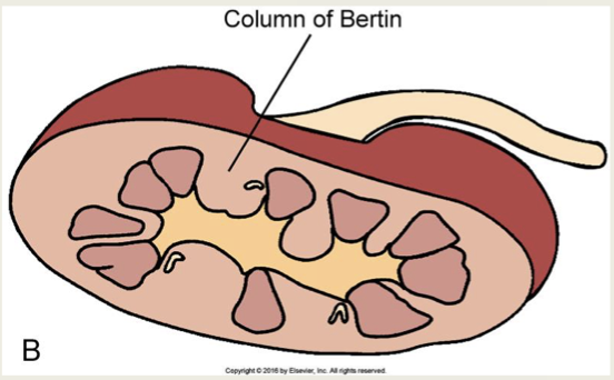
Column of Bertin Location
in the medulla, called pseudotumor

Column of Bertin Sonography
invagination or continuation of cortex within the medulla
extends through medulla into the sinus
same echogenicity as cortex
mimics mass


what is this
Column of Bertin
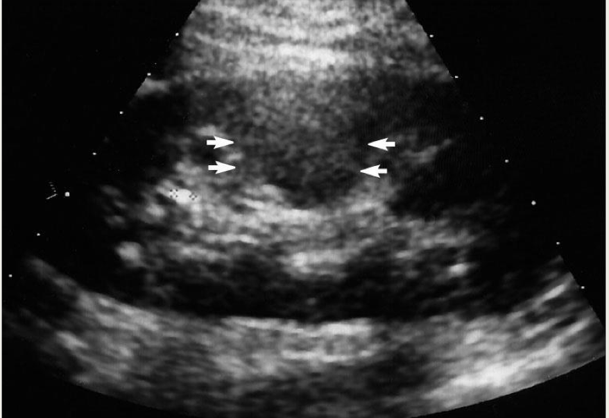
what is this
Column of Bertin
How do you differentiate a true renal mass from a dromedary hump or column of Bertin?
echogenicity is identical/isoechoic to the rest of the renal cortex
look for subtle borders of a mass
compare the vascularity of the mass to the rest of the cortex which should have a similar vascular pattern
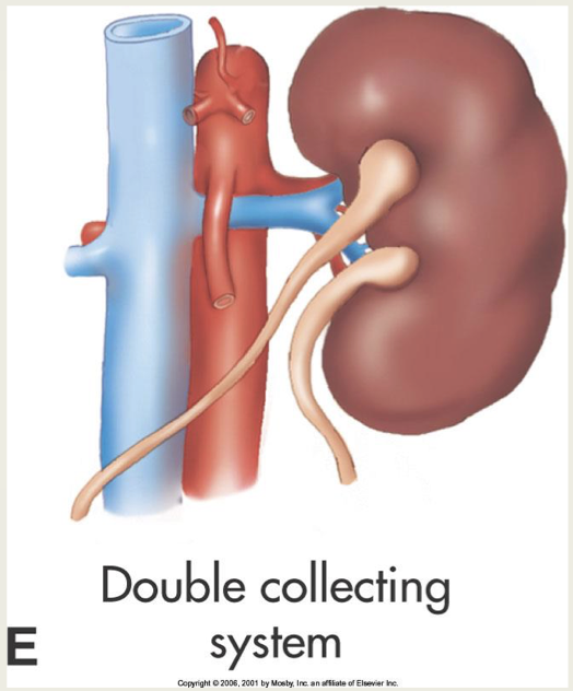
Double Collecting System
occurs when the renal sinus is divided
each sinus has a renal pelvis
Bifid (double) ureter may also be present

Horseshoe Kidneys
occur when kidneys are connected, usually at the lower poles
connection (isthmus) is isoechoic to the normal renal cortex
connection visualized anterior to the aorta, IVC, and spine

Horseshoe Kidney Location and Definition
fusion of (usually) lower pole of kidneys during fetal development
Horseshoe Kidney Sonography
Abnormal renal tissue extending across the midline
Kidneys are more medial and anterior to the spine
inferior poles lie more medial
Isthmus is more anterior to the spine
may mimic a solid pelvic mass
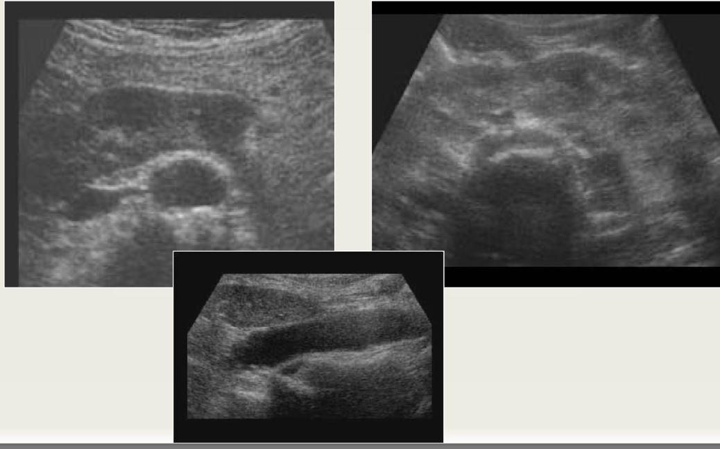

what is this
Horseshoe Kidney
Cake and Sigmoid (S-shaped) kidneys are variants of
a horseshoe kidney
Cake
fusion of entire length of medial surfaces of kidneys
does not ascend normally, found in pelvis
anterior malrotation of renal pelvis
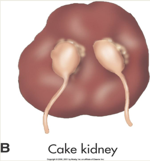
Sigmoid
upper pole of one kidney is fused to the lower pole of the other
anterior malrotation of pelvis

what is this
Cross Fused
Renal Ectopia
occurs when one or both kidneys occur outside the normal renal fossa
locations: lower abdominal and pelvic region
other locations are rare (ex: intrathoracic)

Renal Ectopia is an anomaly of
position
failure of kidneys to ascend / migrate to normal area
If kidney is not seen…
the retroperitoneum and pelvis should be scanned
don’t confuse bowel with kidneys
Scan renal fossa carefully to look for
atrophied kidney
Pelvic Kidney
most common may be malrotated, do not confuse with adnexal mass

Crossed Fused
same quadrant, fused together
Thoracic
herniated through diaphragm, excessive cephalic migration during embryology

what is this
Ectopia (no kidney on rt side)
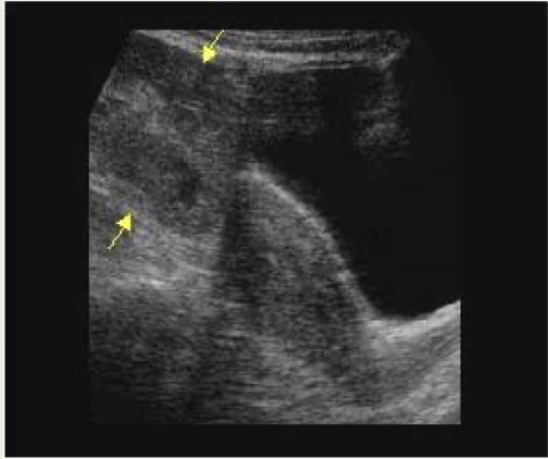
what is this
pelvic kidney

what is this
Calculi in Pelvic Kidney
Renal Malrotation
renal pelvis is medial in the normal kidney
in MALROTATION, the pelvis is directed anteriorly
incidental finding unless obstruction occurs
common with ectopic and horseshoe kidneys


what is this
abnormal rotated kidney

what is this
otherwise normal anteriorly rotated renal pelvis/hilum
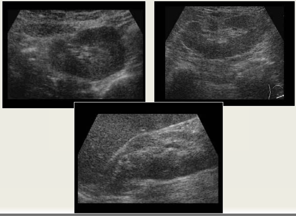
what is this
abnormal rotation

what is this
malrotated duplicated collecting system with anterior pelvis

what Is this
abnormal rotated kidney
Junctional Parenchymal Defect Location and Definition
upper pole of renal parenchyma is a normal extension of renal sinus in cases where there is distinct division between upper and lower poles
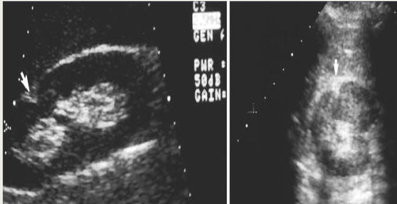
Junctional Parenchymal Defect Sonography
triangular echogenic area in the upper pole
best seen on SAG scans
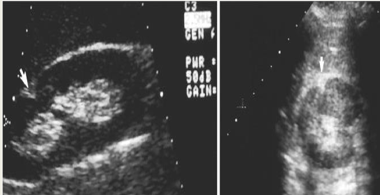

what is this
Junctional Parenchymal Defect

what is this
Junctional Parenchymal Defect
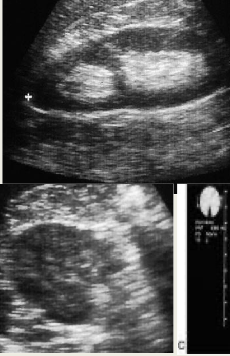
Duplicated Collecting System
common normal variant

Duplicated Collecting System Location
central renal sinus
LP is often larger than UP due to cleft of cortex

Duplicated Collecting System Sonography
two echogenic regions separated by a hypoechoic renal cortex
kidneys may also be large
on TRV view no sinus will be seen where cleft of cortex crosses
in a Duplicated Collecting System, it is difficult to tell if
duplex is partial or complete due to difficulty of ureter visualization on US

what is this
Duplicated Collecting System
Duplication of Ureters
Uni or bilateral
complete with seperate ureters draining the upper and lower poles of the kidneys and enter the bladder seperately
incomplete when the ureters join together to join the bladder as one
what is this
Duplicated Collecting System
Extrarenal Pelvis Location
normal renal pelvis is triangular and lies almost completely within the confines of the central renal sinus
An extra renal pelvis is usually longer and is outside the renal sinus
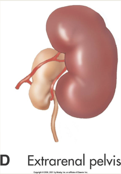
Extrarenal Pelvis Sonography
central cystic area partially inside or outside of the medial border of the kidney
not to be confused with dilated proximal ureter or hydronephrosis
Fetal Lobulation
lobulations seen in cortex with indentation into between the calyces
can give mass effect
failure of lobules to fuse in development

When there is mass effect in Fetal Lobulation…
evaluate the echogeniicity of the cortex
if mass is ISOECHOIC to the rest of the cortex, it is most likely NOT a mass

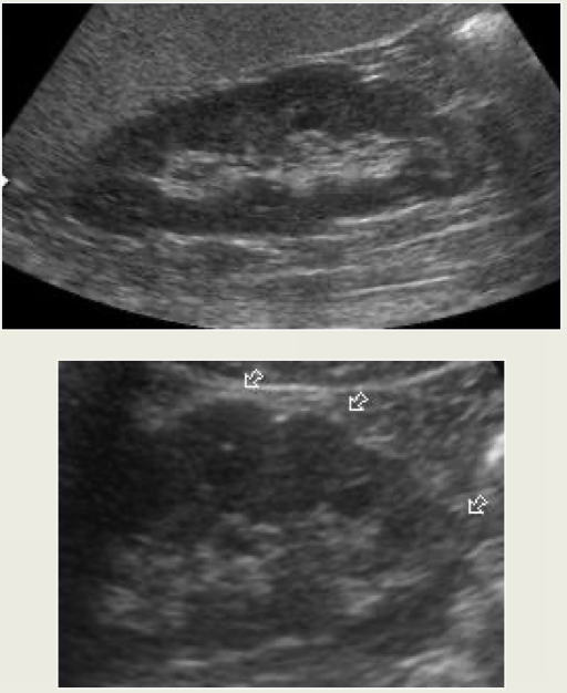
what is this
fetal lobulation
Renal Agenesis
renal anomaly
total failure of kidney and ureter to develop
most common in males
can be uni or bilateral (bilateral is fatal)
no kidney is seen in renal fossa
For Renal Agenesis, look for
a small nonfunctioning echogenic kidney before determining a unilateral kidney
contralateral kidney becomes
larger
Renal Agenesis is associated with
genital anomalies
Dysgenesis
defective embryonic development
Hypoplastic Kidney
renal anomaly
small kidney
has normal function which is proportionate to its size
contralateral hypoplastic kidney becomes
enlarged
Hypoplastic Kidney is difficult to distinguish from
atrophy of any etiology (vascular or infection)
Bilateral Hypoplastic Kidney will have
renal insufficiency
Suprarenal Kidneys
renal anomaly
very rare
complete duplication of kidney
Sinus Lipomatosis
deposition of moderate amount of fat in the renal sinus
occurs with age
large and echogenic sinus
seen with thinning cortex
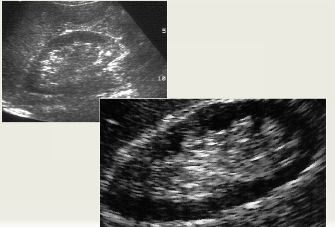

what is this
Sinus Lipomatosis