Physics Chapter 10 and 11
 Call Kai
 Learn
 Practice Test
 Spaced Repetition
 Match
 Flashcards
 Knowt Play
1/78

There's no tags or description
Looks like no tags are added yet.
79 Terms
Resolution
the ability to create accurate images
(image accuracy is the essence of ultrasound)
two forms of resolution
spatial and temporal
Spatial resolution
ability to distinguish two points as separate in space
• Axial
• Lateral
• Elevational (talk about this in later chapters)
Temporal resolution
ability to precisely position moving structures from instant to instant
• Accuracy in time
Axial Resolution
The ability to distinguish two structures that are close to one another and parallel to the sound beam’s main axis.
Axial resolution is AKA
LARRD:
• Longitudinal resolution
• Axial resolution
• Range resolution
• Radial resolution
• Depth resolution
Axial resolution units, determination, adjustability
• Units: of distance (mm)
• Determined by: the spatial pulse length (medium and source)
• cannot be adjusted since the SPL cannot be adjusted
• does not vary along the sound path
What type of pulses improve axial resolution?
shorter pulses improve axial resolution
How is axial resolution related to pulse duration ?
short duration pulses also have shorter lengths
_____ numbers mean shorter pulses and ____ image quality
lower numbers mean shorter pulses and better image quality
typical values of axial resolution
0.1-1.0 mm

black: whats in the body
whats showing up on the image:
green: longer SPL
red: shorter SPL
solving for axial resolution
axial resolution is ½ the SPL
SPL equations
SPL = #cycles x 1.54/frequency
SPL = #cycles x wavelength
Example: If the pulse length is 1mm, what is the axial resolution ?
1/2=0.5
so, structures need to be 0.5 mm or more apart from each other to be seen as two separate structures
Axial resolution in soft tissue equation
axial resolution = 0.77 x #cycles in a pulse / frequency
Why do some transducers have better axial resolution ?
axial resolution is determined by pulse length:
shorter pulses = better image
How are short pulses created?
• Decreased ringing
• Higher frequencies
• Add damping material
Less Ringing
short pulses means there are few cycles in a pulse
Transducers often have pulses of
transducers often have pulses of 2 or 3 cycles because dampening the ringing after the elctrical signal excited the crystal making the pulse
(fewer pulses mean better axial resolution)
Higher Frequency
• Shorter wavelengths mean higher frequency sounds (wavelength and frequency are INVERSELY RELATED)
What frequency of pulses have better image quality ?
pulses of higher freqnecies have better image quality
Better Axial Resolution
• Shorter spatial pulse length
• Shorter pulse duration
• Higher frequencies (shorter wavelengths)
• Fewer cycles per pulse (less ringing)
• LOWER numerical values
Worse Axial Resolution
• Lowest Frequency
• Greatest number of pulses
• HIGHER numerical values
Lateral Resolution
the ability to distinctly identify two structures that are very close together when they are perpendicular to the sound beam’s main axis
how well can you tell two structures apart when they are side by side?
What is the minimum distance that two side by side structures can be from one another and still be identified as two separate structures?
Lateral Resolution
• Small is better- the smaller the number, the better the image.
• Units: of DISTANCE (mm)
• Determined by: width of the sound beam (Narrower beam widths = better resolution)
Lateral resolution AKA
LATA
• Lateral
• Angular resolution
• Transverse resolution
• Azimuthal resolution

• The green dots represent the actual reflectors along the path of the sound beam.
• The black represents what shows up on the image.
• Notice: where the beam is narrowest- the reflectors can be differentiated.
Lateral Resolution and Width of Sound Beam
• Width of sound beam determines the lateral resolution
• Diameter of sound beam varies depending on the depth
• The narrower the sound beam, the better the lateral resolution
• The beam is narrowest at the focus
What width of the beam has the better lateral resolution ?
The narrower the sound beam, the better the lateral resolution
What do you think happens when two structures are side by side and closer to one another than the width of the sound beam?
They will be represented on the image as one structure.
What do you think happens when two structures are side by side and farther from one another than the width of the sound beam?
They will be appropriately represented on the image as two separate structures
What is lateral resolution approximately equal to ?
lateral resolution is approximately equal to the beam diameter:
lateral resolution = beam diameter
Since we know beam diameter changes with depth…
lateral resolution also changes with depth
two structures lying side by side must be at least _____ to be images as two separate structures
a beam widths apart
Where is lateral resolution the best?
Lateral resolution is best in the focal zone where beam diameter is the narrowest
Lateral vs. Axial
Lateral Resolution is not as good in clinical imaging as Axial resolution.
Why??
Pulses are shorter than the beams width
• Pulse duration determines axial resolution
• Beam width determines lateral resolution
Slice Thickness Resolution
• Measured perpendicular to the imaging place
• AKA: elevational axis, Z-axis, section thickness, and out of plane focusing
• Accomplished using lens with fixed focus- curve of lens goes from front to back
How can we improve elevational resolution?
switch to a 1.5 D transducer
Focusing
• Concentrates the sound beam into a narrower beam.
• improves lateral resolution.
• Internal and External can be used on single element transducers
Three methods
1. External focusing
2. Internal Focusing
3. Phased Array focusing
Phased array focusing can only work for …
array transducers (multiple active elements)
Fixed Focusing
• AKA conventional or mechanical focusing
• Includes both internal and external focusing techniques;
The focal depth is determined by….
the manufacture and cannot be changed by the sonographer (unless they change the transducer)
External Focusing
The lens
• A lens is placed in front of the piezoelectric material (fixed focus)
• The amount of curvature in the lens and therefore the focusing, is
predetermined by the manufacturer
The more curved the lens…
the greater the degree of focusing, creating a narrower beam width at the focus.
• Remember narrower beam width means improves lateral resolution

lens (external focusing)
Internal Focusing
• Curvature of the actual piezoelectric crystal
• This is the most common form of FIXED focusing
• Again, the greater the curvature, the greater the degree of focusing, creating a narrower beam width at the focus.
• The amount of curvature in the piezoelectric element, and therefore the focusing, is predetermined by the manufacturer

PZT (internal focusing)
Electronic Focusing
• Used in phased array transducers (phased array literally means adjustable or multiple focus)
• Array transducers have multiple elements
• Electronics within the system focus the sound beam
• Focus can be adjusted by the sonographer
• Cannot be used on single element transducers
Change #1 that the beam undergoes
Beam diameter in near zone and focal zone narrows
Change #2 that the beam undergoes
The focus is moved closer to the transducer- this reduces the length of the near zone
Change #3 that the beam undergoes
Beam diameter beyond focal zone widens
The focusing improves lateral resolution in the near zone and focal zone BUT degrades lateral resolution in all areas beyond focal zone
Change #4 that the beam undergoes
The size of the focal zone is reduced
Display Modes
the way the ultrasound system interprets and displays ultrasound information on the screen.
Amplitude Mode (A-mode)
• One dimensional display or image
• Appears as upward spikes
• Displayed spikes is a relative indication of the intensity or strength of the returning echoes
• Returned reflection processed with upward deflections graphs proportional to the amplitude (bigness) of the echo.
Tall spikes =
strong reflectors
short spikes =
weak reflectors
no spike =
no reflector
x-axis in A-mode represents
depth
• Distance along baseline the corresponds to reflector depth
Y-axis in A-mode represents
amplitude of echoes
• Height= amplitude or strength of the echoes
What is amplitude used for ?
• to accurately determine depth of a structure or thickness of structures
• Commonly used in ophthamology
• Best to use when needing very precise measurements
• No grayscale image given, just spikes

amplitude mode
Brightness mode (B-mode)
• Grey scale mode
• Pulse emitted from transducer (scan line), once echo returns, a brightness is assigned to the dot/pixel (Dot on screen corresponds to transducer position)
• shade of gray corresponds to the strength or amplitude of each echo
• Creates 2-D image
In original mode, pixels could be on or off. What were on and off displayed as?
On: white
off: black
Brighter dot =
stronger reflector
weaker reflection =
dark grey dot
What can brightness mode also be desscribed as?
a line of dots of varying brightness
x-axis in B-mode =
depth (found by time of flight)
z-axis in B-mode =
amplitude/brightness
• An oscilloscope isused to measure this (an instrument that can measure constantly varying signals – amplitude!)
What was the first type of gray scale imaging ?
B-mode
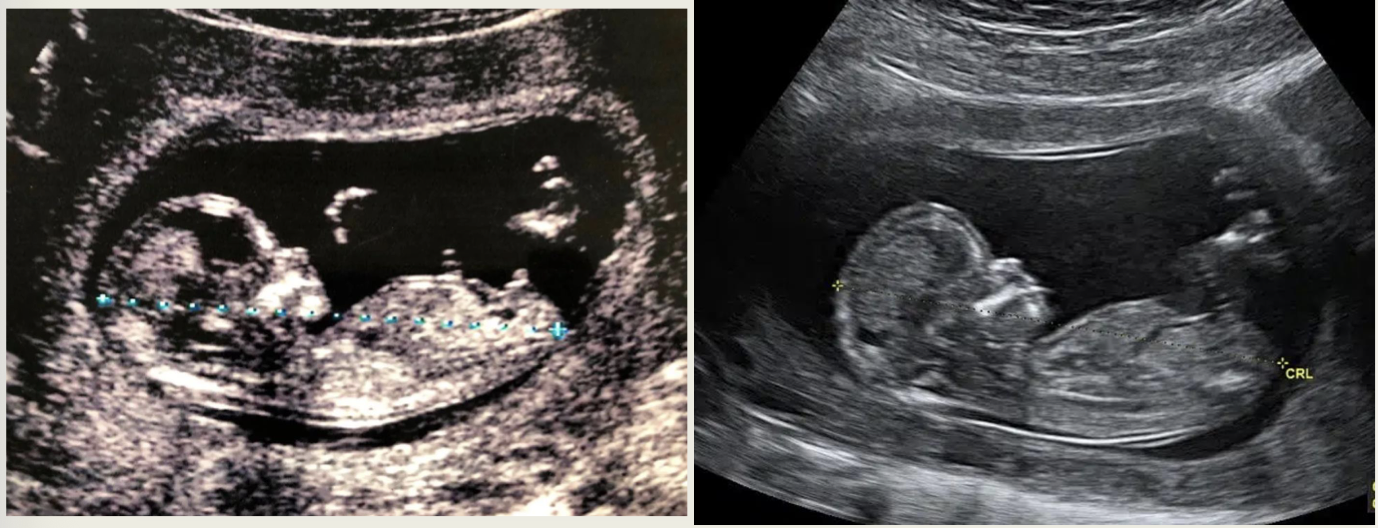
B-mode (left is old machine, right is new)
Motion mode (M-mode)
• Depicts motion over time
• Appears as a group of horizontal wavy lines.
• When a pulse is emitted, reflections move at a constant pace from right to left on the display screen.
• Gives information about an objects (reflectors) changing location with respect to time.
What do the lines in M-mode represent?
• “squiggly” lines represent the changing depths of the reflectors
• As the line moves up and down, M-Mode is showing that a structure is moving nearer then farther from the transducer.
• A horizontal straight line indicates a stationary structure.
the x-axis in M-mode =
time
the y-axis in M-mode =
depth
very high sampling rate =
to PRF of the system
What is M-mode usually used for ?
• mostly for cardiac evaluation, to document viability of the fetus and evaluate lung motion
• No grayscale image

M-mode