UNIT 5 BIOLOGY B1 (CARDIOVASCULAR SYSTEM)
 Call Kai
 Learn
 Practice Test
 Spaced Repetition
 Match
 Flashcards
 Knowt Play
1/47

There's no tags or description
Looks like no tags are added yet.
48 Terms
The human cardiovascular system is a ?. This means that for every complete circulation in the body, the blood passes through the heart ?
The human cardiovascular system is a DOUBLE CIRCULATORY SYSTEM. This means that for every complete circulation in the body, the blood passes through the heart TWICE.
What are the features of the blood vessel known as an Artery?
Thick walls containing lots of thick smooth muscles & elastin fibres to withstand high blood pressure from LEFT VENTRICLE
Elastin allows recoil after pulsation of blood as pressure changes
Small (narrower than vein) Lumen
What are the features of the blood vessel known as a Vein?
Thin walls as blood has low pressure & thin muscle and elastic layer
Largest lumen to reduce friction to blood flow
Semi-lunar valves present to prevent backflow of blood
What are the features of the blood vessel known as a Capillary?
Links arteries and veins through tissues
Site of gas exchange of materials (o2 & co2) between the blood and tissues by diffusion
Walls are one endothelial cell thick, giving a short diffusion pathway
Why is it important that the capillary walls are very thin?
short diffusion pathway
What is the role of the elastic tissue in arteries?
Allows arteries to stetch so it can withstand blood pressure
Recoils to return to their original size
Why are older people more likely to get high blood pressure?
Their arteries are less stretchy due to the loss of elastin
So blood pressure rises
What is the role of the smooth muscle tissue?
Allows vasoconstriction and vasodilation
This allows blood to be directed to specific tissues
What antigens are present on the surface of the red blood cells for the following groups:
A
B
AB
O
A antigens
B antigens
AB antigens
No antigens
What antibodies are present on the surface of the red blood cells, who can they donate to & receive from? for the following groups:
A
B
AB
O
Anti-B antibodies, can donate to A,AB, can receive from A,O
Anti-A antibodies, can donate to B,AB, can receive from B,O
NO antibodies, can donate to AB, can receive from A,O,AB,B
Anti-B & Anti-A antibodies, can donate to A,AB,B,O can receive from O

If a person is rhesus positive, who can they send and receive blood from?
If a person is rhesus negative, who can they send and receive blood from?
rhesus positive- they can send to Rh+ and receive from Rh- & Rh+
rhesus negative- they can send to Rh+ & Rh- and receive from Rh-
Combining the ABO and the Rh systems gives 8 possible blood groups- what are they?
A⁺
A⁻
B⁺
B⁻
AB⁺
AB⁻
O⁺
O⁻
Who can the 8 blood groups receive blood from?
O⁻
O⁺
A⁻
A⁺
B⁻
B⁺
AB⁻
AB⁺
O⁻= O⁻
O⁺= O⁻, O⁺
A⁻= O⁻, A⁻
A⁺= O⁻, O⁺, A⁻, A⁺
B⁻= O⁻, B⁻
B⁺= O⁻, O⁺, B⁻,B⁺
AB⁻ = O⁻, A⁻, B⁻,AB⁻
AB⁺ = O⁻, O⁺, A⁻, A⁺, B⁻, B⁺, AB⁻, AB⁺
Which blood group is the universal donor and why?
Which blood group is the universal receiver and why?
Blood group O⁻ because there are no antigens on the surface
Blood group AB⁺ because it has A, B and Rh antigens on its red blood cells.
Explain the following:
Why AB⁺ cannot give blood to A⁺?
Why AB⁺ cannot give blood to AB⁻ ?
Why B⁻ cannot give blood to A⁻ ?
Why B⁺ cannot give blood to AB⁻ ?
The A⁺ would start producing anti-B antibodies
The AB⁻ would produce anti-D antigens
The A⁻ would produce anti-B antibodies
The AB⁻ would produce anti-D antibodies
What is the myogenic muscle?
What is the septum?
Muscles that are made to contract due to signals from the heart itself, NOT from the brain
The dividing wall between the left and right side
What is the role of the coronary artery?
Delivers blood containing oxygen & glucose to the heart muscle
This is needed for aerobic respiration where it produces ATP for the heart to contract
Why is the muscle of the left hand side thicker than the right hand side?
The blood on the left side of heart travels to tissues, so during contraction a high force must be generating
However, blood from the right side of heart is only going to the lungs so the force generated does not need to be as high
What is the function of the Vena cava?
What is the function of the pulmonary artery?
Carries de-oxygenated blood from tissues of head and body to the right atria.
Carries de-oxygenated blood from right ventricle to lungs, to become oxygenated
What is the function of the pulmonary vein?
What is the function of the aorta?
Carries oxygenated blood from the lungs to the left atrium of the heart
Carries oxygenated blood from the left ventricle to the tissues of the head and body
What happens when there is a high pressure behind a valve?
What happens when there is a high pressure in front of a valve?
Causes it to open
Causes it to close
Where is the Bicuspid valve located?
When will it open?
When will it close?
Between the left ventricle and left atrium
When pressure is high in left atrium & low in left ventricle
When pressure is lower in left atrium & higher in left ventricle
Where is the Tricuspid valve located?
When will it open?
When will it close?
Between the right ventricle and right atrium
When pressure is high in right atrium & low in right ventricle
When pressure is lower in right atrium & higher in right ventricle
Where is the Semi Lunar valve located?
When will it open?
When will it close?
Between the ventricles & blood vessels (aorta and pulmonary artery)
When pressure is high in ventricles & low in blood vessels
When pressure is lower in ventricles & higher in blood vessels
What is the function of the bicuspid valve?
Allows blood to flow from the left atrium into the left ventricle when the valve is open.
Prevents backflow of blood from the left ventricle into the left atrium when the left ventricle contracts.
What is the function of the tricuspid valve?
Allows blood to flow from the right atrium into the right ventricle when the valve is open.
Prevents backflow of blood from the right ventricle into the right atrium when the right ventricle contracts.
What is the function of the aortic valve?
Allows blood to flow from the left ventricle into the aorta when the valve is open.
Prevents backflow of blood from the aorta into the left ventricle when the left ventricle relaxes.
What is the function of the pulmonary valve?
Allows blood to flow from the right ventricle into the pulmonary artery when the valve is open.
Prevents backflow of blood from the pulmonary artery into the right ventricle when the right ventricle relaxes.
What happens in the diastole of a cardiac cycle?
(All chambers are relaxed)
Atria & ventricles relax, so pressure is higher in arteries than ventricles
This closes the SL valves in artery as blood pushes valves, so atria fills with blood from vein
This causes AV valves to open
What happens in the Atrial systole of the cardiac cycle?
(All chambers contract)
Ventricles relax, atria contracts
increases pressure in atria, this forces blood through the AV valves & into ventricles
What happens in the Ventricular systole of the cardiac cycle?
(All chambers contract)
Atria relaxes, ventricles contract
Increasing the pressure in the ventricles above the atria and arteries
This closes the AV valves and opens the SL valves, allowing blood to be pumped into arteries
What is the equation for cardiac output?
CO (L/min or dm3/min) = SV (ml/beat) X HR (beats/min)
Describe the Sino-Atrial Node
Found in the wall of the right atrium
Has a pacemaker which initiates heartbeat & sets heart rate at 70-80 beats/min
Generates electrical impulse that moves across both atria walls, causing them to contract at the same time
Describe the Atrio-Ventricular Node
Electrical impulse from SAN is picked up by the AVN in the septum
AVN imposes a slight delay in sending the wave of excitation to ventricles
Allowing atria to finish contracting & to fully empty of blood before ventricles contract
AVN passes signal to the Bundle of His which is also found in septum
Describe the Bundle of His & Purkyne fibres
Bundle of His conduct electrical impulse down the septum down the apex
The purkyne fibres then spread up to the ventricle walls, transmitting the electrical impulse & causes contraction of ventricles from apex upwards.
This pushes the blood up + out of heart
What does the P wave do?
Shows when atrial systole is happening, so shows the start of atrial contraction in Atrial systole
What does the QRS complex do?
Shows when ventricular systole is happening so shows the start of ventricular contraction in ventricular systole
At the same time, atrial diastole is happening but is hidden by QRS complex
What does the T wave do?
shows when ventricular repolarisation is happening, so shows when the heart is relaxing in cardiac diastole

What is Tachycardia showing?
Increased heart rate
Peaks of QRS complex are regular but closer together
Typically over 100bpm

What is Bradycardia showing?
Decreased heart rate
Peaks of QRS complex are regular but further apart
Slower than 60bpm
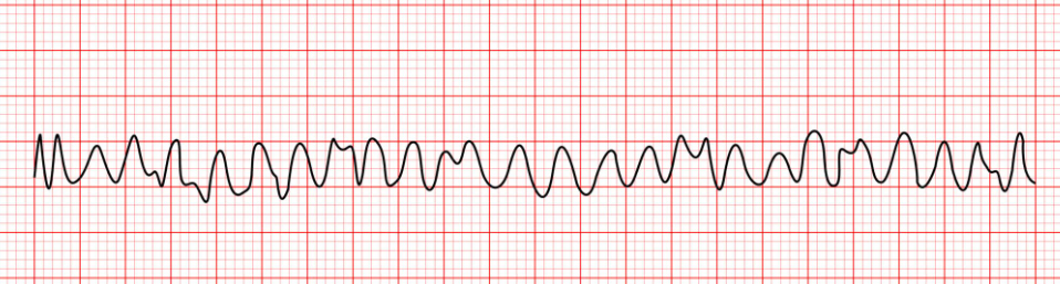
What is Ventricular Fibrillation showing?
No PQRST visible
Contraction is not controlled
Can lead to cardiac arrest

What does Sinus Arrythmia show?
Irregular heart rate
Peaks of QRS complex are not regularly spaced
Linked with breathing- faster when breathing in, slower when breathing out
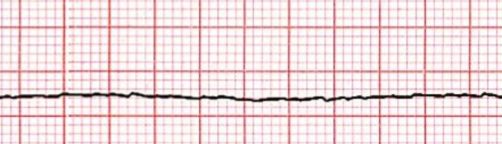
What does Asystole/Flat Line show?
No PQRST visible as no electrical activity in the heart
Heart not beating
Increasing a patients stroke volume increases their cellular respiration enabling them to be more active.
Explain why.
More blood to carry more oxygen & glucose
More aerobic respiration, so more ATP produced
So muscles contract more
What are some examples of Cardiovascular diseases?
Heart attack
Hypertension
Stroke
Transplantation is a type of treatment for CVD, describe what it is, the benefits & risks
What is it?
Used if a person has coronary heart disease/heart failure
Benefits?
Can prolong life
Can be effective if tissue match is found & has long lasting effect
Risks?
Can cause infection from bacteria
Failure to pump
Must take immunosuppressants for rest of life
Must find a donor & a match
Antihypertensives are a type of treatment for CVD, describe what it is, the benefits & risks
What is it?
Ace inhibitors (relaxes blood vessels)
Diuretics (removes excess water from body)
Calcium channel inhibitors (prevents calcium entering heart)
Benefits?
Lowes BP
Reduces risk of CVD
Risks?
Have to keep taking them for life
Causes excess fluid loss & impotence
Raises sodium/blood sugar levels
Statins are a type of treatment for CVD, describe what it is, the benefits & risks
What is it?
Lowers LDL levels in blood
Reduce formation of fatty plaques/blood clots
Benefits?
Cheap & easy to take
Prevents heart disease getting worse
Risks?
Have to keep taking them for life
Increases risk of diabetes & cannot cure CVD
Causes side effects such as nose bleeds and sore throat