Restrictive Lung Dysfunction and Obstructive Lung Disease
 Call Kai
 Learn
 Practice Test
 Spaced Repetition
 Match
 Flashcards
 Knowt Play
1/195

There's no tags or description
Looks like no tags are added yet.
196 Terms
Trachea
Primary bronchi
Secondary bronchi
Tertiary bronchi
bronchioles
Terminal bronchioles
Respiratory bronchioles
Alveolar ducts
Alveoli
What is the path of air flow from the trachea to the alveoli
to help with airway clearance of foreign substances (such as mucus)
What is the functional purpose of the orientation of the tertiary bronchioles
760 mmHg
What is normal atmospheric pressure
760 mmHg
What is the normal intrapulmonary pressure in the lungs
756 mmHg
What is the normal intrapleural pressure in the lungs
fluid in the plural space to reduce friction and allow for smooth breathing (too much fluid is bad)
What is found within the intrapleural space/cavity of the lungs… and what is that substances purpose
parietal pleura
what do we call the outer layer of the pleural cavity
visceral pleura
what do we call the inner layer of the pleural cavity
diaphragm
What is the primary muscle of inspiration
Boyal's Law
What law of physics is able to explain the inverse relationship between pressure and volume that drives respiration
- increase in volume
- decrease in pressure
What happens to intrathoracic volume and pressure when breathing in
- decrease in volume
- increase in pressure
What happens to intrathoracic volume and pressure when breathing out
sternocleidomastoid and scalene muscles
What are the 2 accessory muscles of inspiration
- external intercostals
- abdominals
What 2 muscles engage in active breathing
vital capacity + residual volume
Total lung capacity =
IRV + tidal volume + ERV
Vital capacity =
tidal volume + inspiratory reserve volume
Inspiratory capacity =
expiratory reserve volume + residual volume
Functional residual capacity =
- inspiratory reserve volume
- tidal volume
- expiratory reserve volume
- residual volume
what are the 4 main components of the breathing cycle
21% = 160 mmHg
What percent of the air we breathe is O2
Some O2 is lost on the way to the lungs due to humidification adding moisture to the air
Why is the pressure of O2 lower in the lungs compared to atmospheric pressure of O2 in the air
CO2 in the alveoli is higher than outside since it is getting ready to exit from the blood
Why is the pressure of CO2 in the alveoli greater than the pressure of atmospheric CO2
80-100 mmHg
What is normal PO2
35-45 mmHg
What is normal PCO2
95 mmHg
What is normal PO2 in the arteries
40 mmHg
What is normal PCO2 in the arteries
40 mmHg
What is normal PO2 in the veins
45 mmHg
What is normal PCO2 in the veins
- decreased temperature
- decreased PCO2
- increased pH
What are 3 reasons that oxygen affinity will increase promoting O2 binding to Hgb
- increased temperature
- increased PCO2
- decreased pH
- exercise state
What are 4 reasons that oxygen affinity will decrease promoting O2 unloading from Hgb
Apnea
absence of breathing
Dyspnea
shortness of breath
VC
ventilatory/vital capacity
Paradoxical breathing
inward abdominal or chest wall movements with inspiration and outward movement with expiration
penetrating wounds
What can cause paradoxical breathing
RA
room air
Trendelenburg
head down, feet up
Cor Pulmonale
Right sided HF (RHF)
FiO2
fraction of inspired oxygen
ABG
arterial blood gas
total lung capacity
TLC
restrictive lung disease
What type of lung disease is characterized by a reduction in volume
obstructive lung disease
What type of lung disease is characterized by a reduction in flow
To measure lung volumes and capacities.
What is the purpose of spirometry
FEV1
Forced exhalation volume in 1 second
FVC
Amount of air you can exhale after a max inhale
>0.7 (70%)
Normal FEV1/FVC
FEV1/FVC
This is the percentage of the vital capacity which is expired in the first second of maximal expiration. In healthy patients the FEV1/FVC is usually around 70%.
Normal flow volume loop

- Exhalation curve looks normal but is smaller
- Percentage/ratio is the same but flow volume/capacity is decreased
How does restrictive lung dysfunction alter the flow-volume loop
Restrictive Flow-volume Loop
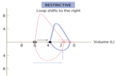
90%
Restrictive FEV1/FVC
- Initially seen with exercises
- Progresses to dyspnea at rest
How does Dyspnea present in patients with restrictive lung dysfunction
due to the alveoli trying to open up during inspiration
Why do we hear dry inspiratory crackles when auscultating a patient with RLD
- Interstitial Lung Disease (ILD)
- Atelectasis
- Pulmonary Edema
- Aging
- Musculoskeletal Causes
- Trauma
- Surgical Causes
What are Causes of Restrictive Lung Dysfunction
Interstitial Lung Disease (ILD)
broad group of inflammatory lung disorder character by acute, subacute, or chronic inflammatory infiltration of alveolar wall by cells, fluid, and connective tissue.
- Sarcoidosis
- Idiopathic Pulmonary Fibrosis (IPF)
- Bronchiolitis Obliterans
What are the different types of Interstitial Lung Disease
Idiopathic Pulmonary Fibrosis (IPF)
What form of Interstitial Lung Disease (ILD) is most common and has the worst prognosis
Bronchiolitis Obliterans
What form of Interstitial Lung Disease (ILD) is Often seen in transplanted patients due to organ rejection
- environmental exposure
- autoimmune processes
- genetics or unknown
what are causes of Interstitial Lung Disease (ILD)
- Medication
- supplemental O2
- lung transplant
What does treatment of Interstitial Lung Disease (ILD) entail
lung fibrosis
What happens as a result of Interstitial Lung Disease (ILD)
Atelectasis
- "Incomplete expansion"
- Umbrella term for a Collection of disorders that cause the lung to be collapsed
leaky
What does pulmonary edema indicate about capillary beds
- L sided HF
- ARDS (acute respiratory distress syndrome)
What are 2 most common causes of pulmonary edema
Decrease cardiac preload
what is the primary treatment for pulmonary edema
Lungs fill with fluid due to fluid leaking from blood vessels into the alveoli
What is the primary cause of ARDS (acute respiratory distress syndrome)
- Supplemental O2
- mechanical ventilation
- prone positioning
What does treatment for ARDS (acute respiratory distress syndrome) entail
fluid in the lungs
What does pulmonary edema indicate
- Diaphragmatic Paralysis
- Kyphoscoliosis
- Ankylosing Spondylitis
What are Musculoskeletal Causes of RLD
Ankylosing Spondylitis
A chronic inflammatory disease of the *spine and pelvis that causes sacroiliitis leading to fusion* of the affected joints.
- Rib fracture
- Lung contusion
- Penetrating wounds
- Burns
What are some Traumatic Causes of RLD
5th-9th more common
A fracture of which ribs are likely to result in RLD
Causing microvascular damage → leaking
Why may lung contusions lead to RLD
Inhalation burns and burns to the chest
What types of burns can lead to RLD
- Spinal Cord Injury
- Amyotrophic Lateral Sclerosis (ALS)
- Poliomyelitis
- Guillain-Barre Syndrome
- Myasthenia Gravis
- Tetanus
- Duchenne Muscular Dystrophy
What types of Neuromuscular diagnoses can cause RLD
- Rheumatoid Arthritis
- Systemic Lupus Erythematosus (SLE)
- Scleroderma
What types of Connective tissue disease diagnoses can cause RLD
Obesity
What type of metabolic diagnoses can cause RLD
- Coal workers pneumoconiosis
- Silicosis
- Asbestosis
What types of environmental disease diagnoses can cause RLD
Severe Delayed Desaturation
a medical phenomenon where a patient's blood oxygen levels drop to a dangerously low level (severe hypoxemia) hours after an initial assessment where their oxygen levels appeared normal or stable
<70%
What is FEV1/FVC for Obstructive lung dysfunction
- Expiration phase is longer and slower
- Inspiratory capacity is decreased
How does obstructive lung disease alter the Flow-Volume loop
by FEV1
How is Obstructive lung dysfunction classified
- COPD
- Asthma
- Bronchiectasis
- Cystic Fibrosis
What are different types of Obstructive Lung Dysfunctions
- Chronic Bronchitis
- Emphysema
What are the 2 primary types of Chronic Obstructive Disease
Chronic Bronchitis
- Hypersecretion of mucus
- "Blue Bloater"
- a long-term inflammation of the breathing tubes (bronchi) that causes the airways to swell and produce excessive mucus
- excess body fluids
- chronic cough
- SOB on exertion
- increased sputum
- cyanosis
What are the clinical manifestations associated with Chronic Bronchitis
Emphysema
- destruction of alveolar walls + loss of elasticity
- "Pink Puffer"
- Spirometry (Pulmonary function test) = Gold Standard for COPD
- Arterial Blood Gases: PaO2, PaCO2
- Auscultation
- Chest x-ray
- GOLD Stages of COPD
What are tests used to help diagnose COPD
Chronic cough +/- sputum
What symptoms of COPD are associated with stage 1: mild
- Chronic cough +/- sputum
- Dyspnea
What symptoms of COPD are associated with stage 2: moderate
- Chronic cough +/- sputum
- Dyspnea
What symptoms of COPD are associated with stage 3: severe
- Chronic cough +/- sputum
- Dyspnea
- Respiratory or R HF
- Weight loss
What symptoms of COPD are associated with stage 4: very severe
<0.7
What is the FEV1/FVC for all stages of COPD
>80%
What is the predicted FEV1 for stage 1 of COPD
50 - 80%
What is the predicted FEV1 for stage 2 of COPD
30 - 50%
What is the predicted FEV1 for stage 3 of COPD
<30%
What is the predicted FEV1 for stage 4 of COPD
BODE Index
What tool/outcome do we use to help determine the Prognosis of COPD
Survivability
What type of information about a patient with COPD can we learn from the BODE index