Quiz 3 - Circulartory System
 Call Kai
 Learn
 Practice Test
 Spaced Repetition
 Match
 Flashcards
 Knowt Play
1/36

There's no tags or description
Looks like no tags are added yet.
37 Terms

What does #1 show
right external jug-ular vein
location: head and neck
Drains blood from head/neck toward heart
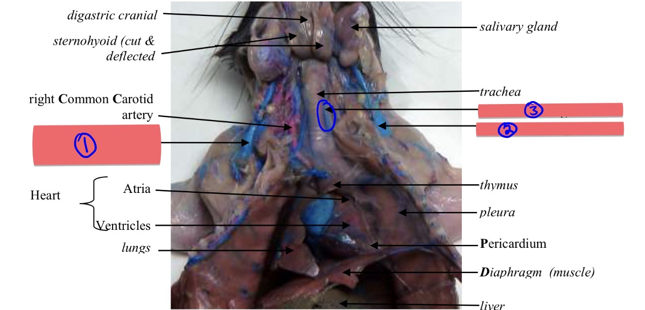
what does #2 show
left external jug-ular vein
location: head and neck
Drains blood from head/neck toward heart

what does #3 show?
left internal jug-ular vein
location: head and neck
Drains blood from brain/deep head and neck

what does #4 show?
right common car-o-tid art-ery
location: head and neck
Supplies oxygenated blood to head/neck

what does #5 show?
right internal jugular vein
location: head and neck
Drains blood from brain/deep head and neck

what does #6 show?
brachi-o-cep-ha-lic ar-tery
fucntion: Branches into the right subclavian artery and the right common artery
Supplies blood toward right head/neck and right forelimb
location: ANTERIOR REGION CIRCULATION

what does #7 show?
left common carotid artery
location: Thoracic Region

what does #8 show?
sub-cla-vian artery right and left
(forms like a V shape near the werid looking tube)
location: Thoracic Region
function: The first branch off the brachiocephalic artery

what does #9 show?
Thoracic Aorta
location: Thoracic Region
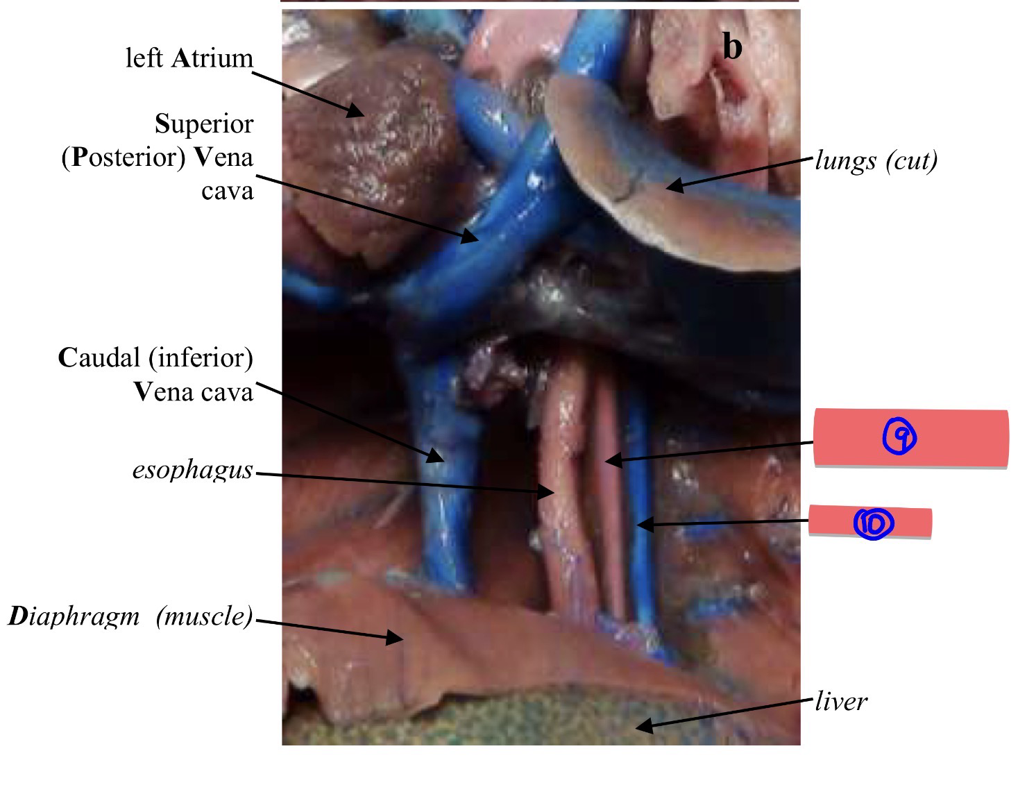
What does #10 show?
Azygos
Location: Thoracic Region
function: Drain directly into the inferior vena cava
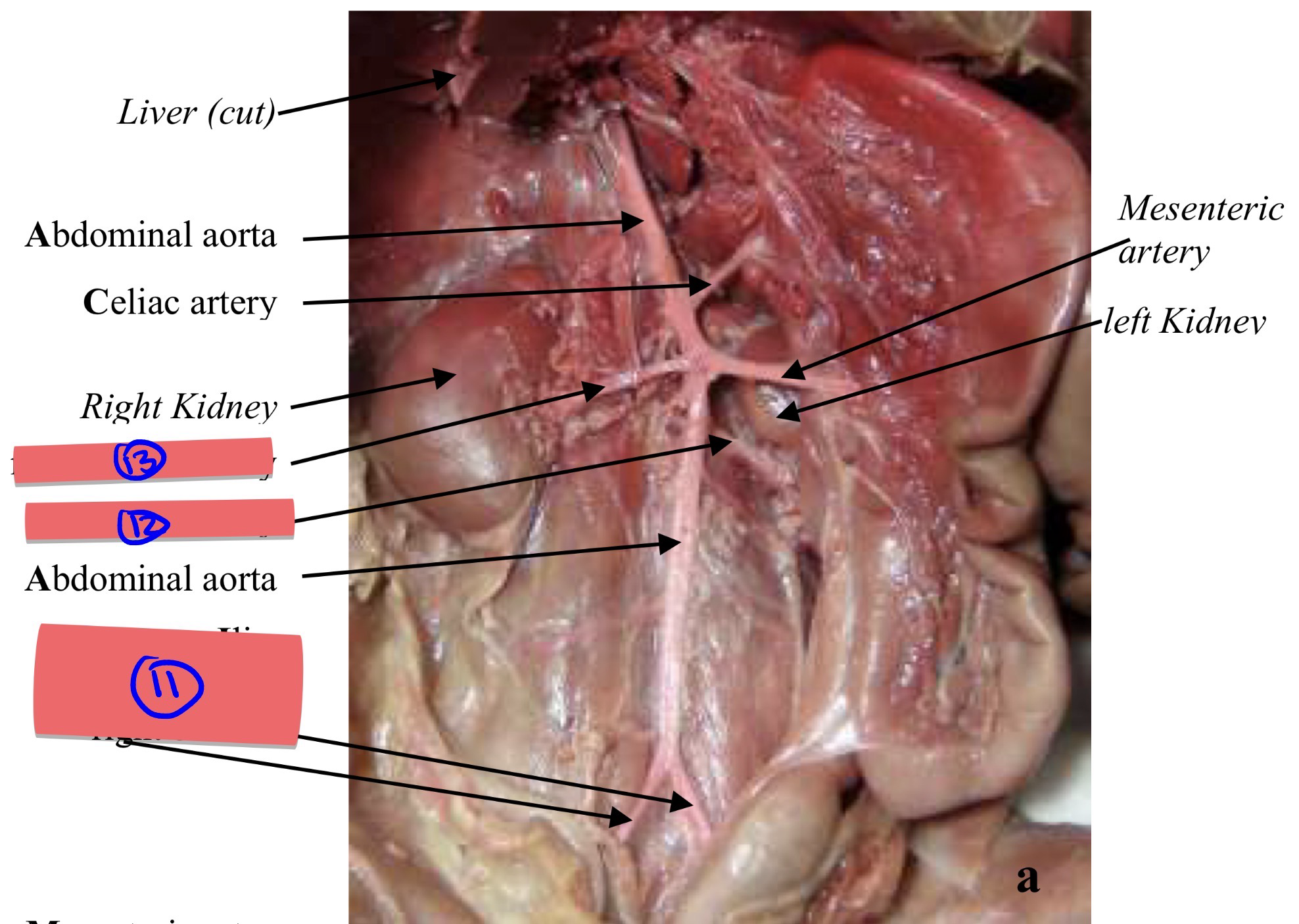
What does #11 show?
common Il-iac art-er-ies right and left
look like upside down V
location: Abdomen & Hind Leg

what does #12 show?
left renal artery
close to left kidney
Location: Abdomen & Hind Leg

what does #13 show?
right renal artery
close to right kidney
Location: Abdomen & Hind Leg

what does #14 show?
renal vein
location: Abdomen & Hind Leg

what does #15 show?
right fe-moral vein
location: Abdomen & Hind Leg

what does #16 show?
right fe-moral artery
literally red part ontop of blue
Location: Abdomen & Hind Leg
function: Branches off the iliac going along the femur bone

what does #17 show?
posterior inter-ven-tri-cular sul-cus
look for lining
btw this posterior view b/c pulmonary artery not showing
Location: Heart
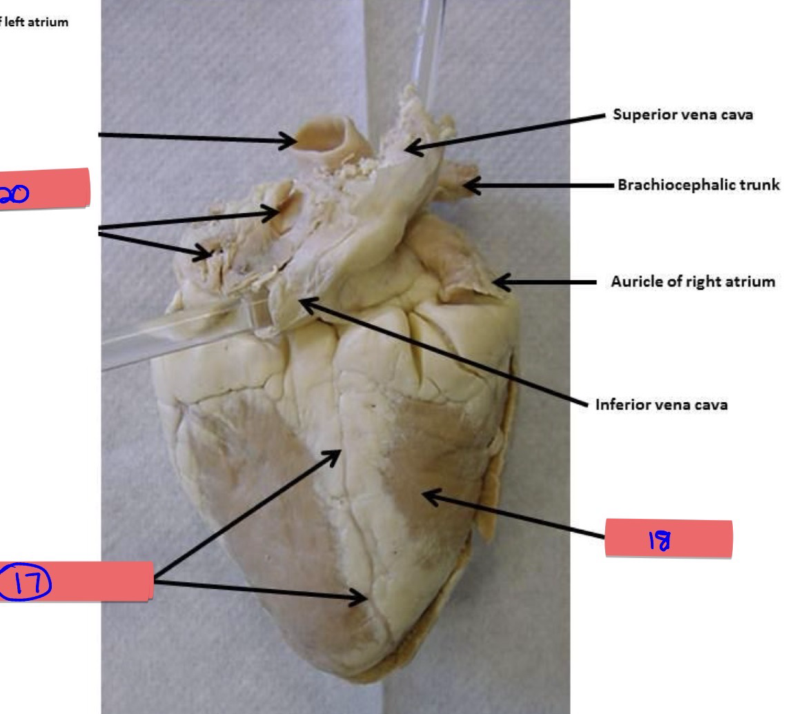
what does #18 show?
right ven-tri-cle
posterior so right = my right
function: pumps deoxygenated blood into the pulmonary trunk.

what does #19 show?
right ven-tri-cle
bc its anterior since u see pulmonary artery right = my left
function: Pumps deoxygenated blood into the pulmonary trunk.

what does #20 show?
anterior inter-ven-tri-cul-ar sulcus
near gaint crack
function: divides the right and left ventricles.

what does #21 show?
chordae tendineae of tricuspid
note: for right ventricle with pulmontary trunk
function: Prevent tricuspid valve from prolapsing backward

what does #22 show?
papillary muscle
function: Small bundles of cardiac muscles projecting from the myocardial wall.

what does #23 show?
interventricular septum
function: Divides the ventricles into right & left

what does #24 show?
chordea tend-i-neae of bi-cus-pid
function: Prevent bicuspid/mitral valve from prolapsing backward
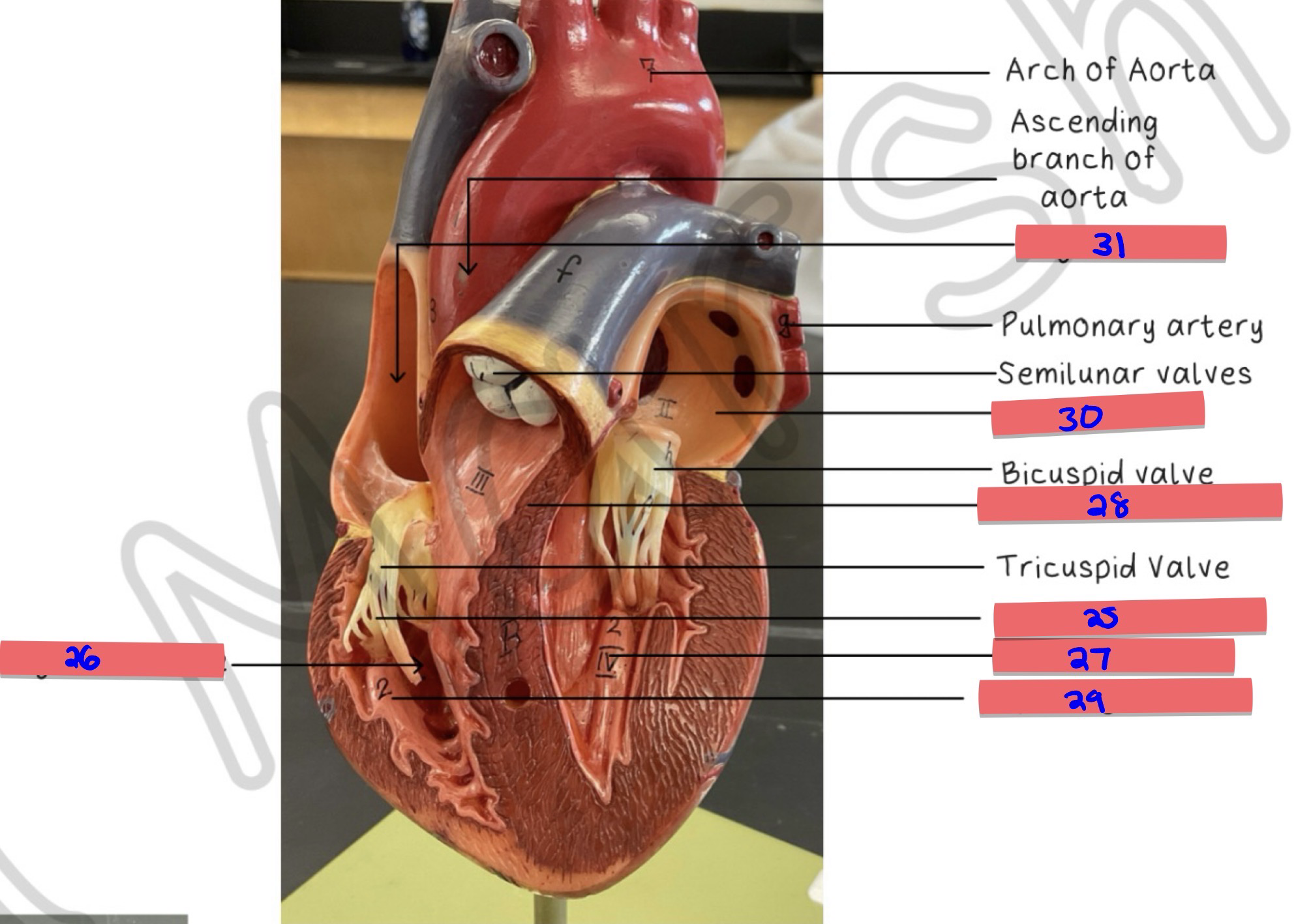
what does #25 show?
chordea tendineae
function: Chords holding the valves. Originate from the papillary muscles.

what does #26 show?
right ventricle
function: Pumps deoxygenated blood to lungs

what does #27 show?
left ventricle
function: Pumps oxygenated blood to body

what does #28 show?
interventricular septum
function: Separates ventricles; prevents blood mixing

what does #29 show?
papillary muscles
function: Tighten chordae to prevent AV valve prolapse

what does #30 show?
left atrium
function: Receives oxygenated blood from lungs

what does #31 show?
right atrium
function: Receives deoxygenated blood from body

what does #32 show
right auricular appendage
function: Increases capacity of right atrium

hepatic portal vein
under werid looking ear like structure + looks like V shape
function: Carries nutrient-rich blood to liver for processing

splenic vein
little piece sticks out
function: Drains blood from spleen

inferior messenteric vein
skinny vein inside
function: Drains blood from distal large intestine
superior messenteric vein
next to big vein
function: Drains blood mainly from small intestine
anastomosis
When blood flow to particular areas of the body are crucial, many of the arteries connect with one another so that when
one vessel is hindered, the other may still function to continually supply blood.