7. Staging & Grading
 Call Kai
 Learn
 Practice Test
 Spaced Repetition
 Match
 Flashcards
 Knowt Play
1/28

There's no tags or description
Looks like no tags are added yet.
29 Terms
workup
series of diagnostic exams performed once tumour is detected
goal: to determine general health status of patient + info about tumour
informs oncologist on most appropriate treatment
specifics depend on cancer type suspected + symptoms experienced by patient
test based on tumour characteristics
early stage tumours require different tests than late stage, eg. bone scan
PET often used to determine
site of primary disease
areas of secondary disease not detectable by other imaging modalities
recurrence post-treatment
tumour info
type
location
size
degree tumour has invaded normal tissue
amount of cell differentiation
presence/absence of spread to distant sites
lymph node involvement
amenability to specific treatment regimens
tumour stage
defines tumour size + extension at time of diagnosis
many common tumour sites have own unique systems
purpose
helps determine best treatment
helps predict prognosis
provides means for continued research
universal staging system
aids clinician in planning of treatment for each patient
assists in evaluation of treatment modalities
facilitates exchange of info b/w treatment centres worldwide
evaluates outcome of treatment, ie. survival
estimates prognosis or outcome by stage + treatment
includes
TNM staging
TNM staging
T → T0-T4, Tx
size/extent of invasion of primary tumour
N → N0-N4, Nx
status of lymph nodes → positive or negative
extent of lymph node involvement
N1-N3 most commonly seen
M → M0-M1, Mx
presence + extent of metastasis
may have subcategories
c = clinical
p = pathologic
T
size/extent of primary tumour
T0
no primary tumour
eg. patient w/ elevated PSA can have prostate cancer but hasn't developed a mass yet
T1
small and/or confined to small area
T4
extremely large and/or extends to other tissues
Tx
inability to obtain info to make determination
N
status of lymph nodes (positive/negative) + extent of lymph node involvement
N0
no lymph node involvement
good prognostic factor
N1
positive nodes close to site of primary tumour → local, regional
N4
positive nodes at more distant sites
Nx
nodal status was not assessed
M
presence + extent of metastasis
M0
no evidence of metastasis found
M1
disease present away from primary tumour
Mx
absence/presence of metastatic disease not assessed
R (residual disease status)
absence or presence of residual tumour after treatment
Rx
presence of residual tumour cannot be assessed
R0
no residual tumour
R1
microscopic tumour
R2
macroscopic residual tumour
clinical staging
subcategory of TNM staging - based on physical exam + imaging
eg. T1cN0M0
pathologic staging
subcategory of TNM staging - clinical staging w/ additional info from surgical removal of all or part of tumour, biopsy or removal of lymph node
eg. T2pN0M0
common to see in breast patient after node biopsy
offers most accurate info about tumour + extent of disease spread
non-invasive approaches can stage tumours but leave lymph node status + microscopic spread unknown
amount of tumour left behind post-surgery important for treatment + prognostic info
helps determine if targeted therapies can be used
post-therapy staging
determined after 1st course of treatment to see how treatment has impacted cancer
eg. post chemotherapy, hormone therapy, radiation therapy
could lead to downstaging
downstaging
preoperative treatment to shrink tumour to make it more surgically accessible
often determined after post-therapy staging
eg. short course rectum RT
T2 ot T3 → shrink down to T1 to make it easier to remove
NOTE: does not change original assigned stage
eg. initial staging is T3 → stays T3 after shrinking
restaging
performed in cases of cancer recurrence after treatment
grade
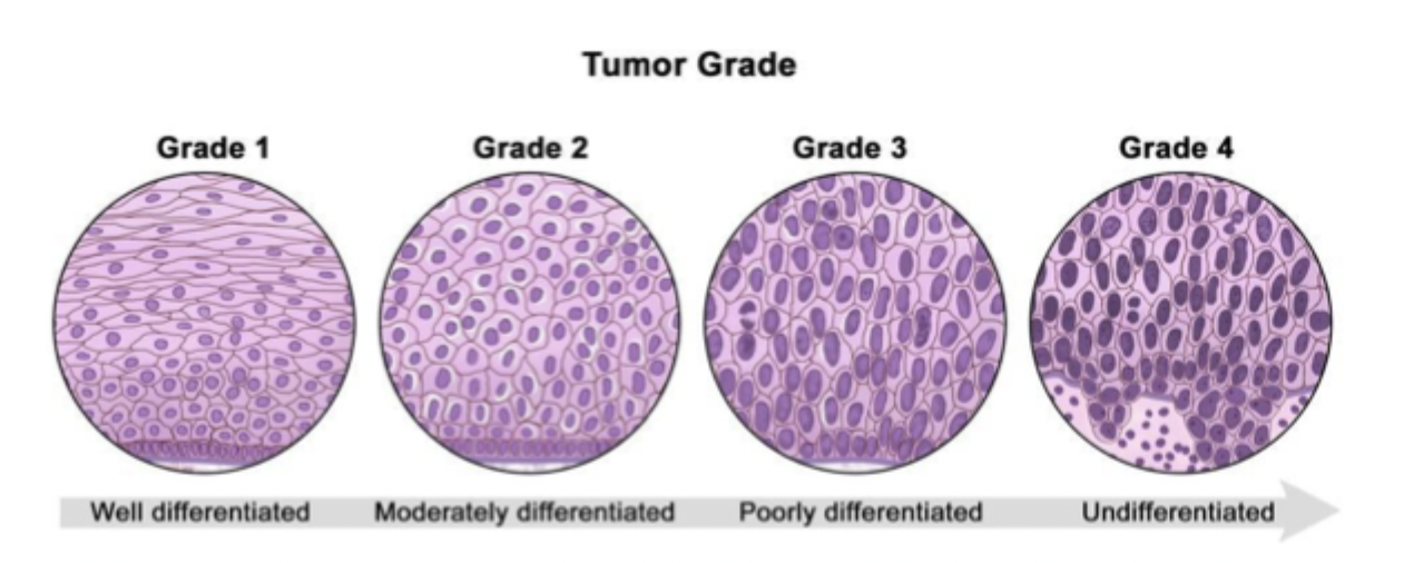
based on differentiation level of tumour cells → info about its aggressiveness
determined by examining cells under microscope → histology
in some cancers = most important prognostic indicator
endometrium
primary CNS
prostate
sarcoma
breast
prognostic factors
stage + grade
patient age + health before diagnosis
patient response to treatment
statistics collected over many years about people w/ same type of cancer
several types of statistics used to estimate prognosis
prognosis
affected by
cancer type + location in body
stage of cancer
size of tumour
presence of spread
grade
how abnormal cancer cells look under microscope
provides clues about how quickly cancer is likely to grow + spread
cancer-specific survival
aka. disease-specific survival
% of patients w/ specific type + stage of cancer who have not died from their cancer during certain period of time after diagnosis
period of time may be 1/2/5 years, etc.
5 years most often used
relative survival
% of cancer patients who have survived for certain period of time after diagnosis compared to people who do not have cancer
how long you survive compared to rest of population
estimates cancer-specific survival that does not use info about cause of death
overall survival
% of people w/ specific type + stage of cancer who have not died from any cause during certain period of time after diagnosis
disease-free survival
% of patients who have no signs of cancer during certain period of time after treatment
ie. recurrence-free or progression-free survival
important for high disease recurrence
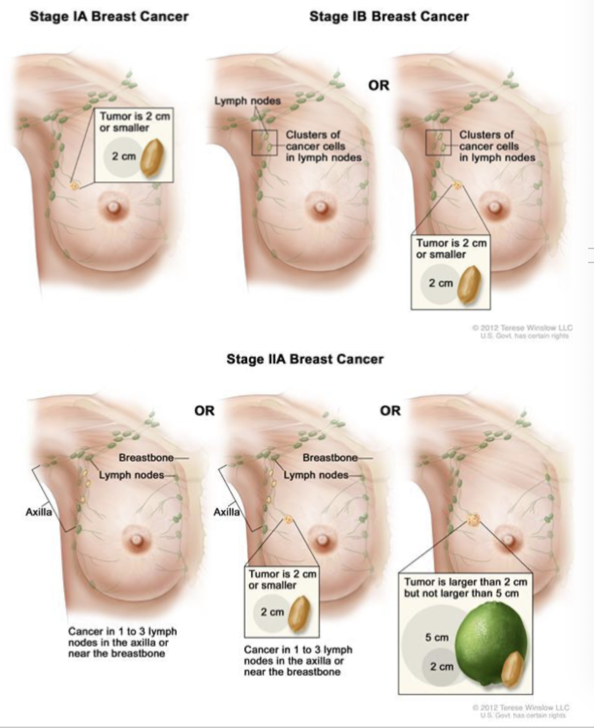
breast staging
clinical staging (cTNM) → physical exam + imaging studies
pathologic staging (pTNM) → detailed pathologic exam of primary tumour + regional lymph nodes
stage
1A = small primary + no lymph node involvement
1B = small primary + lymph node involvement
IV = metastatic, any T + any N
TNM → T
T1 = <2cm
T2 = 2-5cm
T3 = >5cm
T4 = of any size w/ direct extension to chest wall/skin
extension = advanced disease
T4d = inflammatory breast cancer
specific type of breast cancer involving skin
prostate staging
majority = adenocarcinomas
staging incorporates Gleason score = grade looking at cell differentiation → strong correlation to prognosis
low score = slowly growing + non-invasive
high score = more invasive/metastatic
have areas w/ different levels of differentiation
pathologist evaluates predominant + less frequent level of differentiation → assign grade
if tumour is all same grade, value repeats
eg. 3+3=6
Gleason score
2 grades from 1-10 added = Gleason score
1st number = grade most common in tumour
eg. 3+4=7 → most of tumour = grade 3 + less = grade 4
written as
Gleason 7/10
Gleason 7 (3+4)
combined Gleason grade of 7
TNM staging
T1 = not palpable or visible by imaging
T2 = confined within prostate
T3 = extends through prostatic capsule → into seminal vesicle
T4 = invades adjacent structures beyond seminal vesicle
M1 = distant metastasis = determines type of treatment → different sites respond to different treatments
M1a = non-regional lymph nodes
M1b = bone
M1c = other sites

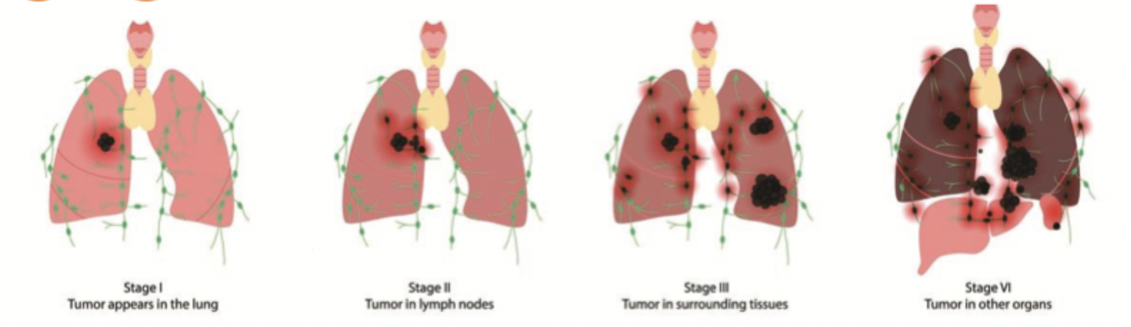
respiratory staging
based on 4 factors
location of primary
tumour size
lymph node involvement
presence/absence of distant metastasis
2 predominant categories of lung cancer
NSCLC - non-small cell lung cancer
SCLC - small cell lung cancer → more aggressive + natural history highly suggestive of spread
further stages as
limited stage = only on 1 side of chest + can be treated w/ definite radiation therapy (stage 1/2)
extensive stage = cancer spread widely throughout lung/to other body parts
TNM staging
T1a = ≤2cm
T1b = >2-3cm
T2a = >3-5cm
T2b = >5-7cm
T3 = >7cm, invasion + same lobe nodules
T4 = extension + ipsilateral lung
M1a = pleural effusion + contralateral lung
M1b = distant
CNS staging
no universal staging system in use
GTM = Grade, Tumour, Metastasis
grade has prognostic significance - G1 → G4
histopathological grading
GX = grade cannot be assessed
G1 = well differentiated
…
G4 = undifferentiated
supratentorial
T1 = one side ≤ 5cm
T2 = one side > 5cm
T3 = invades/encroaches ventricular system
T4 = opposite side, invades infratentorial
infratentorial
T1 = one side ≤ 3cm
T2 = one side > 3cm
T3 = invades/encroaches ventricular system
T4 = opposite side, invades supratentorial

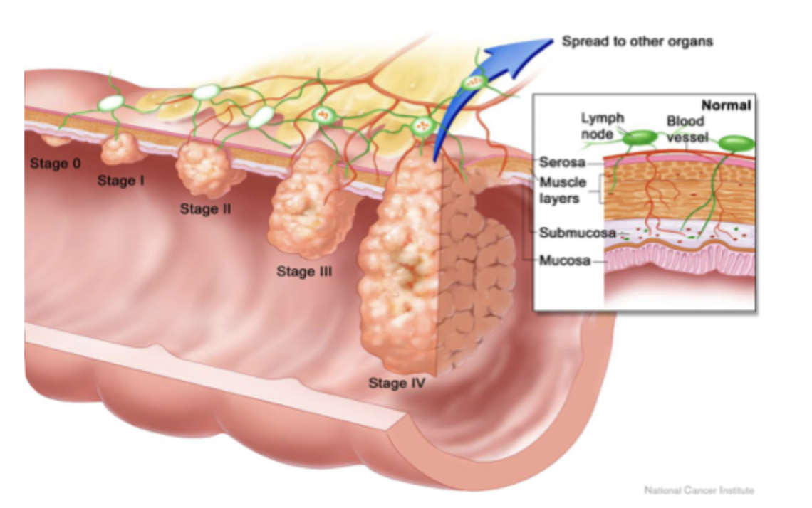
GI staging
TNM staging
Tis = carcinoma in situ → intraepithelial/invasion of lamina propria
T1 = invades submucosa
T2 = invades muscularis propria
T3 = invades through muscularis propria into subserosa OR non-peritonealized pericolic/perirectal tissues
T4 = directly invades other organs/structures +/ perforates visceral peritoneum
T4a = perforates visceral peritoneum
T4b = directly invades/adheres to other organs/structures
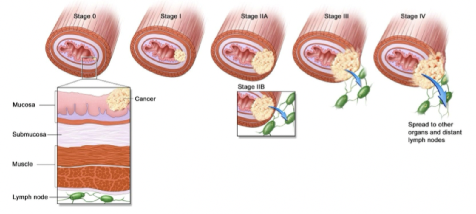
esophagus staging
tumour w/ epicentre within 5cm of esophagogastric junction + extends into esophagus → classified + staged using esophageal scheme
tumour w/ epicentre in stomach ≥5cm from esophagogastric junction w/o extension in esophagus→ classified + staged w/ gastric carcinoma scheme
gyne staging
uses International Federation of Gynecology and Obstetrics (FIGO) + TNM staging system
major difference = FIGO includes stage 0
for cervix → stage most important prognostic indicator

head and neck staging
staging criteria site specific
