Digestive System Anatomy: Oral Cavity, GI Wall & Swallowing
 Call Kai
 Learn
 Practice Test
 Spaced Repetition
 Match
 Flashcards
 Knowt Play
1/72

There's no tags or description
Looks like no tags are added yet.
73 Terms
GI tract components
Mouth → pharynx → esophagus → stomach → small intestine → large intestine.

Accessory digestive organs
Salivary glands, liver, gallbladder, and pancreas; they help digestion with secretions.
Main role of lips
Orbicularis oris lets the lips act like a sphincter at the oral opening.

What structures make up the lips?
Orbicularis oris, connective tissue, skin, sensory receptors, and blood vessels.
Oral cavity boundaries
Cheeks = lateral; hard palate = ceiling; palatine arches/uvula = posterior boundary.

Soft palate
Contains skeletal muscle and participates in the gag/swallowing response.
Intrinsic tongue muscles
Interwoven skeletal muscle within tongue; change the tongue's shape.
Extrinsic tongue muscles
Attach tongue to other structures; change tongue position. Examples: hyoglossus, styloglossus.

Papillae
Small raised tongue areas; some contain taste buds, others increase friction.
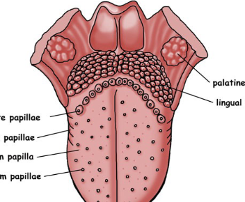
Lingual tonsils
Lymphoid tissue on posterior tongue.

Frenulum
Fold under tongue that restricts tongue movement.

Is the classic tongue taste map correct?
No. Taste receptors are distributed more evenly across the tongue.
Umami
The fifth taste; savory.
Tooth crown
Crown is exposed

tooth root
root anchors the tooth.

Periodontal ligament
Connective tissue that anchors a tooth.
Gingiva
The gums; mucous membrane around teeth.

Alveolus of a tooth
The bony socket that holds the tooth.

Enamel
Outer tooth material made mostly of hydroxyapatite.

Dentin
Bone-like tissue deep to enamel; more dynamic than enamel.

Dental pulp
Contains nerves and blood vessels.

Root canal
Canal within the root where pulp is found.

What is saliva mostly made of?
About 99% water.
Mucus in saliva
Lubricates food/oral surfaces.
Lysozyme in saliva
Helps destroy bacteria.
Digestive enzymes in saliva
Begin digestion while food is in the oral cavity.
Salivary salts
Help maintain proper pH for digestive enzymes and help with taste.
Parotid gland location/drainage
Inferior/anterior to ear; drains near the 2nd upper molar.

When is the parotid gland especially active?
It kicks in with chewing.
Submandibular gland location/drainage
Just inside mandible; empties into floor of mouth.

Sublingual gland location/drainage
Under mucosa in floor of mouth; empties into floor of mouth.

Which salivary glands are active all the time?
Submandibular and sublingual glands.
Mumps and salivary glands
Mumps virus can infect salivary glands and cause painful swelling/parotitis.

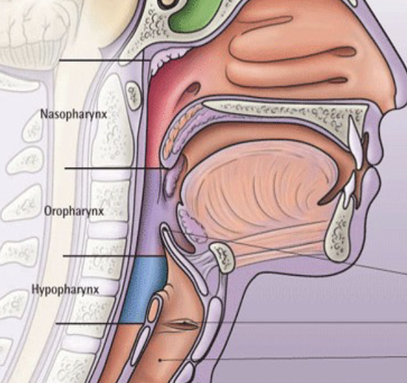
Pharynx epithelium
Oropharynx + laryngopharynx = stratified squamous; nasopharynx = respiratory epithelium.
Other pharynx features
Mucous membrane provides lubrication; pharynx contains skeletal muscle.
How does swallowing begin?
Voluntarily; initiating it triggers a swallowing reflex.
What happens during the swallowing reflex?
Openings coordinate so the bolus is directed into the esophagus, not nasal cavity/airway.
When does swallowing become completely involuntary?
By about the middle of the esophagus.
Who controls movement after voluntary swallowing ends?
Autonomic nervous system plus enteric nervous system.
Four GI wall layers: inside → outside
Mucosa → submucosa → muscularis externa → serosa.
Mucosa
Innermost layer: epithelium plus connective tissue immediately deep to it; contains many glands.
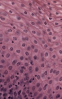
GI regions with stratified squamous epithelium
Mouth, pharynx, esophagus, and anal canal.
GI regions with simple columnar epithelium
Stomach, small intestine, and large intestine.
Easy GI epithelium pattern
Beginning/end = stratified squamous; middle = simple columnar. Think SS → SC → SS.
Why do glands in the mucosa matter?
They produce region-specific GI secretions; the type varies by location.
Submucosa
Connective tissue layer with rich blood/nerve supply plus collagen and elastic fibers.

Why are blood vessels near the mucosa important?
GI epithelium is avascular, so vessels are supplied from deeper connective tissue.
Why collagen and elastic fibers in submucosa?
They let the GI tract expand and recoil as contents pass.
Muscularis externa: main jobs
Propels material and performs mechanical digestion/mixing.

Muscularis externa layers
Outer longitudinal smooth muscle + inner circular smooth muscle.
Outer longitudinal muscle
Runs along tube length; contraction helps shorten/scrunch the tube.
Inner circular muscle
Wraps around the tube; contraction squeezes/narrows the lumen.
Peristalsis
Movement that propels GI contents along the tract.
Segmentation
Kneading/squishing movement that mixes GI contents.
Serosa
Outermost connective-tissue layer of the GI wall.

Three nervous systems controlling GI tract
Enteric, parasympathetic, and sympathetic nervous systems.
Enteric nervous system
Intrinsic nervous system in GI walls; can carry out gut reflexes independently.
Parasympathetic effect on digestion
Rest and digest: stimulates digestive activity.
Sympathetic effect on digestion
Inhibits digestion, including movement and secretion.
Submucosal plexus: other name/location
Meissner plexus; located in the submucosa.

Submucosal plexus: main job
Controls gland secretion and smooth muscle of the mucosa. Think S = secretion.
Myenteric plexus: other name/location
Auerbach plexus; between circular and longitudinal layers of muscularis externa.

Myenteric plexus: main job
Major motor supply to muscularis externa; controls peristalsis and segmentation.
Three unpaired GI arteries: superior → inferior
Celiac trunk (1) → superior mesenteric artery (SMA, 2) → inferior mesenteric artery (IMA,3).

Celiac trunk: three major branches
Left gastric, common hepatic, and splenic arteries.
Celiac trunk: general territory
Upper abdominal organs: esophagus, stomach, spleen, liver, and gallbladder.
Left gastric artery supplies
Stomach and esophagus.

Common hepatic artery supplies
Liver and gallbladder.

Splenic artery supplies
Spleen.

Superior mesenteric artery supplies
Duodenum, pancreas, jejunum, ileum, cecum, ascending colon, transverse colon.

Easy SMA summary
All small intestine + some large intestine + pancreas.
Inferior mesenteric artery supplies
Transverse colon, descending colon, sigmoid colon, and rectum.

Easy IMA summary
Supplies the remainder/lower portion of the digestive tract through the rectum.