9 & 10 - Gastrointestinal Principles I & II
 Call Kai
 Learn
Learn Practice Test
 Spaced Repetition
 Match
Match Flashcards
 Knowt Play
1/76

There's no tags or description
Looks like no tags are added yet.
Name | Mastery | Learn | Test | Matching | Spaced | Call with Kai | Chat |
|---|
No analytics yet
Send a link to your students to track their progress
77 Terms
What are causes of gastric distention?
Postprandial
Food or foreign material
GDV
Pyloric outflow or proximal duodenal obstruction
Gastric functional ileus
Aerophagia
Gastric distention causes caudal displacement of what 3 things?
Splenic head
Colon
Small Intestines
What radiographic sign is pathognomic for GDV?
Double bubble
Where is the pylorus located with GDV?
Dorsal & to the left
What is seen on radiographs with GDV?
Compartmentalization
Pylorus dorsal & left
Splenomegaly
Megaesophagus
Functional ileus
Thin caudal vena cava
Pneumoperitoneum, peritoneal effusion
What is seen with small intestinal ileus?
Distention of the SI tract
Failure of contents to be transported
What does normal jejunal content look like?
Soft tissue opaque or some gas content
How large is the small intestine normally in a dog?
How large is the small intestine normally in a cat?
What is mechanical small intestinal ileus?
Obstruction
What causes functional small intestinal ileus?
Paralytic blockage of movement of cotent
What are causes of functional ileus?
Enteritis
Periotonitis
Chronic mechanical obstruction
Parasympatholytics/Anesthesia
Ischemia
Dysautonomia
Toxicity
What is seen radiographically with functional ileus?
Generalized, mild dilation
Uniform diameter
What factors effect the radiogrphaic appearance of ileus?
Duration, Location & Type of obstruction
What is seen with mechanical ileus?
Various sizes of interstinal diamerter (mixed population)
2.4xs + the diameter of L5 vetrebral body
Distention proximal to obstruction
Parallell stacking
Radiopaque foreign body
What is parallel stacking?
Gas filled distended segment of bowel
What are the 3 location where mechanical ileus can occur?
Intraluminal
Mural
Extramural
What are examples of things that can cause intraluminal mechanical ileus?
FB
Parasites
What are examples of things that can cause mural mechanical ileus?
Tumor
Granuloma
What are examples of things that can cause extramural mechanical ileus?
Adhesions
Hernia
Tumor
What is done to treat functional ileus?
Medical management
(Sx if chronic or volvulus)
What is done to treat mechanical ileus?
Exploratory laparotomy
Medical management
What can be used for a compression study?
A wooden spoon
T/F - Linear FBs cause vomiting in dogs and cats
True
What is the anchor point of linear foreign bodies in a cat?
Tongue
What is the anchor point of linear foreign bodies in a dog?
Pylorus
Dogs/Cats typically have thin and threadlike material for linear FBs
Cats
Dogs/Cats typically have thicker textile material for linear FBs
Dogs
What is the plication pattern for linear FBs in cats?
Tight
What is the plication pattern for linear FBs in dogs?
Bunched loops
What does intestional torsion & volvulus result in?
Sepsis
Circulatory shock
Death
What is intestinal torsion?
Pathologic twisting of bowel along its longitudinal axis
What is intestinal volvulus?
Twisting of bowel around the mesenterid pedicle
What are general indications for abdominal contrast procedures?
Locate an organ
ID rupture/obsturction
Highligh structures in hollow organs
Evaluate basic functional information
What are 2 types of negative contrast media?
Air
Gas (O2, Nitrous oxide, CO2)
What are 2 types of positive contrast media?
Barium
Iodine
What do negative contrast mediums do?
Makes organ of interest more radiolucent
What are negative contrast studies used for?
Pneumogastrogram
Pneumocystogram
Pneumocolonogram
Double contrast studies
What do positive contrast mediums do?
Make organ of interest more radiopaque
What are barium sulfate contrast studies used for?
Esophagram
Gastrogram
Upper GI
Colonogram/barium enema
How can barium sulfate?
Per annum (anus)
Per os (mouth)
When should barium NOT be used?
If GI perforation is suspected
What are uses of iodinated contrast studies?
Excretory urogram/IV pyelogram
Cystogram
Retrograde Urethrogram/Vaginourethrogram
Fistulogram
Arthrogram
Myelogram
GI obstruction
When should ionic iodinated contrast NOT be used?
Intrathecally
High risk patients
What are the 6 Roentgen signs?
Size
Shape
Location
Number
Opacity
Margination
What are additional considerations with Roentgen signs?
Timing
Degree of opacification
Luminal diameter
Wall thickness
Filling defects
What are indications for upper GI contrast studies
If US & endoscopy is unavialbe
How long does it typically take liquid to be emptied from the stomach?
1-4 hours
Cats/Dogs have a faster gastric emptying
Cats (within 1 hour)
Iodinated contrast empties faster/slower than normal emptying
Faster
Emptying & transit times are faster/slower with barium soaked kibble
Slower
How much contrast should small dogs be given?
8-12 mL/kg
How much contrast should large dogs be given?
5-7 mL-kg
How should contrast be administered?
Orogastric tube
What are indications for a pneumocolonogram?
Differentiate small vs large intestines
Large bowel stricture or masses
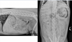
What abnormality is seen in this image?
Gastric distention

What structure is highlighted in pink?
Cecum

What does this indicate?
Gastric Dilation & Volvulus

Which marker is highlighting the abnormality?
Yellow

What is the yellow marker indicating?
Some textile FB in the intestines

What abnormality is seen in this image? Point it out.
FB Obstruction
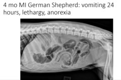
Is this functional or mechanical ileus?
Functional

What is the most likely cause of gas appearing like this? 1 yo FS Min Pin.
Linear foreign body

What is the most likely cause of this apperance? 1 yo FS DSH
Linear foreign body

What is seen in this radiograph generally?
Small intestinal plication

What causes this appearance?
Linear FB
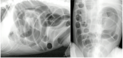
Is this functional/mechanical ileus?
Functional

What is seen here after the barium study?
Chronic colonic torsion

What is seen in this radiograph?
Esophageal obstruction

What type of luminal defect is this?
Intraluminal

What type of luminal defect is this?
Mural

What type of luminal defect is this?
Extramural

What type of luminal defect is this?
Intraluminal
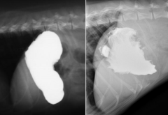
What type of luminal defect is pictured on the right? (Left is normal)
Mural

What type of luminal defect is this?
Extramural (dilation around obstruction)

What type of study is this?
Contrast study

What type of study is this?
Pneumocolonogram