2. Tumour Growth & Metastases
 Call Kai
 Learn
 Practice Test
 Spaced Repetition
 Match
 Flashcards
 Knowt Play
1/29

There's no tags or description
Looks like no tags are added yet.
30 Terms
proto-oncogenes
code for proteins that promote cell cycle progression → mutate to become oncogenes
have potential to cause cancer
stimulate
cell division
cell differentiation
halt cell death
eg. growth factors (receptors), signal transduction proteins, transcription factors, proteins that inhibit apoptosis
tumour suppressor genes (TSG)
normal growth suppressor genes
limit cell division
promote apoptosis (cell death)
repair defective DNA
eg. TP53 - found on chromosome 17
blocks proliferation in G1 (growth 1 - phase of cell cycle)
apoptosis
DNA repair
1 normal gene inherited = predisposed to cancer
two-hit hypothesis
need loss-of-function in both copies of proto-oncogene/tumour suppressor gene to cause cancer
1 functioning copy is enough to present normal phenotype
mutations
translocation
chromosome damaged in one area
fixing machinery swaps damaged areas of 2 different chromosomes
gene amplification
mutation can result in higher amount of proteins than necessary
point mutation
one codon deleted/mutated + replaced by wrong one
can sometimes function normally, other times results in mutated protein
doubling time
time taken for tumour to double its volume
takes about 1-3 months for humans
varies b/w different tumour type
sub-clinical disease
disease that may escape detection (0.5-1cm3) until it causes clinical symptoms
many tumours have long subclinical period
may allow for metastasis prior to detection, eg. small cell lung cancer
early treatment might reduce but not eliminate risk of metastasis → screening
to palpate tumour, must measure at least 1cm3
1 billion cells or 30 doublings
growth fraction
fraction of cells that are proliferating
most tumours contain non-proliferating cells = support cells
some tumour cells may have lost capacity to divide
differentiation
biological change
change in micro-environment (pH, oxygenation, nutrition)
other cells in population continue dividing, while others stop
cell loss
rate at which cells are shed from tumour
necrosis
metastasis
continuous shedding of cells + cells failing to mature + cell death = 80-90% of loss of replicating cell pool
why it may take many years for 1cm mass to form
local spread (direct extension)
direct extension + invasion along tissue planes
common method of spread
invades + destroys local surrounding normal tissue
eg. prostate cancer spread to bladder wall
causes compression, ulceration, pain, bleeding, swelling
surgical implications → if surgery can be performed must include margin of normal tissue to limit local recurrence
transcoelomic spread
cancer cells break out of original site + carried in peritoneal fluid
uncommon method of spread
challenging to treat
disseminated growth (scattered)
overproduction of ascitic fluid
peritoneal cavity
spread across peritoneal surface from one organ to another
eg. stomach → ovary
pleural cavity
spread of primary lung cancer or metastatic cancer across pleura from one lung to another
ascites
collection of fluid in abdomen
contains proteins + growth factor
provides environment for malignant cells to grow w/o attachment = anchorage independent
overproduction can be caused by metastasis → requires draining
in lungs = pleural effusion
lymphatic spread
lymphatic vessels do not have basement membrane → easy to enter + can pass cancer cells to blood stream
most common method of spread
lymph travels via lymphatic vessels to subclavian vein → SVC → bloodstream
cancer cells can break tumour + enter lymphatic vessel → enter bloodstream = hematogenous spread
fixed node
refers to cancer in lymph node that has grown beyond capsule to surrounding tissue
cancer cell has gotten into lymph node → attached to node → grown beyond to surrounding tissue = cancer progression = bad
higher risk of getting cancer to other parts of body
sentinel node
first lymph node in path of lymph drainage from primary tumour
biopsied to assess for lymphatic spread
sentinel lymph node biopsy
radioactive dye injected into area of interest to locate sentinel node
sentinel node removed + examined
negative = no cancer cells → rest of lymph nodes in area unlikely to contain cancer
positive = cancer cells found → more lymph nodes from area may be removed to check for spread
hematogenous spread
blood vessels provide least difficult barrier of entry + exit for cancer cells
common method of spread
capillaries constructed from single layer of endothelial cells
built for passage of materials + fluids
arteries more difficult to spread through b/c of smooth muscle layer
circulation plays role in sites of metastatic growth
liver + lung = common metastasis sites
lung → blood returning to heart from upper + lower body passes lungs for reoxygenation
liver → blood from intestines travel through hepatic portal vein before traveling to heart
leptomeningeal spread
spread through cerebrospinal fluid (CSF)
very uncommon method of spread
CSF surrounds brain + spinal cord
specific to neural tumours (brain + spine)
if tumour from brain/spinal cord were to pass to other organs/parts of body → need to pass BBB
eg. medulloblastomas of brain often seed (spread) into spinal canal via CSF
first-pass organ
first organ encountered by cells frequently supports secondary tumour growth
main organs:
heart
metastasis very difficult → not conducive environment for cancer cell growth
hard for cells to latch on + stay
thick muscle + connective tissue
thicker walls, hard for cells to break through
blood flows from heart to lungs where metastasis frequently occurs
liver
accessible to cancer cells due to lack of barriers b/w sinusoids + hepatocytes
seed and soil theory
metastasis required for cancer cells (seeds) + correct environment (soil) in which to grow
soil = tissue in which appropriate factors could be recognized by cancer cell receptors providing homing mechanism
most cells destroyed in transit or cannot survive new environment
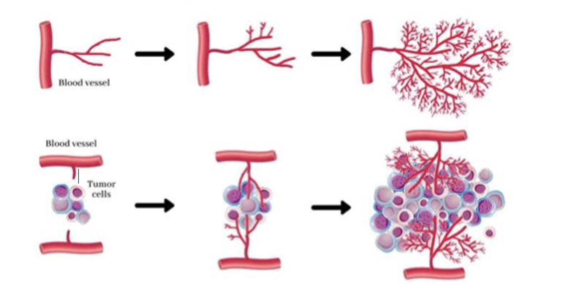
angiogenesis
formation of new blood vessels
initial growth of tumour is close to existing blood vessels (1mm)
tumour grows → more cells required more nutrients + oxygen supplied by blood
success of tumour induced blood vessel formation → dictates if metastasis will remain dormant or grow larger than 1mm
cancer cells secrete signalling molecules → stimulate proliferation of endothelial cells → secrete proteases causing capillary sprouts to form = new capillary bed
liver + lung = common metastasis sites
cancer-induced growth disorganized + not well-structured
leaky + unstable
large tumours can be bleeding
radiation can cauterize bleeding
radical (curative) treatment
potential to completely cure patient of cancer
if cancer is widespread, may need chemotherapy treatment
palliative (symptom management) treatment
deal w/ symptoms
if cancer is widespread throughout body + cannot be cured
lung primary site
common metastatic sites
liver
adrenal gland
bone
brain
breast primary site
common metastatic site
lung
bone
brain
stomach primary site
common metastatic site
liver
anus primary site
common metastatic site
liver
lungs
bladder, cervix, and prostate primary site
common metastatic site
lungs
bone
liver
colorectal primary site
common metastatic site
liver
lung
peritoneum
uterus primary site
common metastatic site
bone
liver
lung
peritoneum
vagina
melanoma primary site
common metastatic site
bone
brain
liver
lung
skin/muscle