Dermis & Subcutis - Structure and Function

0.0(0)
Studied by 0 people
call kaiCall Kai
learnLearn
examPractice Test
spaced repetitionSpaced Repetition
heart puzzleMatch
flashcardsFlashcards
GameKnowt Play
Card Sorting

1/35

encourage image

There's no tags or description

Looks like no tags are added yet.

36 Terms
1
New cards
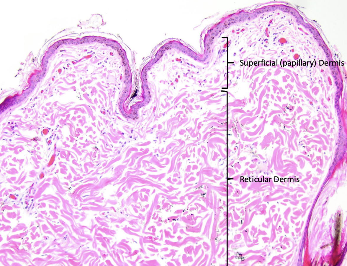
⭐ 1. Compare the papillary and reticular layers of the dermis.
A: The papillary dermis is the superficial dermis composed predominantly of loose connective tissue, while the reticular dermis is the mid-to-deep dermis composed predominantly of dense irregular connective tissue. 🔎 What this means: The papillary dermis has thinner, more loosely arranged collagen with more cells and blood vessels. The deeper reticular dermis contains thicker collagen bundles running in many directions, which provides strength and flexibility. 💡 Think: papillary is superficial and loose; reticular is deeper and dense irregular.
2
New cards
⭐ 2. How do the dermis and epidermis interact to create the unique surface pattern of a dog's nose?
A: Dermal papillae project upward and interdigitate with downward-projecting epidermal ridges or rete pegs to create the dog's unique nasal surface pattern. 🔎 What this means: The epidermis and dermis interlock rather than meeting along a flat line. This arrangement is especially prominent in high-friction nonhaired skin such as the nose, and the resulting pattern is unique to each dog like a human fingerprint.
3
New cards
  1. How is the reticular dermis different from reticular fibers?


A: The reticular dermis is the deep layer of the dermis and is composed mainly of dense irregular type I collagen, while reticular fibers are thin type III collagen fibers present in much smaller amounts. 🔎 What this means: The word reticular is used in two different ways here. Do not assume the reticular dermis is made predominantly of reticular fibers; its major collagen is still type I.
4
New cards
⭐ 4. What are the resident and transient cell populations of the dermis, and what does their presence indicate?
A: Resident dermal cells normally include fibroblasts, histiocytes, mast cells, and T lymphocytes, while transient cells include monocytes, circulating lymphocytes, neutrophils, and eosinophils recruited during inflammation. 🔎 What this means: Resident cells are expected in normal skin. Increased transient inflammatory cells in a biopsy suggest an inflammatory process, and the specific population present can help characterize that inflammation.
5
New cards
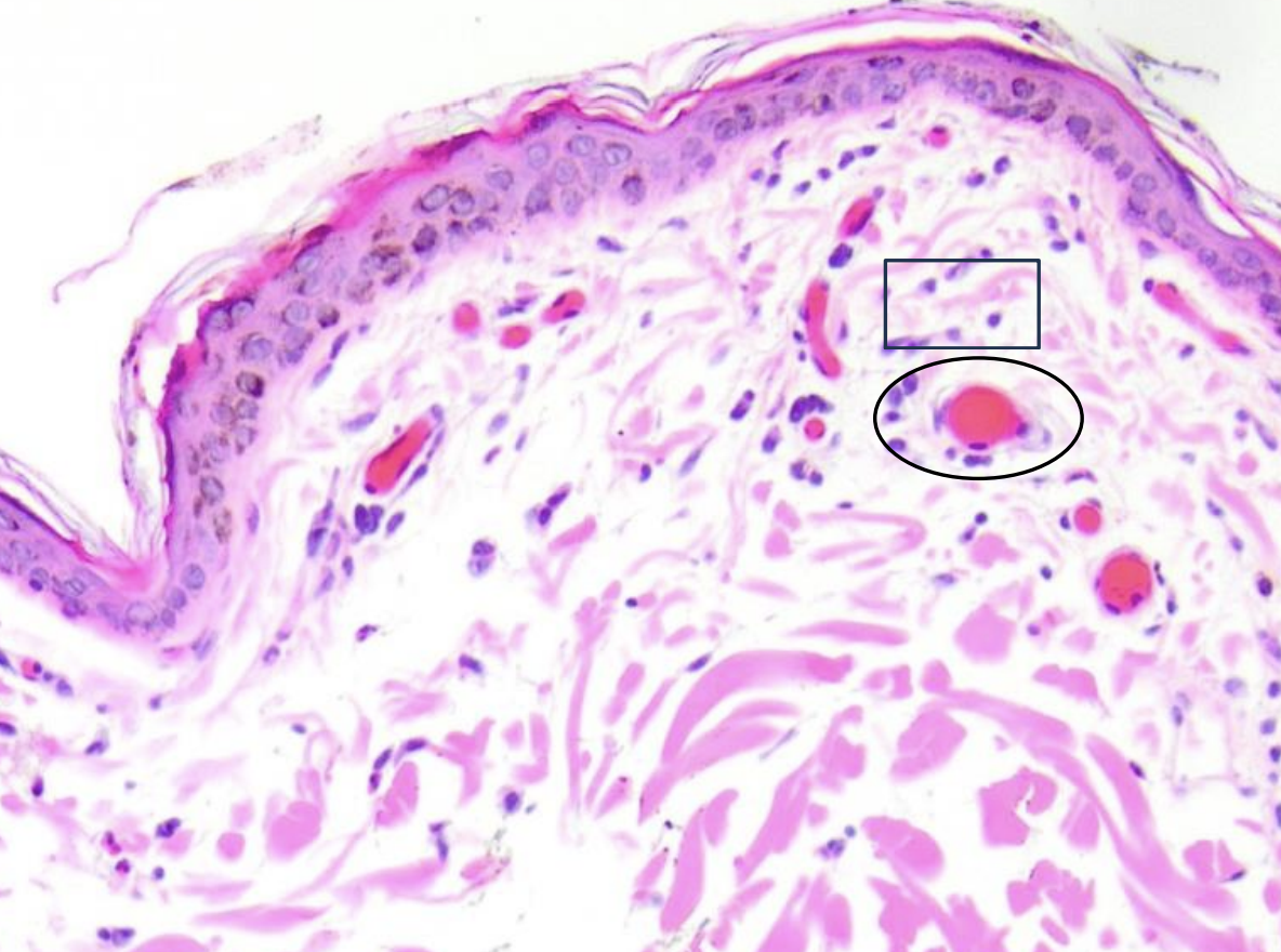
⭐ 5. What is the major role of fibroblasts in the dermis, and how can they be recognized histologically?
A: Fibroblasts produce collagen and much of the extracellular matrix and appear as small, thin, spindle-shaped or elongated cells between collagen fibers. 🔎 What this means: Fibroblasts occur in both dermal layers but are more numerous in loose connective tissue. A relatively small fibroblast can produce a large amount of surrounding extracellular matrix.
6
New cards
⭐ 6. What is a myofibroblast, and what is its important role in skin?
A: A myofibroblast is a contractile cell that can develop from a fibroblast and helps contract the wound bed during healing. 🔎 What this means: Fibroblasts are normal resident dermal cells, but when activated they can acquire muscle-like properties. Myofibroblasts can also arise through other pathways, but the major point for this lecture is their role in wound contraction.
7
New cards
🔄 UPDATED/REPLACEMENT 7. What are the fibrous and nonfibrous components of the dermal extracellular matrix?
A: The fibrous component contains collagen, reticular, and elastin fibers, while the nonfibrous ground substance contains glycosaminoglycans and multiadhesive glycoproteins. 🔎 What this means: This expands the earlier general ECM material specifically to the dermis. GAG-containing structures such as proteoglycans and hyaluronan contribute hydration and shock absorption, while multiadhesive glycoproteins help stabilize the ECM to surrounding cells.
8
New cards
⭐ 8. What collagen types and locations in the skin were emphasized in this lecture?
A: Type I collagen is the predominant collagen of the dermis, type III collagen forms reticular fibers especially in loose connective tissue and early wound healing, type IV collagen is associated with the lamina densa, and type VII collagen forms anchoring fibrils. 🔎 What this means: Type I provides most of the dermis's strength. Type III is present in much smaller amounts and is later replaced by type I during wound healing. Types IV and VII were introduced here but will be covered more fully with the basement membrane.
9
New cards
⭐ 9. What is the sequence of collagen synthesis from preprocollagen to a collagen fibril?
A: Collagen synthesis begins intracellularly with a preprocollagen alpha chain, followed by vitamin C-dependent hydroxylation of proline and lysine to form procollagen, glycosylation that permits triple-helix formation, secretion of the triple helix, extracellular removal of its terminal ends to form tropocollagen, and copper-dependent lysyl oxidase crosslinking of tropocollagen into collagen fibrils. 🔎 What this means: The early modification and triple-helix steps occur inside the fibroblast. Once the molecule is secreted, its frayed ends are removed and mature tropocollagen molecules are cross-linked outside the cell. 💡 Think: modify inside, trim and cross-link outside.
10
New cards
⭐ 10. What roles do vitamin C and copper play in collagen synthesis?
A: Vitamin C is required for hydroxylation of proline and lysine early in collagen synthesis, while copper is required by lysyl oxidase for extracellular collagen crosslinking. 🔎 What this means: Both are needed at different stages. Without adequate vitamin C, collagen does not form properly and becomes fragile. Without proper lysyl oxidase activity, tropocollagen cannot be effectively cross-linked into strong fibrils.
11
New cards
⭐ 11. What consequences can result from a defect in collagen synthesis?
A: Defective collagen synthesis can produce fragile connective tissue or abnormally hyperelastic skin depending on which step is disrupted. 🔎 What this means: Vitamin C deficiency impairs hydroxylation and produces weak collagen, so tissues and vessel walls can break and hemorrhage in areas under stress. Other abnormalities in collagen processing can produce unusually stretchy skin, as illustrated by the HERDA example. The important skill is predicting how abnormal collagen structure changes tissue strength rather than memorizing the disease details.
12
New cards
12. How is elastin produced, and what important enzyme does elastogenesis share with collagen synthesis?
A: Fibroblasts produce tropoelastin, which associates with fibrillin-1 and is cross-linked by copper-dependent lysyl oxidase to form elastic fibers. 🔎 What this means: The professor emphasized much less detail for elastin synthesis than collagen synthesis. The major relationship to remember is that lysyl oxidase participates in crosslinking both collagen and elastin and that elastic fibers allow skin to stretch.
13
New cards
🟡 LOWER PRIORITY 13. What is solar elastosis, and what does it demonstrate about abnormal elastic fibers?
A: Solar elastosis is abnormal accumulation and disordered assembly of elastin associated with excessive ultraviolet exposure. 🔎 What this means: In affected skin, abnormal elastin may become visible between normal dermal collagen and can be highlighted strongly with a Verhoeff-Van Gieson stain. The example illustrates that abnormal ECM production can alter normal skin structure.
14
New cards
14. What happens to dermal structure when excessive hyaluronic acid accumulates in the ground substance?
A: Excess hyaluronic acid increases ground substance and separates the normal collagen bundles, producing a more hydrated, thickly spaced or wrinkled appearance without necessarily increasing the actual amount of skin. 🔎 What this means: The Shar Pei example demonstrates the structural effect of excess ground substance. Their dermal collagen remains present, but abundant hyaluronic acid creates large clear spaces between it. The professor emphasized understanding this structure-function relationship rather than memorizing the disease state.
15
New cards
⭐ 15. How is the vasculature of the dermis and subcutis organized into plexuses?
A: Cutaneous vessels form a deep or subdermal plexus near the dermis-subcutis interface, a middle dermal or cutaneous plexus around the level of hair follicles and sebaceous glands, and a superficial or papillary plexus just below the epidermis. 🔎 What this means: Direct cutaneous arteries and veins arise from deeper tissue and repeatedly branch as they travel toward the surface. The epidermis itself has no vessels and depends on the superficial dermal circulation for oxygen and nutrients. 💡 Think: deep feeds middle, middle feeds superficial, superficial supports epidermis.
16
New cards
⭐ 16. Why are basal epidermal cells especially vulnerable when dermal blood flow is compromised?
A: Basal keratinocytes are mitotically active regenerative cells with a high oxygen demand, so reduced dermal blood supply causes them to become injured and die early. 🔎 What this means: The epidermis is avascular and depends on the dermal vasculature. When blood flow is compromised, the highly active basal cells cannot meet their metabolic demands and may undergo apoptosis before more superficial viable keratinocytes.
17
New cards
⭐ 17. What are the major functions of the subcutis, and what other names are used for it?
A: The subcutis, also called the hypodermis or panniculus, provides insulation, physical protection, connection to the underlying fascia of muscle or bone, and an energy reserve. 🔎 What this means: The subcutis is more than simply a layer of fat. Its adipose, connective tissue, vessels, and muscle contribute to cushioning, attachment, energy storage, and temperature regulation.
18
New cards
⭐ 18. How is adipose tissue organized microscopically within the subcutis?
A: Adipocytes are arranged into lobules called the lobular panniculus and are separated and connected by fibrovascular connective tissue septa called the septal panniculus. 🔎 What this means: Lipid is removed during routine histologic processing, so adipocytes often appear as large clear spaces. The connective tissue septa carry supporting fibrous and vascular components between adipose lobules.
19
New cards
⭐ 19. What is the panniculus muscle, and what is its function?
A: The panniculus muscle is skeletal muscle located within the subcutis that allows movement or twitching of the skin. 🔎 What this means: On a sufficiently deep histologic section, it can appear as a band of skeletal muscle beneath or among the subcutaneous adipose tissue. It provides a mechanism for moving the skin independently of the deeper body musculature.
20
New cards
⭐ 20. What are the major functions of white adipose tissue?
A: White adipose tissue stores energy as triglycerides and also synthesizes and secretes adipokines including hormones, growth factors, and inflammatory cytokines. 🔎 What this means: White adipose tissue is metabolically active rather than simply passive fat storage. Leptin was given as an example of an adipose-derived hormone involved in the sensation of satiety.
21
New cards
⭐ 21. What is the major function of brown adipose tissue, and where is it particularly important?
A: Brown adipose tissue primarily produces heat and is especially important in neonates and hibernating animals. 🔎 What this means: Brown adipocytes contain many mitochondria and use uncoupling protein 1, or UCP1, to favor heat production rather than conventional energy capture. This produces nonshivering thermogenesis.
22
New cards
⭐ 22. How can white and brown adipose tissue be distinguished histologically and functionally?
A: White adipocytes appear dominated by a large clear lipid space with the nucleus pushed to the edge and specialize in energy storage and signaling, while brown adipocytes have more visible pink or flocculent cytoplasm because they contain many mitochondria and specialize in heat production. 🔎 What this means: Lipid washes out during histologic processing in both tissues, but brown fat retains a more protein-rich appearance because of its abundant mitochondria. 💡 Think: white stores; brown burns for heat.
23
New cards
⭐ 23. iClicker: The bulk of the dermis is made up of what type of connective tissue?
A: The bulk of the dermis is dense irregular connective tissue. 🔎 What this means: The reticular or mid-to-deep dermis occupies most of the dermis and contains thick type I collagen bundles running in multiple directions, allowing the skin to resist stress from different orientations.
24
New cards
⭐ 24. iClicker: Which resident cell of the skin produces the extracellular matrix of the dermis?
A: Fibroblasts produce the extracellular matrix of the dermis. 🔎 What this means: Fibroblasts synthesize dermal collagen and other extracellular matrix components. Histiocytes, T lymphocytes, and mast cells have immune rather than primary matrix-producing roles.
25
New cards
⭐ 25. iClicker: Which listed component is a nonfibrous component, or ground substance, of the dermal extracellular matrix: elastin, collagen, or GAGs?
A: Glycosaminoglycans, or GAGs, are nonfibrous components of the dermal extracellular matrix. 🔎 What this means: Collagen and elastin are fibers, whereas GAGs contribute to the hydrated ground substance between the cells and fibers.
26
New cards
⭐ 26. iClicker: Which vitamin is a necessary cofactor for collagen synthesis?
A: Vitamin C is a necessary cofactor for collagen synthesis. 🔎 What this means: Vitamin C is required for hydroxylation of proline and lysine during the intracellular portion of collagen synthesis. Without this step, collagen becomes structurally weak.
27
New cards
⭐ 27. iClicker: Where does lysyl oxidase-mediated crosslinking of tropocollagen occur?
A: Lysyl oxidase-mediated crosslinking of tropocollagen occurs in the extracellular space within the dermis. 🔎 What this means: The collagen triple helix is secreted from the fibroblast before its terminal regions are removed to form tropocollagen. Tropocollagen molecules are then cross-linked extracellularly by copper-dependent lysyl oxidase to form collagen fibrils.
28
New cards
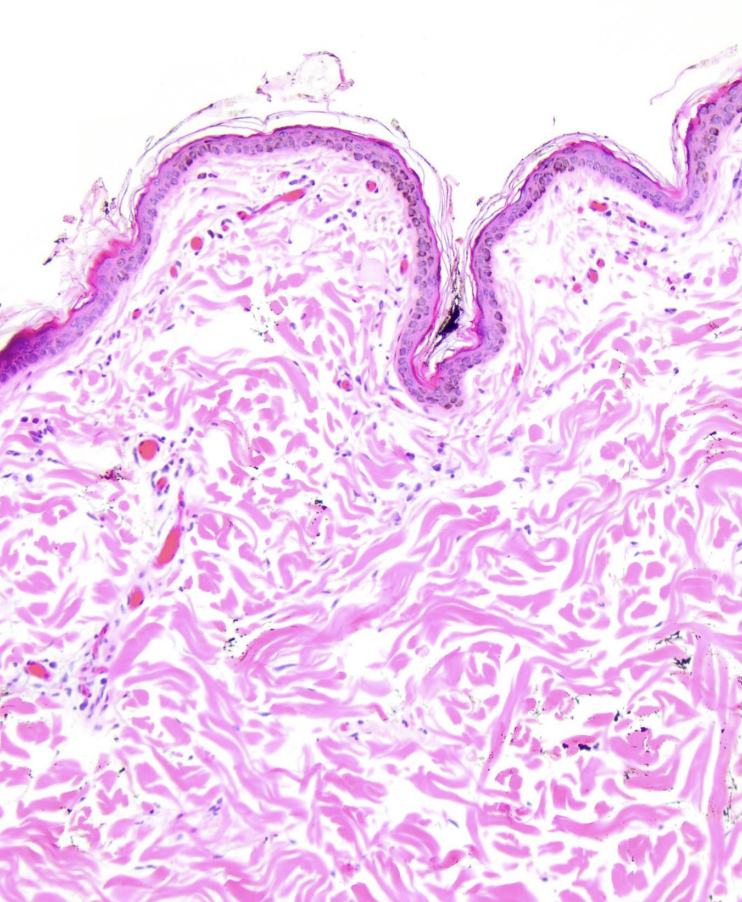
⭐ 28. [ATTACH SLIDE/PAGE 9] Identify the papillary and reticular dermis in this histologic image and state the connective tissue type of each.
A: The superficial papillary dermis is loose connective tissue and the deeper reticular dermis is dense irregular connective tissue. 🔎 What this means: Look for thinner, more loosely arranged collagen near the epidermis and thicker multidirectional collagen bundles deeper in the section.
29
New cards
⭐ 29. [ATTACH SLIDE/PAGE 8] Identify the dermal papillae and epidermal ridges or rete pegs in this canine nose section.
A: Dermal papillae are projections of dermis extending upward, while epidermal ridges or rete pegs are projections of epidermis extending downward. 🔎 What this means: Their interdigitation creates the distinctive surface pattern of the canine nasal planum.
30
New cards
⭐ 30. [ATTACH SLIDE/PAGE 13] Identify the fibroblasts in this dermal histologic image.
A: Fibroblasts are the thin, elongated or spindle-shaped cells lying between the pink collagen fibers. 🔎 What this means: Their nuclei look much smaller than the large amount of extracellular matrix they produce. Do not try to identify the various round inflammatory cells individually because the professor specifically said that was not required histologically.
31
New cards

⭐ 31. In collagen-synthesis, identify which processes occur intracellularly and which occur extracellularly.
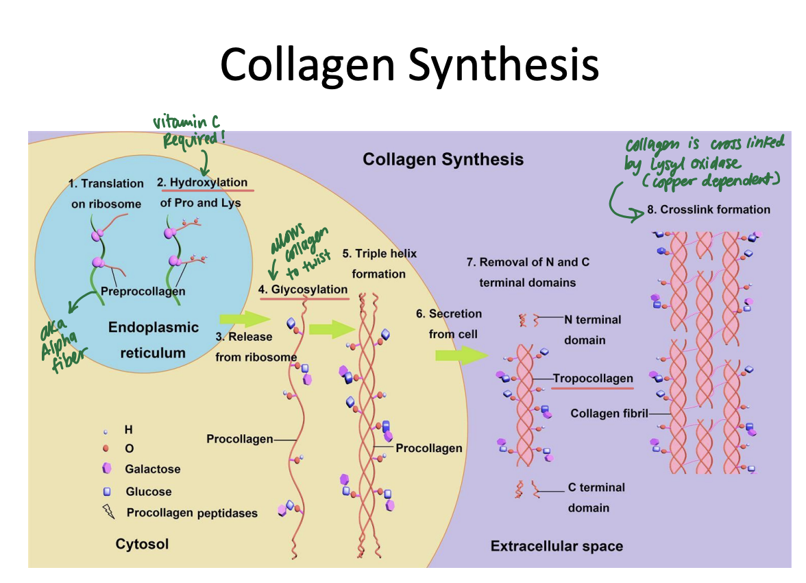
A: Hydroxylation, glycosylation, and triple-helix formation occur intracellularly, while terminal cleavage to form tropocollagen and lysyl oxidase-mediated crosslinking occur extracellularly. 🔎 What this means: The cell builds and modifies procollagen first, then secretes it so final processing and fibril assembly can occur outside the fibroblast.
32
New cards
⭐ 32. [ATTACH SLIDE/PAGE 43] Identify the superficial, middle, and deep dermal vascular plexuses in this histologic section.
A: The superficial plexus lies closest to the epidermis, the middle plexus lies deeper around follicular structures, and the deep plexus lies near the dermis-subcutis interface. 🔎 What this means: The three vascular levels progressively connect the deeper cutaneous blood supply with the superficial dermis that nourishes the avascular epidermis.
33
New cards
⭐ 33. [ATTACH SLIDE/PAGE 49] Identify the lobular panniculus, septal panniculus, and panniculus muscle in this section of subcutis.
A: The lobular panniculus consists of adipose lobules, the septal panniculus consists of fibrovascular connective tissue separating those lobules, and the panniculus muscle is the band of skeletal muscle within the subcutis. 🔎 What this means: Together these structures show that the subcutis contains organized adipose, connective tissue, vessels, and skeletal muscle rather than being an unstructured fat layer.
34
New cards
<p><span data-name="star" data-type="emoji">⭐</span> 34. Identify what type of tissue this is and why</p>

⭐ 34. Identify what type of tissue this is and why

This is brown adipose tissue, because it has more pink, internally textured cytoplasm because of its abundant mitochondria.

🔎 What this means:

The histologic appearance reflects function:

white adipose primarily stores energy, whereas mitochondria-rich brown adipose specializes in heat production.

35
New cards
<p><span data-name="star" data-type="emoji">⭐</span> 35. Identify what type of tissue this is and why</p>

⭐ 35. Identify what type of tissue this is and why

This is white adipose tissue because it has large clear lipid-dominated cells with peripheral nuclei

🔎 What this means:

The histologic appearance reflects function:

white adipose primarily stores energy, whereas mitochondria-rich brown adipose specializes in heat production and is more pink in color

<p>This is white adipose tissue because it has large clear lipid-dominated cells with peripheral nuclei<br><br><span data-name="mag_right" data-type="emoji">🔎</span> What this means: </p><p>The histologic appearance reflects function: </p><p>white adipose primarily stores energy, whereas mitochondria-rich brown adipose specializes in heat production and is more pink in color</p>
36
New cards