ANPS Exam 3
 Call Kai
 Learn
 Practice Test
 Spaced Repetition
 Match
 Flashcards
 Knowt Play
1/104

There's no tags or description
Looks like no tags are added yet.
105 Terms
Skeletal Muscles are connected to the CNS by peripheral nerves:
-To understand the control of skeletal muscles, we need to examine the organization of sensory and motor components of the peripheral nervous system (PNS) and their connections to the central nervous system (CNS)

Review : Structure of a Peripheral Nerve
-A peripheral nerve is a collection of axons in the PNS. (outside brain + spinal chord)
-Layers of connective tissue in a peripheral nerve:
Endoneurium is a connective tissue layer surrounding a single axon and its myelin sheath
Perineurium is a connective tissue layer surrounding a fascicle (bundle of axons)
Epineurium is a connective tissue layer surrounding an entire nerve
Blood vessels travel between fascicles to supply oxygen and nutrients
-All nerves have a specific name/job, we will learn some of the many

Sensory Neurons:
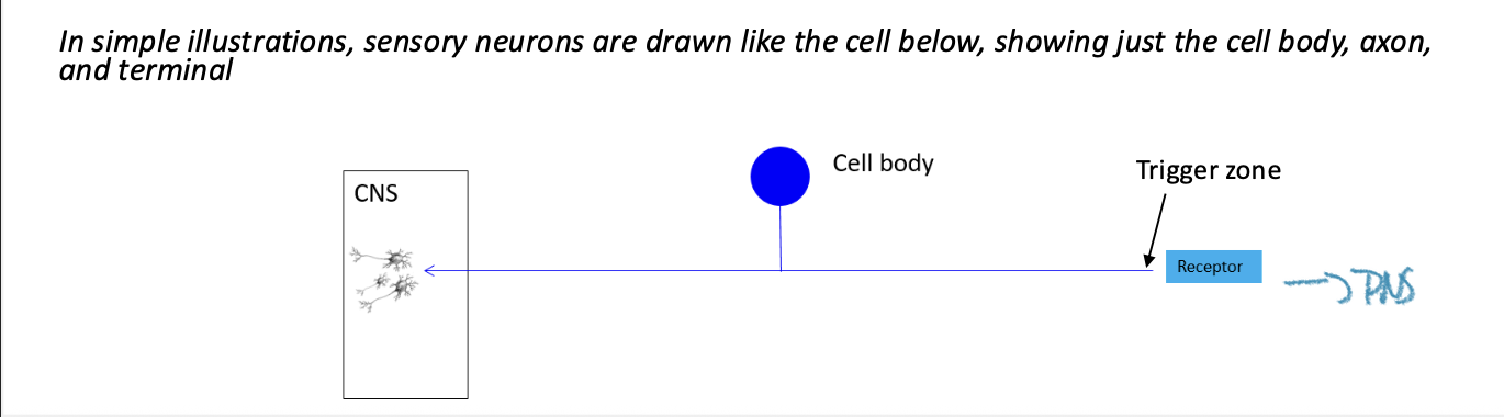
-Some axons in a peripheral nerve belong to sensory neurons
Sensory neurons are pseudounipolar in shape
The cell body is outside the CNS in a ganglion
-Sensory neurons collect information from the periphery and carry it into the CNS
Information is collected by a specialized sensory receptor, which stimulates action potentials in the sensory neuron.
Sensory neurons send signals to the CNS and ultimately the brain through an afferent pathway.
Motor Neurons:
-Another type of axon in a peripheral nerve belongs to a motor neuron
Motor neurons are multipolar in shape
The cell body and dendrites are in the CNS (spinal cord)
Motor neurons send signals from the CNS to targets in the “periphery” (outside of the CNS). Target cells include skeletal muscle, smooth muscle, and glands.

Skeletal Muscles are Innervated by Branches of Peripheral Nerves:
-Somatic sensory and somatic motor axons are bundled together to innervate skeletal muscle
Somatic sensory neurons collect information from muscle receptors about muscle activity (stretch, contraction) and convey it to the CNS
Somatic motor neurons send commands telling the skeletal muscle cells to contract; they release acetylcholine at the neuromuscular junction on individual cells

Cranial and Spinal Nerves Connect the Periphery to the CNS:
-31 pairs of spinal nerves connect the spinal cord to the periphery (limb/trunk region)
-12 pairs of cranial nerves connect the brain to the periphery (head/ neck region)
-named similar to vertebrae: 2 come out of each segment
*Spinal nerves become peripheral nerves
Spinal Nerves Connect to the Spinal Cord through Dorsal and Ventral Roots:
-Axons of sensory neurons enter the spinal cord through the dorsal root, which enters the dorsal portion of the spinal cord.
-Cell bodies of sensory neurons are clustered in a dorsal root ganglion which lies just outside the vertebral column (spine).
-Axons of motor neurons leave the ventral portion of
the spinal cord through the ventral root. Cell bodies of
motor neurons are in the spinal cord. (Ventral horn)
**The dorsal (back) root carries sensory information into the spinal cord; the ventral (front) root carries motor information out of the spinal cord.
-Spinal nerves forms when the dorsal root and ventral root merge and thus contain both sensory and motor axons.

Spinal Nerves Connect to the Spinal Cord through Dorsal and Ventral Roots:

The Central Nervous System has an internal pattern of gray and white matter:
-In cross sections of the spinal cord, you can see a central grey area (butterfly shape) surrounded by a white area.
-The white area is called white matter (myelinated axons), and the darker central area is called gray matter (unmylinated axons, cell bodies, dendrites).
-Although only the spinal cord has a central gray butterfly shape, anywhere we look in the CNS we see areas of gray matter and areas of white matter.
Understanding Gray Matter and White Matter:
-The key to understanding gray and white matter is to recall the structure of a typical neuron.
-Dendrites, cell body and terminals have no myelin- are found in grey matter .
-Myelinated axons and oligodendrocytes (CNS) are found in white matter. Myelin has a white color in unstained tissue due to its high lipid content (lipid content gives it color)
[The spinal cord connects the “periphery” to the brain]- Picture

Internal Anatomy of the Spinal Cord : White Matter:
-The white matter of the spinal cord contains tracts - collections of myelinated axons traveling in the longitudinal plane (vertically).
-Some groups of axons carry information up to the brain; we call these sensory tracts (ascending / afferent)
-Other groups of axons carry information down from the brain; we
call these motor tracts (descending / motor)

nternal Anatomy of the Spinal Cord : Gray Matter
-The gray matter of the spinal cord is also divided into functional areas
-Sensory axons synapse in gray matter in the dorsal half of the spinal cord; this gray matter is called the dorsal horn. Dorsal horns contain large and small neurons, lots of astrocytes, and relatively few axons with oligodendrocytes. (bc grey matter)
-Motor neurons reside in gray matter in the ventral half of the spinal cord; these are the ventral horns. Ventral horns contain large motor neurons, many small local neurons, lots of astrocytes, and a few axons with oligodendrocytes. (bc grey matter)
-Between dorsal and ventral horns is an intermediate zone; we will learn later that autonomic nervous system neurons are located in this area. (covering somatic rn)
*Note the symmetry; there are left and right spinal nerves, so there are left and right dorsal roots and ventral roots, left and right dorsal horns and ventral horns.

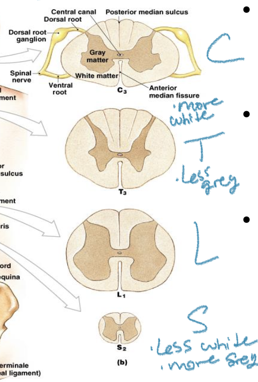
Different regions of the spinal cord have different ratios of white and gray matter:
* whitematter: descending/ascending tracks, don't need as many in lumbar or sacrum because impulses already arrived at upper limb
-The overall size (cross-sectional) of the spinal cord decreases from cervical to sacral levels.
-The volume of white matter decreases from cervical to sacral levels.
-The shape of the gray matter changes at each level, with greatest volumes of dorsal and ventral horn in the areas communicating with limbs
Cervical enlargement for upper limb
Lumbosacral enlargement for lower limb
* need more more neurons the further down you go: ventral horns, and gray matter get bigger (motor)
The CNS is Surrounded and Protected by the 3 layers of meninges:
-There is no connective tissue within the CNS to hold tissue together
-Support for CNS derives from 2 tissue layers outside CNS
-Pia mater is adhered to surface of brain and spinal chord; it cannot be peeled off- microscopic, not seen with naked eye
-Arachnoid mater is transparent, delicate layer external to pia and separated from the pia by a small space (subarachnoid space).
-This subarachnoid space is filled with a fluid called cerebrospinal fluid (CSF).
-Dura mater is a tough collagen rich tissue layer external to the arachnoid; because of its toughness, it provides the most protection for the CNS tissue.
**Dura transitions to Epineurium at the spinal nerve
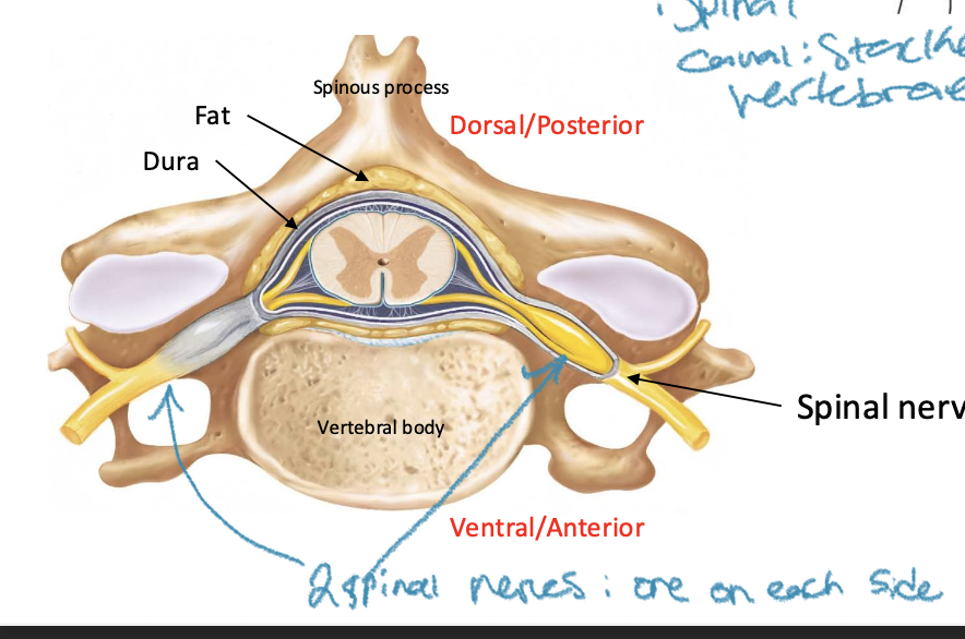
The Spinal Cord is Protected by Bone:
-The spinal cord lies in the vertebral canal, surrounded and protected by bone
-Fat fills the space between the dura and the bone, providing cushioning
-Spinal nerves exit between bones at the intervertebral foramina
-Spinal canal": stacked vertebrae
There is segmental organization to most of the nervous system:
-Early in embryonic life, the body has visible segments, small repeating blocks of tissue (somites)
-Each segment contains some CNS tissue innervating skin and muscle cells developing adjacent to it
-These segments underlie the organization of the adult spinal cord and spinal nerves
-The adult spinal cord is simply repeating units called spinal segments; each spinal segment forms a pair of spinal nerves (where a group of spinal nerves exits from vertebrae
There are 31 pairs of spinal nerves, so there are 31 spinal segments:
-Each spinal nerve is named relative to the intervertebral vertebra it exits.
Examples: The C3 spinal nerve exits between C2 and C3 vertebrae
Spinal nerves C8 and below exit below their numbered vertebra- Example : The T7 spinal nerve exits between the T7 and T8 vertebrae
* there are eight cervical nerves instead of seven because first one is on top, from then on all below the vertebrae
-Spinal segments are named for the spinal nerves they form
Example: the T7 spinal segment is the segment of spinal cord that the T7 spinal nerves attach to

Each Spinal Segment Forms a Pair of Spinal Nerves (right and left):

Each Spinal Segment Innervates (to supply something w/ nerves) a Dermatome and a Myotome:
-Each spinal segment innervates skin, muscle and joints that developed adjacent to it in early development (somites)
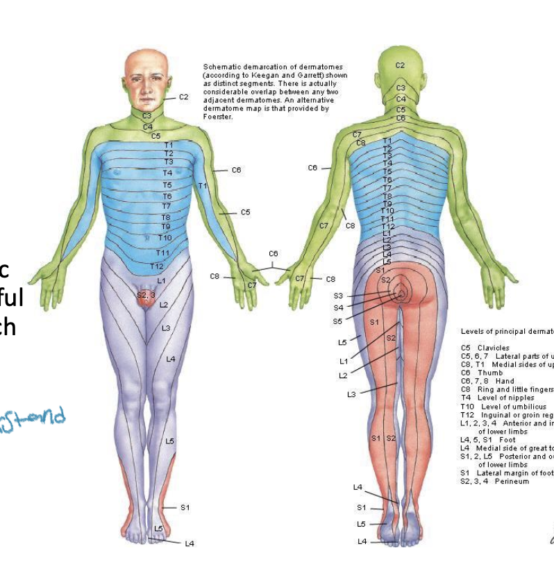
-The band of skin innervated by a pair of spinal nerves is called its dermatome
-The skeletal muscle cells innervated by a pair of spinal nerves is called its myotome

Dermatome Maps are Useful in Helping Diagnose the Location of an Injury:
-A dermatome is a patch of skin innervated by sensory neurons at a specific spinal segment.
-Clinical relevance: Impaired sensation in specific parts of the body can be useful clinically in determining which part of the spinal cord is damaged. (when something hurts or tingles, could have a deeper connection to spinal chord issue)
Myotomes are also used Clinically:
-A myotome is a group of muscles innervated by motor neurons at a specific spinal segment.
-Ex. The triceps muscle is innervated by motor neurons at C6,C7, and C8 segments. (C 6,7,8 control triceps)
-Myotomes are also useful clinically to understand he root of pain
The Adult Spinal Cord Ends at the L1 Vertebra:
-The adult spinal cord is shorter than the vertebral column because the spinal cord stops growing first
-In the adult, only cervical level dorsal and ventral roots exit straight laterally from their spinal segment. At all other levels, dorsal and ventral roots run inferiorly before forming spinal nerves: In adults, cervical roots join to form spinal nerves directly by exiting straight out to the sides of the spinal cord, while roots at all other levels travel downward before joining to form spinal nerves.
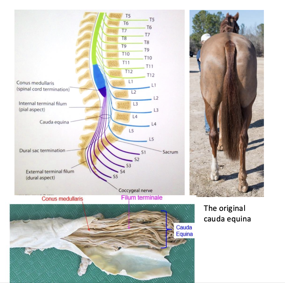
-The collection of dorsal and ventral roots below the L1 vertebra is called the cauda equina
**As a person grows, the vertebral column grows longer than the spinal cord, so the spinal cord ends higher up in the back. The lower spinal nerves must travel downward inside the vertebral column to reach the openings where they exit.

The Cauda Equina is the Collection of Dorsal and Ventral Roots at the end of the Spinal Cord
-The end of the spinal cord tapers to a point; this region is called the conus medullaris which contains sacral and coccygeal spinal segments
-Dorsal and ventral roots leaving the lower spinal cord region create a mass of stringy nerves called the cauda equina (from the latin “horses tail”)
-The cauda equina is bathed in a large pool of CSF inside the subarachnoid space; we can collect CSF from here without damaging the spinal cord tissue. (important for clinical testing)
-A strand of pia mater called the filum terminale extends from the tip of the conus medullaris to the bottom of the dura in the sacrum. This helps anchor the spinal cord to the coccyx.
Spinal Segments and Nerves Innervate Different Regions of the Body:
**There is longitudinal organization of innervation of skin and muscle by spinal nerves
-Cervical spinal nerves primarily innervate the and upper limb. primarily innervate skin and muscle in neck region and upper limb
-Thoracic spinal nerves primarily innervate the chest, abdomen, and spine. primarily innervate the chest, abdomen and spine.
-Lumbar spinal nerves primarily innervate the lower limb.
-Sacral spinal nerves innervate the lower limb and muscles of the pelvic floor.
**Innervation means to supply with nerves: Ex. Lumbar and sacral spinal nerves travel to the skin and muscles of the lower limb, supplying these parts of the body

The Cervical and Lumbosacral Enlargements are the Regions Controlling Limbs:
-All along the length of the spinal cord gray matter process information to and from the core of the body
-The gray matter in the lower cervical region also processes information for the upper limb; the extra gray matter creates a swelling the spinal cord called the cervical enlargement
-The gray matter in the lumbar and sacral regions processes lower limb information; this swelling is called the lumbar or lumbosacral enlargement.
Most Spinal Nerves Contribute to a Nerve Plexus
-Axons from spinal nerves merge with adjacent segments to form a nerve plexus
-There are 4 plexuses: cervical, brachial, lumbar and sacral
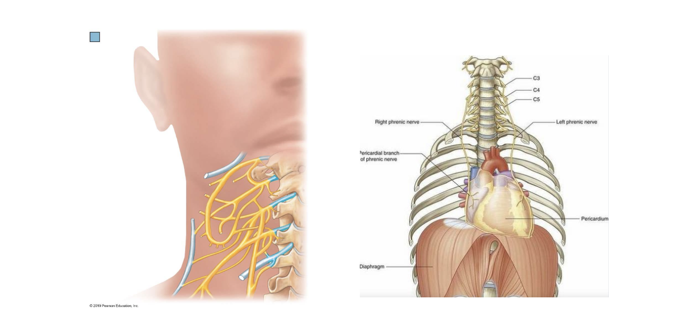
The Cervical Plexus:
-Formed from axons in C1-C4 spinal nerves
-Innervates skin and anterior spine muscles of neck
-Most important branch is the phrenic nerve, from C3-5 levels to the diaphragm “C3,4,5 keeps the diaphragm alive”
The Brachial Plexus:
-Formed from axons in C5-T1 spinal nerves
-Plexus forms under clavicle where it can be damaged by broken bone
-Branches of plexus innervate skin and muscles of upper limb (except trapezius)
Forms 5 major nerves of limb:
Axillary nerve innervates 2 muscles of shoulder
Musculocutaneous nerve innervates anterior arm muscles
Median nerve innervates most of anterior forearm and thumb muscles
Ulnar nerve innervates muscles in anterior forearm and most muscles within hand
Radial nerve innervates all muscles in posterior arm and forearm

The Lumbar Plexus:
-Formed from axons in L1-L4 spinal nerves
-Plexus forms in abdominal cavity on posterior wall
-Branches of plexus innervate skin and muscles of anterior and medial thigh and skin of medial leg
Forms 2 major nerves of limb:
Femoral nerve to anterior thigh
Obturator nerve to medial thigh

The Sacral Plexus (lumbosacral plexus):
-Formed from ventral rami of L4-S3 spinal nerves
-Plexus forms in pelvis on anterior side of sacrum
-Branches of plexus innervate skin and muscles of gluteal region, posterior thigh, leg and foot
Forms 2 major nerves of limb:
Gluteal nerves to gluteal muscles (butt)
Sciatic nerve to remainder of limb muscles via its 2 branches:
Tibial nerve to posterior thigh and posterior leg, foot
Common fibular nerve to lateral and anterior leg
Joints (Articulations) allow for movement in an otherwise rigid skeleton:
-Joints: The point of contact between two bones, holding the skeleton together
-Two main functions: stability and mobility
*Inverse relationship: As stability increases, mobility decreases
*Mobile joints are the weakest part of the skeleton

Joints can be classified structurally as: Fibrous, cartilaginous, and synovial
3 structural Categories:
Little to no movement:
Fibrous: bones linked by collagen fibers of connective tissue (bones fused by dense regular CT)- No or Little Movement\
Cartilaginous: bones linked by cartilage (Kind of movable. Ex. vertebrae, kind of movable but some parts not)
Movement:
Synovial: ends of bones not connected by connective tissue; separated by fluid filled joint cavity
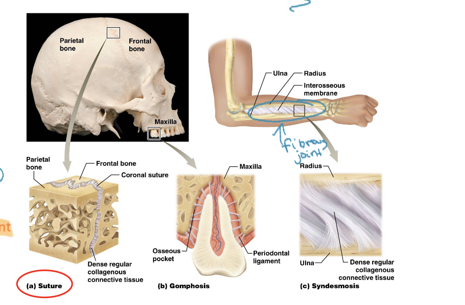
Fibrous Joints:
-hold bones together with dense regular connective tissue rich in collagen fibers
**Know: suture joints in skull
(Fibrous= collagen fibers, dense regular CT)
Cartilaginous Joints:
-Bones held together by cartilage
-Balance between mobility and stability
2 types of cartilaginous joints:
Bones linked by Haylie cartilage
Bones linked by fibrocartilage
(see image for what I need to know)

Synovial Joints:
-separate articulating bones with a fluid filled joint cavity
Components common to all synovial joints:
Articular cartilage: hyaline cartilage at ends of bones that absorb compression
Joint cavity: potential space containing lubricating synovial fluid
Articular (Joint) capsule: two-layer fibrous capsule reinforcing joint
Fibrous layer continuous with bone periosteum
Synovial membrane inner capsule lining; produce synovial fluid
Tendons and Ligaments Support Synovial Joints: (Dense regular CT proper)
-Ligaments:
Connective tissue bands connecting bone to bone
Limit bone movement in some directions
-Tendons:
connective tissue from muscle to bone
cross over joints and stabilize
muscle/tendon unit creates movement at joint

Fibrocartilage (type of cartilage) can provide extra support within a synovial joint:
*Just like tendons and ligaments, fibrocartilage (articular discs and labrum) can provide extra support
Articular disc:
fibrocartilage disc to absorb joint shock and help joint fit
Examples : meniscus in knee
Labrum:
fibrocartilage ring that deepens a synovial joint to help keep bones in alignment
Found at shoulder and hip joints
Hold ball in socks more tightly
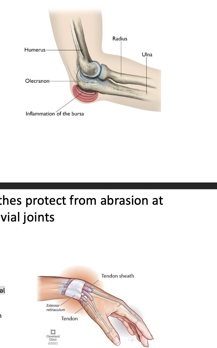
Bursa and Tendon Sheathes protect from abrasion at synovial joints:
*Protects against ‘banging up’ of highly movable joints (because synovial joints are mobility as tradeoff for stability)
1. Bursa:
Connective tissue sac containing synovial fluid
Provides padding and reduces friction between two structures that rub at a synovial joint
Clinical problems: Bursitis (inflammation)
2. Tendon Sheathes:
Connective tissue sac containing synovial fluid
Wraps around tendon at area of abrasion (so tendons don’t rub up on each other - > inflammation)
Clinical problems: Tendinitis and Tenosynovitis
The shape of the bones at the synovial joint dictate the direction and degree of movement:
Hinge joints:
Ex. Knee and elbow
Uniaxial: can move one direction or another
Flexion or extension
Ball and socket joint:
Shoulder and hop joints
Multiaxial movement: can move in many directions
Flexion/extension, abduction/adduction, rotation
Main Joints of the Knee: Hinge joint
-A hinge joint between the femur and tibia
***There is a separate joint between femur and patella

Knee joint stabilizers
-Anterior cruciate ligament (ACL) and posterior cruciate (PCL) ligament within joint limit anterior-posterior movement of femur over tibia
-Medial collateral ligament (MCL) and lateral collateral (LCL) ligament limit side to side movement of femur over tibia
-Medial and lateral meniscus cartilages are c-shaped rings of fibrocartilage (Type of articular disk- supports synovial joints) helping femoral condyles stay oriented over articular surfaces of tibia

Stabilizing muscles around Knee Joint: (connected via tendons)
-Quadriceps: anteriorly (Knee extension)
-Hamstring group: posteriorly (Knee flexion)
Injury to the Knee Joint: The Unhappy Triad
**Injuries to the knee joint often damage more than one stabilizing structure

Injury to the knee joint: degeneration
-Without correct alignment of surfaces, cartilage wears away and bone spurs form
-Inflammation accelerates the process


Shoulder (Glenohumeral) Joint: Ball and socket joint
-A ball and socket joint between the rounded head of humerus and glenoid fossa of scapula
-Multiaxial joint with great range of movement (at the expense of stability)
**Glenoid Labrum and Glenohumeral Ligaments add Stability:
Glenoid labrum (Supportive fibrocartilage from previous slide): is a ring of fibrocartilage around edge of glenoid fossa that deepens the cup for the head of the humerus
Thickenings of the connective tissue form glenohumeral ligaments, supporting the position of the head of humerus
Stabilizing muscles around Shoulder Joint: 4 Rotator Cuff Muscles
-Muscles acting at the shoulder must cross the shoulder joint
-The 4 muscles of the “rotator cuff” surround the shoulder joint capsule and their tendons fuse with the capsule to further stabilize the joint
Supraspinatus
Infraspinatus
Teres Minor
Subscapularis
*Rotator cuff tears destabilize the entire joint and make it more susceptible to further injury

Movements at Synovial Joints:
-First recall anatomical body planes: Sagittal, coronal/frontal, transverse/horizontal
Review anatomical portion:
-Standing erect
-Facing forward
-Feet together
-Palms forward
*Motion terms relate to this position

Movements Allowed by Synovial Joints: ANGULAR Movements- Flexion and Extension
-Increase (extension) or decrease (flexion) the angle between two bones in the sagittal plane
**Hyperextension is merely extension past anatomical position
-Can flex and extend elbow, knee, hip, wrist, fingers, toes

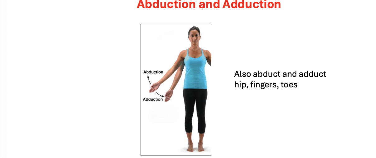
Movements Allowed by Synovial Joints: ANGULAR Movements- Abduction and Adduction
-Increase or decrease the angle between two bones in the coronal (frontal) plane:
-Can abduct and adduct arm, hip, fingers, toes
Movements Allowed by Synovial Joints: ANGULAR Movements- Circumduction
-Increase or decrease the angle between two bones
-(shoulder and hip joints)

Movements Allowed by Synovial Joints: ANGULAR Movements- lateral & medial rotation
-Increase or decrease the angle between two bones in the longitudinal axis
-Have this at head, shoulder
*Often called external and internal rotation


Movements Allowed by Synovial Joints: Special Movements- Radius & ulna
-performed by a select few joints, in this case radius and ulna
-Movements within forearm : Pronation and Supination
-Pronation: Radius rotates over ulna (palms down)
-Supination: Radius and ulna are paralell (palms up)
Movements Allowed by Synovial Joints: Special Movements: ankle joint
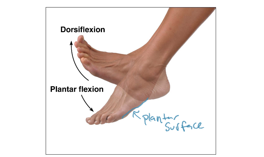
-Movements at ankle joint: Dorsiflexion and Plantarflexion
-Dorsiflexion: foot pointing up
-Plantarflexion: foot pointing down
Movements Allowed by Synovial Joints: Special Movements- tarsal bones of foot
-Movements within tarsal bones of foot: Inversion & Eversion
-Inversion: Coming towards the midline
-Eversion: coming away from midline

Movements Allowed by Synovial Joints: Special Movements- Jaw/scapula
-Movements of jaw or scapula: Protraction and Retraction
-Protraction: moving forwards
-Retraction: Moving back
-Movements of jaw or scapula: Elevation and Depression
-Elevation: moving/looking up
-Retraction: moving/looking down

Movements Allowed by Synovial Joints: Special Movements- Thumb and pinky
Movements of thumb and pinky: Opposition & Reposition
Next lecture transition:
-3 systems makes up movement: nervous, skeletal, muscular- neurons control muscles, which pull on bones for movement

Muscle is intimately associated with connective tissue (fascia):
-Muscle is encased in deep fascia for structural support, force transmission, & compartmentalization
Force transmission: fascia, attaches, attendance, so they can all transmit force at the same time
Muscle compartments: groups of muscle developed deep in fascia; contains muscles that share similar actions – (ex. interior compartment of thigh, has four different muscle muscles, but they all have the same function, to help extend knee joint)
-Connective tissues surrounds entire muscle, fascicles, and individual cells within muscle (Epi, peri, and endomysium)
-Connective tissues continue into tendon and attach to periosteum of bone → movement of body from muscle pulling on bone
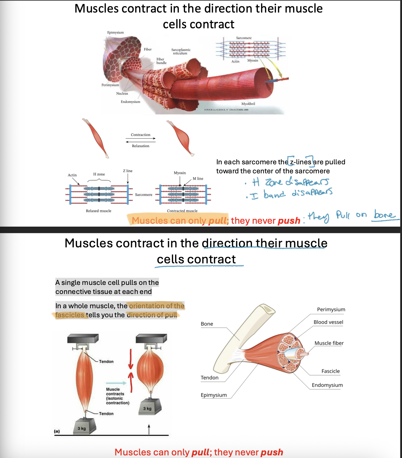
Muscles contract in the direction their muscle cells contract:
-In each sarcomere the z-lines are pulled toward the center of the sarcomere
-H zone disappears, I band does too
**Muscles only PULL they can NEVER push (they pull on bone)
-A single muscle cell pulls on the connective tissue at each end. In a whole muscle, the orientation of the fascicles tells you the direction of pull
Usually, one end of a muscle moves more easily than the other end: Origins and insertions
-Muscles have origins and insertions (Sites where tendons attach)
Origin: attachment on fixed or less moveable bone
Insertion: attachment on the more moveable bone
Ex. Biceps- Organ is shoulder/scapula; Insertion is Forearm/radius
Fascicle arrangements determine skeletal muscle shape, function, and overall strength:
*By identifying the fascicle arrangement, you can infer the direction in which the muscle contracts and the type of movement it produces

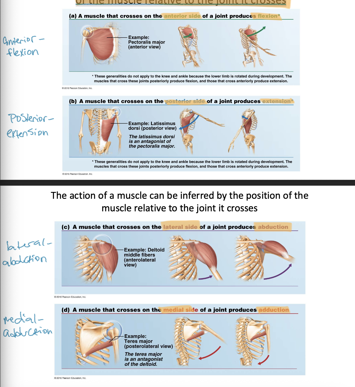
The action of a muscle can be inferred by the position of the muscle relative to the joint it crosses:
-If muscle crosses on anterior side of joint → flexion
-If muscle crosses on posterior side of joint → extension
-If muscle crosses on lateral side of joint→ abduction
-If muscle crosses on medial side of joint → adduction
Summary : Things to think about when viewing a muscle
1. What joints does a muscle cross? It can have one or more actions at each joint.
2. Which side of the joint does the muscle cross? This helps with actions.
3. What is the orientation of the muscle fascicles within the muscle? This helps with direction of movement, helping name the action.
Muscles can be classified into four functional groups:
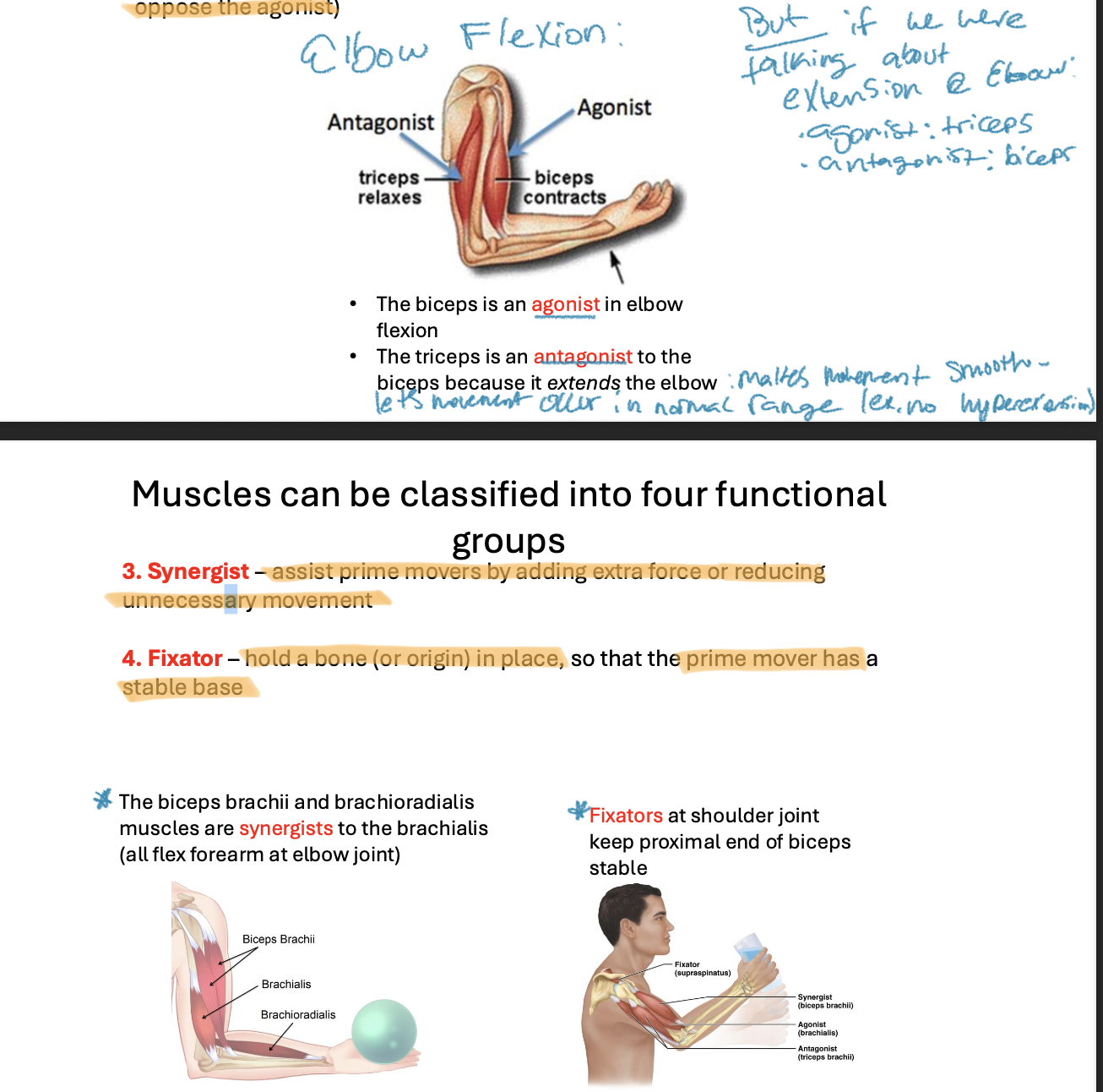
Agonist: (prime mover) muscle that has the major responsibility for
producing a specific movement
Antagonist: muscles that oppose or reverse a specific movement (i.e.
oppose the agonist)
Relative to what movement you are looking at (see photo on first slide)
Synergist: assist prime movers by adding extra force or reducing
unnecessary movement
The biceps brachii and brachioradialis muscles are synergists to the brachialis (all flex forearm at elbow joint)
Fixator: hold a bone (or origin) in place, so that the prime mover has a
stable base
Fixators at shoulder joint keep proximal end of biceps stable
There are four groups of axial muscles: (overview)
-Associated with bones of axial skeleton, most move the axial skeleton, some move contents of cavities
Muscles groups of head
Facial expression
Eye movements
Jaw movements
Tongue, pharynx, palate (speech, swallowing)
Muscles of vertebral column
Stabilize, flex, extend, or rotate the vertebral column
Muscles of thorax & abdomen
Breathing, Core movements
Muscles of pelvic floor
Span pelvic outlets
Support pelvic organs
Muscle Groups of the Head: Muscles of Facial Expression
-Small, thin
-Insert into skin of face to change expression
-Innervated by a cranial nerve (umbrella); facial nerve (cranial nerve VII) (specific name)
*Cranial nerves: originate in brainstem, controls head and neck

Muscle Groups of the Head: Extrinsic Eye Muscles (6)
-Located in the orbit but outside the eyeball
-Insert into outer surface of eye to move the eyeball
-Innervated by 3 different cranial nerves- Cranial nerves 3,4, and 6 (need all three because we need precise control over our eye- constantly scanning for movement)
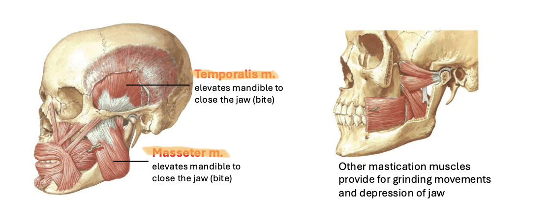
Muscle Groups of the Head: Muscles of Mastication
*Think Mastica = Chew
-Insert on the mandible (lower jaw)
-Perform chewing movements
-Innervated by a cranial nerve
Temporalis M: Elevates mandible to close jaw
Masseter m: elevates mandible to close the jaw
Muscle Groups of the Head (and Neck): Muscles of Swallowing and Speech
Swallowing:
-Located in the oral cavity and throat
-Direct movement of food toward esophagus
-Innervated by a cranial nerve
Speech:
-Located in the larynx (voice box)
-Control vocal cords
-Innervated by a cranial nerve
Muscles of the Neck: Head Movement
-Anterior neck:
Sternocleidomastoid muscle
Bilateral - head flexion (looking down)
Unilateral - rotation of head to opposite side (turn head)
-Posterior neck:
Trapezius and many muscles deep to trapezius that stabilize, laterally flex, and rotate head
Muscles of the Posterior Vertebral Column: Erector spinae group
-Prime mover of back extension and maintenance of posture
-Contribute to lateral bending and rotation of vertebral column
-Regions in neck assist head extension and rotation
-3 columns (spinalis group, longissimus group, iliocostalis group)
-Innervated by cervical, thoracic and lumbar nerves: spinal because we are below the head and neck

Deep Muscles of the Thorax: Respiratory muscles
-Diaphragm: prime mover of inspiration; contraction expands thoracic cavity (muscle flattens and moves toward abdomen)
Controlled by phrenic nerve in cervical plexus
-Intercostal Muscles: move ribs to expand or reduce chest volume

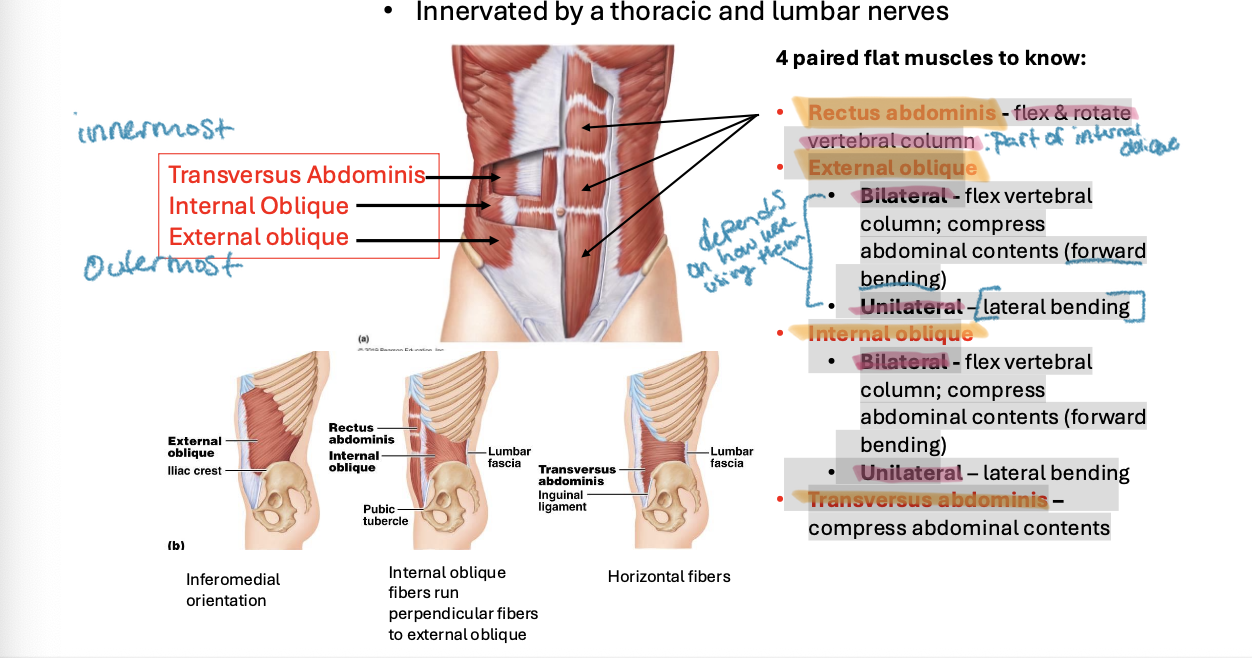
Muscles of the Abdominal Wall:
-Support and protect abdominal contents
-Innervated by the thoracic and lumbar nerves (spinal)
4 Paired plat muscles to know:
Rectus abdominis - flex & rotate vertebral column
External oblique
Bilateral - flex vertebral column; compress abdominal contents (forward bending)
Unilateral: lateral bending Internal
Internal oblique:
Bilateral: flex vertebral comlumn, compress abdominal contents (forward bending)
Unilateral: lateral bending
Transversus abdominis: compress abdominal contents
Pelvic Floor Muscles:
-Span the pelvic outlet
-Support pelvic organs and regulate urination and defecation
-Innervated by a sacral nerves (specifically ones in the caudal equine because spinal chord ends around L1)
The appendicular skeleton:
-The appendicular muscles work in coordination with the peripheral nerves and the skeletal system to stabilize, position, and support the limbs, enabling controlled and efficient
-Most muscles of lower limb innervated by lumbar or sacral plexus
-Most muscles of upper limb innervated by branches of the brachial plexus
What I need to know: key points:
Nerve Innervation:
Every muscle is innervated by a nerve: Every one of these nerves carries both sensory and motor axons
If a limb muscle is innervated by a terminal branch of a plexus, know the highlighted nerve name
Example: The biceps brachii is innervated by the musculocutaneous nerve, a terminal branch of the brachial plexus
highlighted nerve name
Joint Interaction:Distal Movement:
For highlighted muscles, know major actions (movements they create, and at
which joint)
The biceps brachii flexes (incorrect) vs The biceps brachii flexes at elbow joint (correct)
Muscles act at any joint they cross and therefore can have more than one action
Distal movement:
Muscles typically move a part of the limb distal to the muscle itself
Example: arm muscles move the forearm, forearm muscles move the hand
Nerve Supply to Upper Extremity: Brachial plexus
Axillary nerve :
innervates deltoid muscle
nerve ends at shoulder
Musculocutaneous nerve :
innervates elbow flexors in anterior compartment of arm
nerve runs through anterior arm muscle group
Front should-elbow
Median nerve :
innervates wrist and finger flexors in anterior forearm, thenar muscles (thenar=thumb)
nerve runs through anterior elbow, anterior forearm, and carpal tunnel
Front elbow-wrist; thumb
Ulnar nerve:
innervates some flexors in anterior forearm, most small hand muscles
nerve runs through back of elbow - “funny bone”
Radial nerve:
innervates extensor muscles in posterior arm and forearm
Nerve runs through lateral elbow and posterior forearm
Whole back of arm + funny bone
Movement of Scapula (Pectoral Girdle):
-The pectoral girdle consists of the clavicle and the scapula forming the shoulder and providing the attachment point for the upper limb
-Most movements of the pectoral girdle involve moving the scapula:
Elevation and depression
Upward rotation
Protraction and retraction
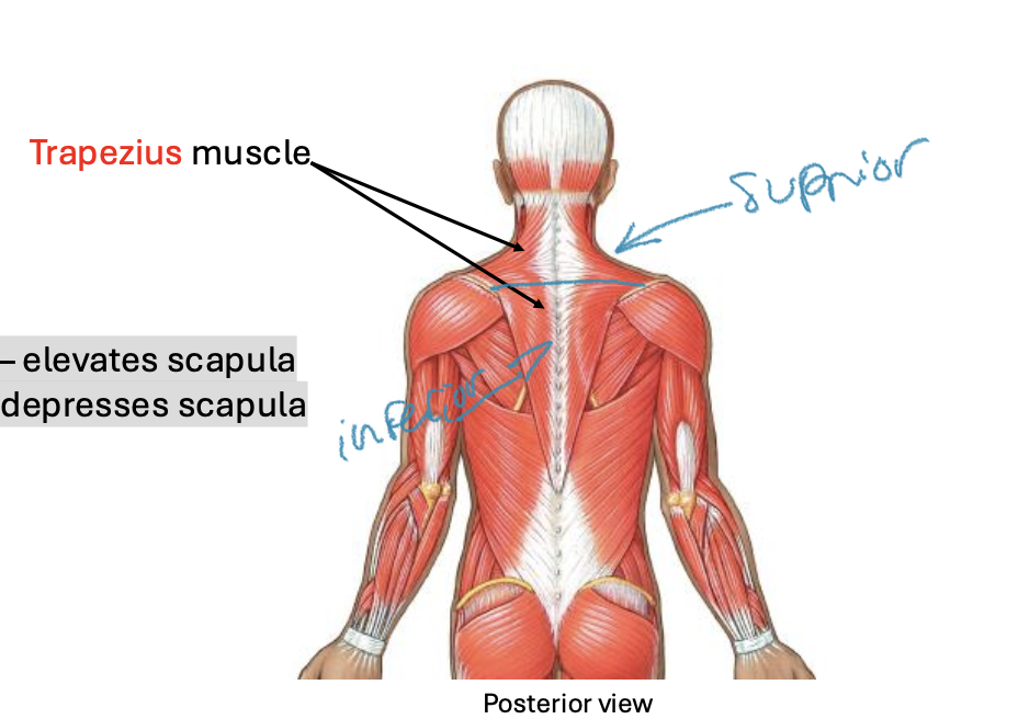
Movement of Scapula : Trapezius muscle:
Major actions:
-Superior fibers elevates scapula
-Inferior fibers depresses scapula
Movement of Scapula : muscles deep to trapezius
Rhomboids (major and minor):
retract scapula

Movement of Scapula : Anterior muscles:
Serratus anterior:
-protraction and upward rotation of scapula
-critical for raising arm above head


Movement of Humerus : Anterior
Deltoid
abducts arm
innervated by axillary nerve
Pectoralis major
flexes, adducts and medially rotates arm
Movement of Humerus : Posterior
Latissimus dorsi
extends, adducts and medially rotates arm
Teres major
extends, adducts and medially rotates arm

Movement of Humerus Summary:

Movement of Humerus : Rotator Cuff Muscles
-4 Rotator cuff muscles: Move humerus AND reinforce joint capsule
Supraspinatus: abducts arm
Infraspinatus: lateral rotation of arm
Teres minor: lateral rotation of arm
subscapularis : medial rotation of arm

Muscles of the Arm : 2 Compartments
Anterior : flex elbow
Posterior : extend elbow
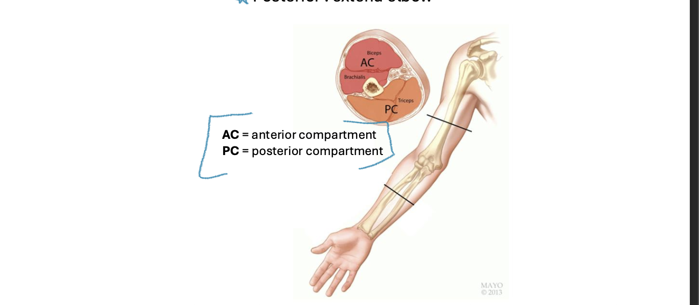
Muscles of the Arm: Movement of arm- Anterior compartment
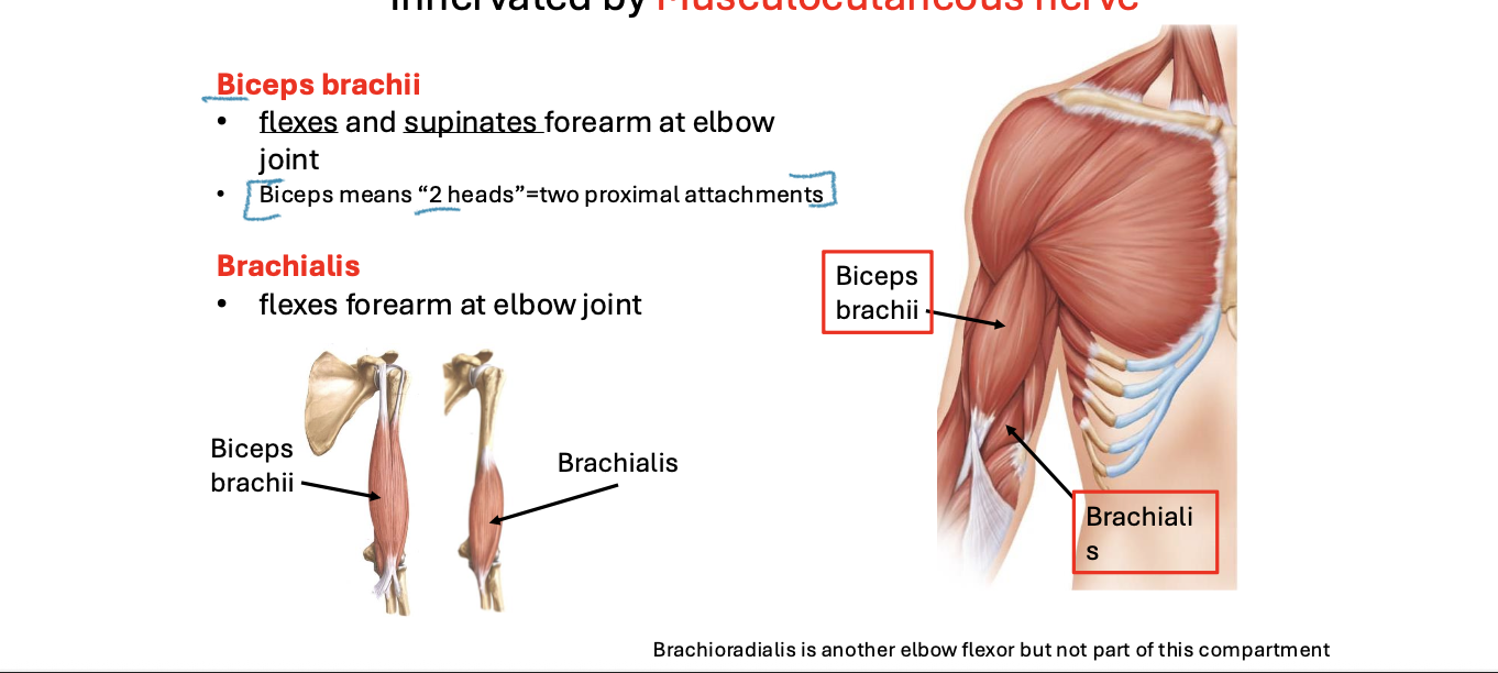
-Innervated by Musculocutaneous nerve
Biceps brachii: flexes and supinates forearm at elbow joint
Biceps means “2 heads” = 2 proximal attachments
Brachialis: flexes forearm at elbow joint

Muscles of the Arm: Movement of arm- Posterior compartment
T-riceps brachii: extends forearm at elbow joint
Triceps means “3 heads” = 3 proximal attachments
Muscles of the Forearm : 2 Compartments
Anterior: flex wrist and digits
Posterior: extend wrist and digits

Anterior Compartment of Forearm:
-Innervated by Ulnar and Median nerves
-Flexion of the Wrist and Fingers
-Some muscles only cross the wrist joint, so only work the wrist; others cross wrist and end on phalanges, so work wrist and digits

The tendons of the finger flexors and the median nerve pass through the carpal tunnel and may become inflamed through repetitive use
Inflammation = Carpal tunnel syndrome
Carpal Tunnel Release surgery: involves cutting through the flexor retinaculum

Posterior Compartment of the Forearm:
-Innervated by the Radial nerve
-Extension of the Wrist and fingers
-Some muscles only cross the wrist joint, so only work the wrist; others cross wrist and end on phalanges, so work wrist and digits

Muscles of the Hand
-Also called intrinsic muscles of the hand because the muscles and tendons are within the hand
Hypothenar group: moves 5th digit (pinky finger)- Innervated by ulnar nerve
Thenar group: moves thumb (1st digit)- Innervated by median nerve

Nerve Supply to Lower Extremity:
**Terminal branches of lumbar and sacral plexuses
Femoral nerve : anterior compartment of thigh and hip flexors
Obturator nerve : medial compartment of thigh (adductors)
Sciatic nerve branches: (posterior)
Tibial nerve : posterior compartment of thigh, leg, foot
Common fibular nerve : anterior & lateral compartments of leg and

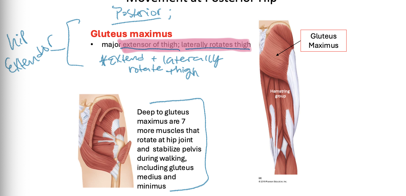
Muscles of the Gluteal Compartment : Movement at Posterior Hip
-Gluteus maximus: major extensor of thigh; laterally rotates thigh
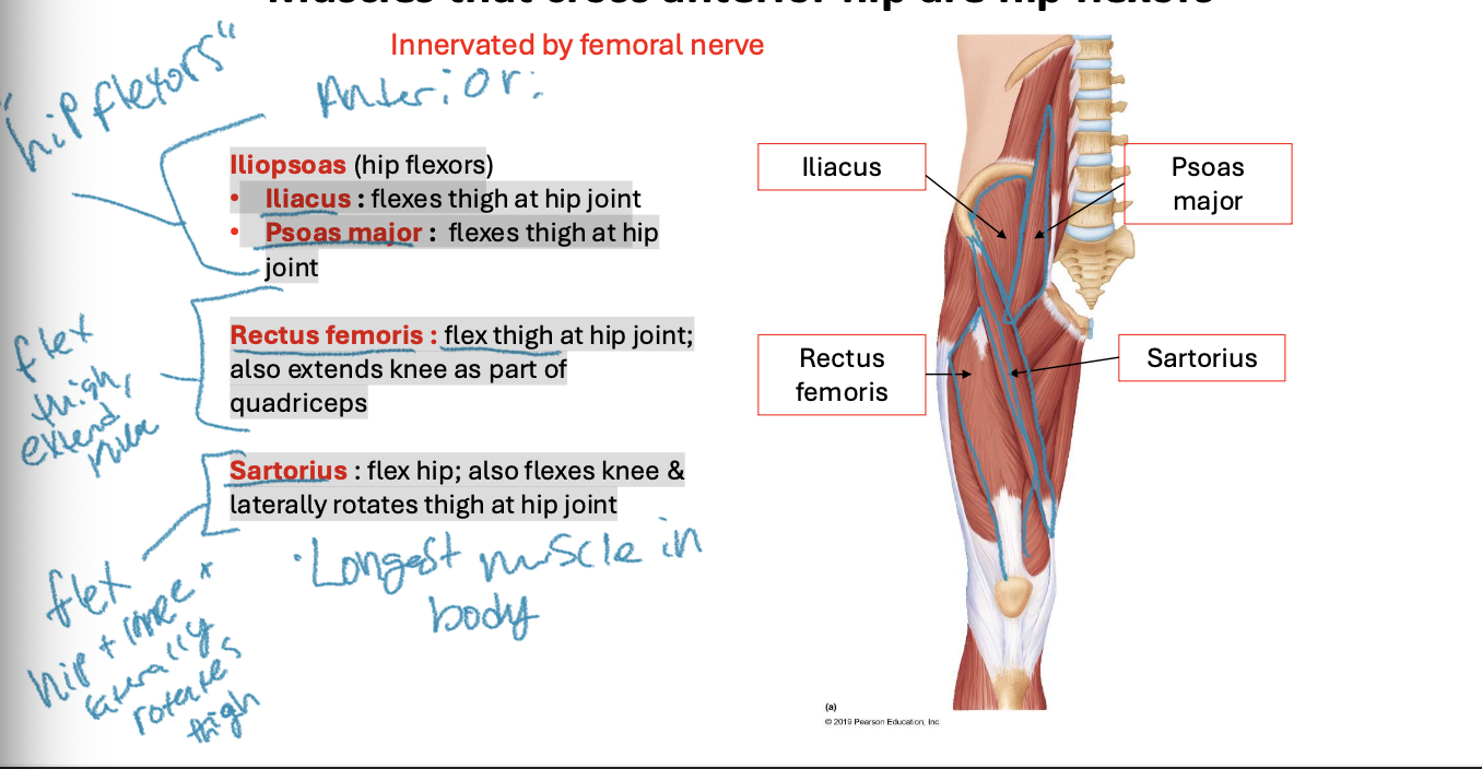
Muscles that cross anterior hip are hip flexors:
**Innervated by femoral nerve
Hip flexors: Iliopsoas
Iliacus : flexes thigh at hip joint
Psoas major : flexes thigh at hip joint
-Rectus femoris : flex thigh at hip joint; also extends knee as part of quadriceps
-Sartorius : flex hip; also flexes knee & laterally rotates thigh at hip joint
Muscles of the Thigh : 3 Compartments:
Anterior : flex hip and extend knee
Medial : adduct hip
Posterior : extend hip and flex knee

Muscles of the Anterior Compartment of the Thigh:
-Movement of hip and Knee
-Innervated by Femoral nerve
Quadriceps femoris : 4 muscles that form a common tendon bulling on the tibia all extend the leg at the knee joint
Rectus femoris: knee extension; hip flexion
Vastus lateralis: knee extension
Vastus medialis: knee extension
Vastus intermedius (deep): knee extension
Sartorius flex leg at knee; flex & lateral rotation of thigh at hip

Muscles of the Medial Compartment of the Thigh:
-5 muscles with main action: Adduction of hip
**Innervated by Obturator nerve

Muscles of the Posterior Compartment of the Thigh:
-Movement at the Hip and Knee
-Innervated by Tibial nerve
Hamstrings: extension of thigh at hip joint flexion of leg at knee joint
Biceps femoris (lateral side)
Semitendinosus (medial)
Semimembranosus (medial)

Tensor Fascia Lata Muscle and Iliotibial Tract:
Tensor fascia lata muscle:
On lateral thigh, but often grouped with gluteal compartment
provides lateral stability through iliotibial tract- “locks” knee in extension
IT tract (also called IT band) often inflamed in runners

Muscles of the Leg : 3 Compartments
Anterior : dorsiflex ankle, extend toes
Posterior : plantarflex ankle, flex toes
Lateral : eversion
