Prep & Reporting
 Call Kai
 Learn
Learn Practice Test
 Spaced Repetition
 Match
Match Flashcards
 Knowt Play
1/89

There's no tags or description
Looks like no tags are added yet.
Name | Mastery | Learn | Test | Matching | Spaced | Call with Kai | Chat |
|---|
No analytics yet
Send a link to your students to track their progress
90 Terms
Which exam would be improved by the administration of Simethicone prior to the ultrasound evaluation?
A. renal artery duplex
B. transcranial duplex
C. carotid duplex
D. lower extremity segmental pressures
A. renal artery duplex
Simethicone is a medication that is used to reduce bowel gas. It may be beneficial when performing abdominal duplex exams.
A patient is referred for a Doppler ultrasound of the IVC and hepatic veins due to an abnormal echocardiogram. The report states that the hepatic veins appear to empty directly into the right atrium with limited visualization of the upper IVC. What are you looking for in the abdomen?
A. situs inversus
B. duplicated IVC
C. portal HTN
D. azygous or hemiazygos IVC
D. azygous or hemiazygos IVC
Interrupted IVC - failure of the IVC to form correctly in the upper abdomen/liver; azygous and hemiazygos veins drain blood from the distal IVC segment; hepatic veins drain directly into the right atrium

You complete a segmental pressure evaluation of an aorta-bifemoral arterial bypass graft and are reviewing the prior report.
The prior exam demonstrates:
Left ABl of 0.95 and the Right ABl of 0.90.
The current exam demonstrates:
Left ABI of 0.88 and Right ABl of 0.95.
How would you report these findings?
A. Normal exam today, no significant change in ABI values
B. Normal left leg, abnormal right leg
C. Normal right leg, abnormal left leg
D. Abnormal change in the ABI values of both legs
A. Normal exam today, no significant change in ABI values
An abnormal exam is indicated when the ABI values drop by more
than 0.15.
What technique is recommended for evaluation of the main portal vein in an obese patient with ascites and significant bowel gas?
A. patient in the supine position with subcostal transducer position
B. patient in the left lateral decubitus or oblique position with intercostal transducer position
C. patient in the left lateral decubitus or oblique position with subcostal transducer position
D. patient in the right lateral decubitus or oblique position with intercostal transducer position
B. patient in the left lateral decubitus or oblique position with intercostal transducer position
The preferred technique for evaluation of the main portal vein is to place the patient in the left lateral decubitus or oblique position with an intercostal transducer position. The intercostal approach from the right side uses the liver as an acoustic window. It also provides a shorter distance from the probe to the MPV than using a subcostal approach on an obese patient with a large abdominal girth. This approach also allows for the use of proper Doppler angles of 60 degrees or less. This is important when evaluating the velocity of flow in the MPV.
A patient presents for a mesenteric Doppler to rule out stenosis. They have history of diabetes, HTN, pain and nausea post-prandially. Which of the following appointment times would be best for this exam to be scheduled?
A. 10am, after light breakfast
B. 1pm, after a good fatty meal
C. 8am, NPO for 6-8hrs prior
D. 230pm because the patient prefers this time
C. 8am, NPO for 6-8hrs prior
Diabetic patients and those for studies that require an NPO status should be scheduled very early in the morning.
A recent CT scan demonstrates an acute infarct in the parietal lobe and a new blockage in the proximal left middle cerebral artery. Which of the following is the most likely source for the embolism?
A. Plaque broke away from the wall of the proximal external cerebral artery
B. Plaque broke away from the wall of the left anterior cerebral artery
C. Plaque broke away from the sclerotic aortic valve
D. Thrombus formation in the left jugular vein
C. Plaque broke away from the sclerotic aortic valve
A sclerotic aortic valve can send an embolus through the aorta into one of the arch branches. If the embolus enters the left carotid system, it could lodge in the left MCA.
Thrombus in the jugular vein would move into the right side of the heart/lungs.
The ECA does not supply flow to the MCA, therefore there is no way for the embolism to enter the circle of Willis (without getting stuck in a smaller ECA branch first).
Flow in the ACA normally moves away from the MCA. An embolus in the ACA will move into smaller branches toward the front of the brain and eye.
Which of the following describes an important reason to verify the US examination that is ordered is the exam that should really be performed?
A. When the suspected abnormality is better visualized by another modality
B. Sonographers should not question the orders written by physicians and perform the exams as ordered.
C. When the suspected diagnosis is not able to be evaluated by the exam that is ordered
D. When the exam is ordered after hours and no reading physician is available
C. When the suspected diagnosis is not able to be evaluated by the exam that is ordered
When the suspected diagnosis is not able to be evaluated by the exam that is ordered, this is an important reason to verify the US examination that is ordered is the exam that should really be performed.
A patient presents for a dedicated transcranial Doppler exam to rule out an AV fistula. What should you do first?
A. Consult the radiologist regarding the inability to visualize the abnormality on TCD.
B. Start with the MCA on the affected side.
C. Send the patient to the MRI department instead.
D. Start with the MCA on the opposite side from the suspected abnormality.
A. Consult the radiologist regarding the inability to visualize the abnormality on TCD.
TCD does not provide an image, only Doppler waveforms. This makes diagnosis of an AV fistula very difficult if not impossible. Anytime the procedure cannot evaluate the suspected disorder, you should clarify the order with the radiologist or referring physician before proceeding.
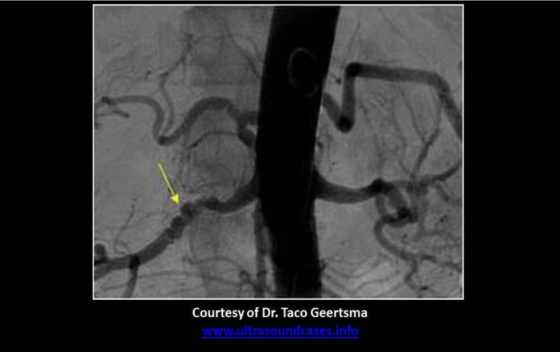
What is the arrow pointing to on the abdominal angiography exam?
A. fibromuscular dysplasia of the left renal artery
B. normal right renal artery
C. fibromuscular dysplasia of the right renal artery
D. normal tortuous splenic artery
C. fibromuscular dysplasia of the right renal artery
FMD demonstrates a bumpy lumen contour like a string of pearls.
A significant stenosis in which of the following arteries would be the most likely cause for an acute infarct of the right frontal lobe seen on a recent CT of the brain?
A. left posterior cerebral artery or left vertebral artery
B. left anterior cerebral artery or left middle cerebral artery
C. right anterior cerebral artery or right middle cerebral artery
D. bilateral anterior cerebral arteries
C. right anterior cerebral artery or right middle cerebral artery
The right frontal lobe is supplied with blood by the right ACA and MCA. Stenosis in either of the two vessels could cause ischemia in the frontal lobe.
You perform a carotid Doppler exam that demonstrates minimal atheroma formation bilaterally. You record waveforms that demonstrate laminar flow with the following velocities:
Right Proximal CCA PSV 145cm/s, EDV 35cm/s
Right Proximal ICA PSV 185cm/s, EDV 50cm/s
Left Proximal CCA PSV 140cm/s, EDV 38cm/s
Left Proximal ICA PSV 190cm/s, EDV 40cm/s
What will you report on your worksheet?
A, Flow velocities are increased bilaterally, which is most likely related to soft plaque formation not visible on 2D and color evaluation
B. Flow velocities are normal bilaterally
C. Flow velocities are increased bilaterally, which is most likely related to stenosis at the aortic valve
D. Flow velocities are increased bilaterally, which is most likely related to increased cardiac output
D. Flow velocities are increased bilaterally, which is most likely related to increased cardiac output
Bilateral increased velocities in the CCA and ICA with no evidence of stenosis is usually related to increased cardiac output. Bilateral CCA velocities can be increased with aortic stenosis also, but the flow would not be laminar in the proximal CCA.

A patient presents for a TCD exam with a recent diagnosis of subdural hematoma. What is the physician looking for on the TCD exam?
A. associated distal carotid stenosis
B. hematoma formation within the cranium
C. vasospasm
D. associated intracranial stenosis
C. vasospasm
TCD does not provide an image and US is not effective for accurate diagnosis of hematoma formation in the cranium.
Intracranial stenosis would most commonly lead to ischemia, not hemorrhage
TCD for Vasospasm is used in patients with subarachnoid hemorrhage, usually caused by a slow bleed. Vasospasm usually occurs within 14 days of the start of the hemorrhage. Flow velocities increase in MCA, ACA and PCA with subarachnoid hemorrhage. Serial evaluations are performed to evaluate the PSV in the MCA. If hemorrhage is present, MCA peak velocity over 80cm/s indicates flow changes due to hemorrhage; MCA peak velocity over 200cm/s indicates severe significant reduction in intracerebral flow.
Hemispheric Index - divide MCA velocity by the distal/terminal ICA velocity; HI > 3 ABNL
A patient presents with dysphagia and hoarseness. The referring physician suspects Ortner syndrome. What type of exam will you be performing?
A. bilateral carotid duplex exam
B. upper extremity arterial duplex exam
C. transcranial Doppler imaging exam
D. transcranial Doppler exam
B. upper extremity arterial duplex exam
In some patients the right subclavian artery can be a direct branch of the aorta distal to the left subclavian origin. It is called retroesophageal subclavian artery or aberrant right subclavian artery. The artery usually originates from a dilated segment of the proximal descending aorta called the Kommerell diverticulum. The abnormal course of the vessel can cause dysphagia due to compression of the esophagus or the laryngeal nerve. Ortner syndrome - palsy of the recurrent laryngeal nerve that causes changes in the voice, including hoarseness.

According to the findings on this arteriogram, which of the following would NOT be an expected symptom related to the findings?
A. Right leg paralysis
B. Left leg paresthesia
C. Left arm paresthesia
D. Visual disturbances of the left eye
B. Left leg paresthesia
There is a significant stenosis of the left CCA and an occlusion of the proximal left subclavian artery. The left CCA flow velocities will be elevated with diminished flow distally. The left vertebral artery is not demonstrated due to the proximal subclavian obstruction. NO steal is present or these vessels would be identified.
Occlusion of the left subclavian = left arm paresthesia
Diminished left CCA flow = right leg paralysis (left side of brain controls right side of body) and left eye symptoms
There is no explanation for left leg paresthesia on this image. This symptom would require right CCA/ICA blockage if it was related to abnormal cerebrovascular circulation.
***Image Credit Bronchiectasis and hoarseness of voice in takayasu arteritis: a rare presentation. Nd Perera G, C Jayasinghe A, D Dias L, Kulatunga A - BMC Res Notes (2012)
You are performing a left lower extremity venous duplex exam. DVT is identified in the popliteal and femoral veins. Which of the following must also be included in the exam protocol for this patient?
A. flow in the contralateral common femoral vein
B. arterial flow pattern in the arteries associated with the thrombosed veins
C. pulse volume recording of the calf and thigh of the contralateral leg
D. ankle brachial index
A. flow in the contralateral common femoral vein
For all unilateral extremity venous exams, the flow in the contralateral extremity should be documented for comparison. Common femoral vein for the lower extremities and subclavian or internal jugular vein for the upper extremities.
Why are serial exams necessary on patients prescribed Streptokinase?
A. to evaluate for the recanalization of an acute arterial occlusion
B. to evaluate for the reduction of plaque formation in the arteries
C. to evaluate for evidence of resolving arteritis due to Buerger disease
D. to evaluate for the presence of newly formed collateral arterial pathways
A. to evaluate for the recanalization of an acute arterial occlusion
Streptokinase is a lytic agent used to treat an acute arterial occlusion and significant, extensive acute DVT. Serial exams are necessary to evaluate recanalization and tailor the treatment accordingly.

The 2D images displayed are from a normal renal Doppler evaluation in a patient with a history of 4.5cm distal AAA and acute onset of moderate HTN.
What should you do to further evaluate or communicate the findings?
A. Evaluate the AAA for change in size or wall continuity.
B. Include notes in the renal artery Doppler report for the reading physician regarding the presence of free fluid around the kidney that is most likely due to chronic renal failure.
C. Include notes regarding the presence of ascites in the peritoneal cavity in the renal artery Doppler report for the reading physician.
D. Review the chart to confirm the suspected abnormal renal function testing results.
A. Evaluate the AAA for change in size or wall continuity.
The free fluid found in the perirenal space posterior to the kidney (not in peritoneal cavity) can indicate a rupture of the AAA which is the number one complication of AAA. The acute change in blood pressure also indicates a leak because the kidneys are raising the BP due to ischemia. The kidney demonstrates normal size and echotexture with no sign of renal failure.
If an arteriogram of the carotid arteries indicates a 60% stenosis of the left internal carotid artery, which of the following Doppler velocities from the ICA will correlate well with the predicted level of stenosis?
A. PSV 300cm/s, EDV 150cm/s
B. PSV 200cm/s, EDV 80cm/s
C. PSV 125cm/s, EDV 40cm/s
D. PSV 200cm/s, EDV 160cm/s
B. PSV 200cm/s, EDV 80cm/s
Strandness Criteria: 50-79% stenosis = PSV >125cm/s, EDV < 140cm/s
SRU Criteria 50-69% stenosis = PSV 125 - 230cm/s, EDV 40 - 100cm/s
ICAVL Criteria 50-69% stenosis = PSV 180 - 230cm/s, EDV 40 - 100cm/s
When preparing for a patient with suspected thoracic outlet syndrome, what other information should you review for pertinent information?
A. Chest x-ray
B. Lab results for hematocrit levels
C. CT of the brain
D. Aortogram
A. Chest x-ray
A chest x-ray could demonstrate the cervical rib that is a common cause for TOS.
Which of the following acronyms refers to the standard image file storage format used in medical facilities in the United States?
A. MPEG
B. AVI
C. DICOM
D. JPEG
C. DICOM
DICOM - Digital Imaging & COmmunications in Medicine provides a standard for handling, storing, and transmitting information in medical imaging.
A patient presents for a carotid ultrasound. The exam demonstrates significant calcification in the proximal ICA on the left side and the Doppler signal is not obtained. Antegrade flow in the mid ICA has a peak velocity of 30cm/s and end diastolic velocity of 10 cm/s. How will you report these findings?
A. Normal exam with limited evaluation of the proximal ICA.
B. Suspected critical stenosis in the proximal left ICA, but the exam was limited by shadowing from atheroma formation.
C. Normal exam.
D. Critical stenosis proximal left ICA.
B. Suspected critical stenosis in the proximal left ICA, but the exam was limited by shadowing from atheroma formation.
Any limitation in the exam that obscures your view of anatomy should be reported to the physician in the technologist findings.
A patient presents with an order for a lower extremity venous Doppler exam to rule out DVT. This patient should be evaluated in the _____ position in order to _____
A. semi-erect, increase hydrostatic pressure
B. supine, reduce hydrostatic pressure
C. semi-erect, reduce hydrostatic pressure
D. supine, increase hydrostatic pressure
A. semi-erect, increase hydrostatic pressure
The semi-erect position will also increase intraabdominal pressure which slows lower extremity venous return. This will fill the veins of the legs with blood making the US and Doppler evaluation much easier.

What will be included in your preliminary report for this patient?
A. There is a possible stenosis of the right mid brachial artery.
B. There is a possible stenosis of the right and left distal brachial artery
C. There is a possible isolated stenosis of the left axillary artery
D. There is a possible stenosis in the right palmar arch arteries
D. There is a possible stenosis in the right palmar arch arteries
The significant drop in pressure occurs in the right arm between the forearm and the digit cuff. This indicates a possible stenosis of the distal radial or ulnar artery or a stenosis in the palmar arch arteries. A mid brachial artery stenosis will cause a drop in pressure between the upper arm and forearm cuffs. The digit-brachial index should be greater than 0.60. The DBI in the right arm is 70/156mmHg = 0.45. Although there is a drop in pressure between the left forearm and digit, the DBl is >0.60.

The attached image is taken from a lower extremity venous Doppler exam. Which of the following should be included in your preliminary report?
A. The left PTV demonstrates normal flow and response to proximal augmentation.
B. The left PTV demonstrates abnormal absence of spontaneous flow but normal response to distal augmentation.
C. The left PTV demonstrates normal flow and response to distal augmentation.
D. The left PTV demonstrates abnormal absence of spontaneous flow but normal response to proximal augmentation.
C. The left PTV demonstrates normal flow and response to distal augmentation.
The deep calf veins and superficial veins do not normally exhibit spontaneous flow patterns. Augmentation is usually necessary to demonstrate patency of these veins.
A patient presents for a segmental pressure exam. She states she has had DVT in her right leg three times over the last 2 years. Today she complains of right leg pain and mild swelling, but no color change. She states that the doctor could not find the pedal pulses on the right side. What should you review in the patient chart prior to starting the exam?
A. Lab values for hematocrit levels
B. Lab values for lipid levels
C. Medical history for current Coumadin therapy
D. Surgical history for endoluminal graft placement
C. Medical history for current Coumadin therapy
The pressure cuffs can increase the risk for embolization to occur in the right leg. Current Coumadin therapy drastically reduces the ability of the embolism to make it to the lungs if it does form. Hematocrit levels can give you a clue to the blood thickness, but this does not assist you when considering the risk of embolism.
If the velocities in both carotid arteries have increased since last year's exam with no visible change in plaque formation, what should you review in the patient chart as a possible explanation?
A. lab values for decreased hematocrit levels
B. medical history for controlled HTN
C. medical history for polycythemia vera
D. recent echocardiogram for signs of decreased cardiac output
A. lab values for decreased hematocrit levels
Decreased hematocrit will lead to thinned blood with increased bilateral carotid velocities. Polycythemia vera leads to thickening of the blood which would decrease carotid velocities bilaterally. Increased output and uncontrolled HTN would cause increased bilateral carotid velocities.

You perform a bilateral venous Doppler due to unilateral swelling and pain in the right leg. Which of the following should be reported on your technologist worksheet?
A. Probable left common iliac vein thrombosis
B. Probable May-Thurner syndrome
C. Probable IVC thrombosis
D. Probable right common iliac vein thrombosis
D. Probable right common iliac vein thrombosis
Continuous flow in the iliac vein indicates a proximal obstruction. The left EIV waveform is normal but the right EIV waveform is continuous. This indicates a right common iliac obstruction. IVC obstruction would cause bilateral continuous waveforms.
A patient presents for a venous insufficiency exam. The exam demonstrates normal bilateral great and small saphenous veins. The deep system is not identified well in either leg. Both calves have multiple areas of small, clustered veins superficially. What should you review in the patient chart to explain these findings?
A. medical history for Klippel Trenaunay Webber syndrome
B. surgical history for venous ligation of the bilateral GSVs
C. treatment history for RF ablation of the bilateral GSVs
D. medical history for Paget Schroetter syndrome
A. medical history for Klippel Trenaunay Webber syndrome
RF ablation would affect a larger segment of the GSV. Klippel Trenaunay Webber syndrome refers to the congenital absence of the deep venous system of the legs. Paget Schroetter syndrome refers to thrombosis of the subclavian vein due to excessive motion, usually related to TOS.
What lab values may be elevated with renal artery stenosis?
A. prothrombin time
B. hematocrit
C. blood pressure
D. serum BUN and creatinine
D. serum BUN and creatinine
Renal artery stenosis will limit the blood flow through the kidney. This decrease in renal filtration of blood can be demonstrated in lab testing by increased serum levels of BUN and creatinine. Systemic BP will rise with significant RAS, but it is not a lab value.
A patient presents for a venous Doppler evaluation of the left lower extremity. Thrombus is identified in the left leg. What should you include in the preliminary report?
A. Amount of exercise the patient performed before symptoms occurred
B. What leg position causes the most pain
C. Location, estimated age and extent of thrombus
D. The peak velocity of the augmentation response proximal and distal to the thrombus
C. Location, estimated age and extent of thrombus
When reporting on DVT, the location, estimated age (acute or chronic) and extent of thrombus should be provided. Exercise and augmentation will not be performed with patients with suspected DVT.
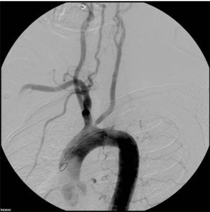
According to the findings on this arteriogram, which of the following symptoms would be explained by the findings?
A. Left leg paresthesia
B. Right arm paralysis
C. Left arm paresthesia
D. Isolated visual disturbances in the left eye
C. Left arm paresthesia
The carotid system appears patent. The left subclavian artery is absent from the image which indicates complete obstruction at the origin, The left vertebral artery normally originates from the proximal subclavian artery and is also absent from the image. Both of these vessels will demonstrate absence of flow on a Duplex exam. The only symptoms listed that would be related to these findings would be related to the left arm.
What vessel(s) should be closely evaluated for a related cause for a new cerebellar infarct seen on a CT of the brain?
A. posterior cerebral artery and carotid siphon
B. anterior and middle cerebral arteries
C. middle and posterior cerebral artery
D. vertebral and basilar arteries
D. vertebral and basilar arteries
The cerebellum is supplied by branches from the vertebral and basilar arteries. A cerebellar infarct would be related abnormalities of the vertebrobasilar circulation.

A patient presents with a 2-year history of a brachiocephalic hemodialysis fistula. She currently complains that her hand goes numb and turns white when she exercises. You are performing an upper extremity arterial exam and the attached images are from her exam. What will you include in your report?
A. The exam is normal and does not explain the patient's symptoms.
B. Steal syndrome is present.
C. The proximal radial artery is occluded
D. There is an obstruction in the palmar arch
B. Steal syndrome is present.
Steal syndrome can occur with a hemodialysis graft. Flow moves down the arm through the ulnar artery, through the palmar arch to the radial artery. Blood flow takes the path of least resistance.
Because the pressure in the outflow vein is so low, flow reverses in the radial artery as the blood is sucked up the arm and into the cephalic vein. When the patient exercises, the demand for blood increases in the hand but the ulnar artery cannot supply enough by itself = symptoms.
Which of the following is a required part of the Sonographer's preliminary report for a lower extremity segmental pressure exam?
A. patient position during the exam
B. peak velocity of the flow in the arteries
C. systolic and diastolic pressure measurements
D. duration of cuff inflation during the exam
A. patient position during the exam
If the patient is unable to be examined in the supine position, this must be documented on the preliminary report for the interpreting physician because the pressures obtained may be artifactually elevated. The systolic pressure measurements are obtained and reported, but no diastolic pressures are obtained in the legs. Flow velocity is not reported on a segmental pressure exam. It is an important part of an arterial duplex exam.
You are performing a left upper extremity venous duplex exam. DVT is identified in the axillary and subclavian veins. Which of the following must also be included in the exam protocol for this patient?
A. bilateral brachial pressures
B. arterial flow pattern in the arteries associated with the thrombosed veins
C. pulse volume recording of the ipsilateral upper arm and forearm
D. flow in the contralateral subclavian vein
D. flow in the contralateral subclavian vein
For all unilateral extremity venous exams, the flow in the contralateral extremity should be documented for comparison. Common femoral vein for the lower extremities and subclavian or internal jugular vein for the upper extremities.
Which of the following is a critical finding?
A. Atherosclerotic occlusion of the superficial femoral artery
B. AV fistula with a volume flow rate of 800 ml/min
C. DVT with a free-floating tail
D. 4cm aortic aneurysm
C. DVT with a free-floating tail
The free-floating tail can be a critical finding because it greatly increases the risk of a pulmonary embolism. Normal VFR for an AV fistula is 500 - 1200 ml/min. Atherosclerotic occlusion is a chronic process that often results in collateral formation. Total occlusion is typically accompanied by an increase in collateral flow. A 5.5cm AAA or larger is considered a critical finding.
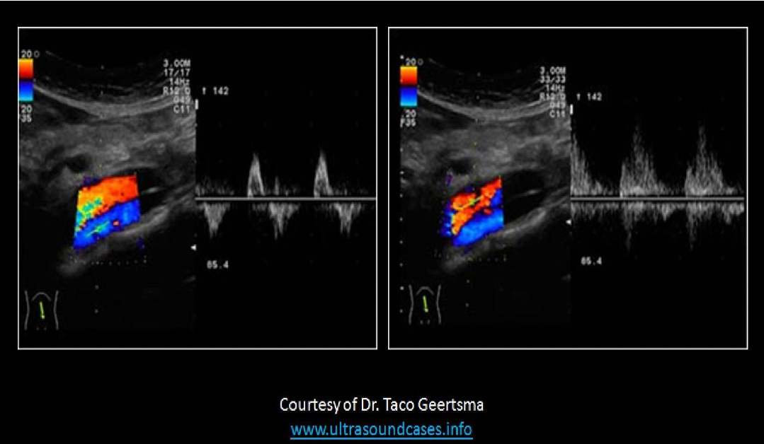
The images displayed are from an aortic Doppler evaluation on an ER trauma patient. What should you do upon completion of the exam?
A. Leave a preliminary report with the nurse describing the dissection and send the exam to the PACS system for a stat interpretation by the radiologist.
B. Leave a preliminary report with the nurse describing the AV fistula and send the exam to the PACS system for a stat interpretation by the radiologist.
C. Leave a preliminary report with the nurse describing the mild atherosclerosis in the distal aorta but an otherwise normal exam.
D. Provide a preliminary report describing normal flow in the aorta and IVC.
A. Leave a preliminary report with the nurse describing the dissection and send the exam to the PACS system for a stat interpretation by the radiologist.
A dissection is a critical finding that requires immediate attention.
A patient presents for a carotid ultrasound. The exam demonstrates significant atheroma formation and string flow in the proximal right ICA.
Which of the following signs or symptoms listed in the patient chart would be related to the findings?
A. right sided hemiparesis
B. left eye visual disturbances
C. right sided bruit
D. left arm and leg paresthesia
D. left arm and leg paresthesia
A critical stenosis that produces only minimal flow at the stenosis will not demonstrate a bruit. The right side of the brain controls the left side of the body. Left side symptoms in the body will be associated with right ICA disease. Visual symptoms occur on the same side as the obstruction. Left eye symptoms will be associated with left ICA disease.
Which of the following is important information to record on the final report for a carotid Doppler exam on a patient with 50-69% ICA stenosis?
A. Mechanical index settings
B. Doppler angles used on the exam
C. Transducer frequency used for the exam
D. Recommendation for TCD exam for associated intracerebral disease
B. Doppler angles used on the exam
A patient with 50-69% stenosis will be followed with serial ultrasound examinations annually or semiannually. The Doppler angles used in this exam should be duplicated on the serial exams for the most accurate assessment of changes in the level of stenosis.
An 80yr old female patient asks you to explain the purpose of a segmental pressure exam of the lower extremities. Which of the following is the best response?
A. Tell her that the pressure in your legs decreases with disease and the doctor wants to see how much her ankle pressures have dropped.
B. Tell her the exam uses blood pressure cuffs to take her blood pressure in her legs to evaluate the circulation to her feet.
C. Tell her the exam uses blood pressure cuffs to take her blood pressure in her legs to see if she needs surgery.
D. Tell her you are going to squeeze her legs for a minute just relax.
B. Tell her the exam uses blood pressure cuffs to take her blood pressure in her legs to evaluate the circulation to her feet.
Always tailor your explanation to your audience. Limit technical details that are not relevant for the patient. You should not suggest that surgery may be necessary or that her ankle pressures are decreased. As a Sonographer, you should always explain the exam and never be condescending or dismissive of a patient's request.
A 65yr old female presents chronic leg pain and swelling, but no brown discoloration is present in the distal calf and ankle area. Which of the following patient history questions would provide an explanation of the symptoms?
A. Does your pain increase with quick dorsiflexion of the left foot?
B. Do you have pain in the legs at night when in bed?
C. Have you had coronary artery bypass surgery?
D. If you elevate the leg, does the pain increase?
C. Have you had coronary artery bypass surgery?
The great saphenous is removed for most CABG procedures. The removal of this vessel can lead to swelling in the leg with the reduced outflow. Brown discoloration is not a common sign of GSV removal because the reduced outflow is in the deep veins and not related to insufficiency. Pain with quick dorsiflexion of the foot is a sign of DVT called Homan's sign. Pain increase with leg elevation or at night would indicate arterial disease, which does not correlate with swelling.
You identify acute thrombosis of the left popliteal and femoral vein, and the proximal end of the clot has a free-floating tail. What should you do next?
A. Check the patient chart for a history of Coumadin therapy
B. Call for a code blue due to the significant increased risk of embolism
C. Check the great saphenous vein for thrombus propagation
D. TeIl the patient to limit all movement because the DVT is unresolved and you need to consult the reading physician immediately
A. Check the patient chart for a history of Coumadin therapy
The free-floating tail can be a critical finding, The recent history of DVT could indicate that the patient is on anticoagulant therapy. If she is on Coumadin, this greatly reduces the risk of a pulmonary embolism from the tail. A preliminary report should be given to the nurse and the exam should be read by a physician immediately. A Sonographer should never give results or medical advice to a patient. Code blue is reserved for patients that are not breathing and in cardiac arrest.
Which of the following is not a patient demographic that would be used to track vascular disease occurrence?
A. Ethnicity
B. Degree of stenosis found on the current exam
C. Geographic location
D. Gender
B. Degree of stenosis found on the current exam
Degree of stenosis on the exam is not a patient demographic.
If a patient forgets to take their hypertension medication before a renal duplex exam, what effect can this have on the findings?
A. overestimation of renal parenchymal thickness
B. underestimation of renal artery stenosis
C. overestimation of renal artery stenosis
D. underestimation of peak renal artery velocity
C. overestimation of renal artery stenosis
Elevated systemic BP can cause increased velocity in the systemic arteries. If the pressure is high, velocity measurements will be overestimated and renal artery stenosis can be overestimated. The patient should be evaluated when the HTN is controlled, whenever possible.

According to the findings on this arteriogram, which vessel will demonstrate absent or abnormal flow pattern on a carotid Duplex exam?
A. R Vertebral and R Subclavian
B. R and L vertebral arteries and R Subclavian
C. R Subclavian and R CCA
D. LCCA, L Vertebral, L Subclavian
D. LCCA, L Vertebral, L Subclavian
There is a significant stenosis of the left CCA and an occlusion of the proximal left subclavian. The left CCA will demonstrate post-stenotic turbulence and elevated velocity in the proximal segment. There is no flow demonstrated in the left subclavian or vertebral artery. NO steal is present or these vessels would be identified on the image.
**Image Credit Bronchiectasis and hoarseness of voice in takayasu arteritis: a rare presentation. Nd Perera G, C Jayasinghe A, D Dias L, Kulatunga A - BMC Res Notes (2012)
When providing a preliminary report for a significant ICA stenosis, what should be included?
A. angles of insonation used to obtain Doppler signals
B. hemispheric index for both carotids
C. peak systolic and end diastolic velocities in the common and internal carotid arteries
D. end systolic velocities in the common and internal carotid arteries
C. peak systolic and end diastolic velocities in the common and internal carotid arteries’
The peak systolic AND end diastolic velocities should be included in the report. The hemispheric index is used in TCD evaluation to compare MCA and ICA velocities. The Doppler angles do not need to be included in the preliminary report provided by the technologist but should be included in the final report. Most ultrasound systems transfer the velocity information on each image and a chart is available at the end of the exam.
A curvilinear transducer can be used to better evaluate:
A. varicosities covering a large area
B. flow in the distal posterior tibial artery
C. flow in the anterior tibial artery
D. deep veins in obese patients
D. deep veins in obese patients
Most DVT exams will be performed with a 5-7MHz linear transducer. A curvilinear transducer may be necessary to evaluate obese patients because of the lower frequency range and wider field of view in the near field compared to a sector array. Superficial structures are best evaluated with a high frequency linear transducer.
When evaluating flow in the superior mesenteric artery, what is the most important history question to ask the patient?
A. Do you have a pain in your abdomen that is associated with breathing motions?
B. When was your last meal?
C. Do you have high blood pressure that cannot be treated with medication?
D. Do you have diabetes?
B. When was your last meal?
In order to evaluate the SMA waveform, it is important to know the activity level in the digestive tract.
Pre-prandial (NPO) = no digestion, high resistance flow with minimal diastolic flow.
Post prandial - digestion increases inflow needs, low resistance flow with mild diastolic flow
Untreatable HTN is associated with renal artery stenosis. Abdomen pain with expiration is associated with celiac artery compression.
An upper extremity venous Doppler that is performed for suspected DVT should be performed with the patient in the _____ position
A. supine
B. Trendelenburg
C. prone
D. standing
A. supine
Patient should be evaluated in the supine position with the head turned slightly away from the affected side; the affected arm should be in the dependent position and externally rotated.
D-dimer levels are most accurate for predicting:
A. the presence of DVT
B. the risk of clot formation
C. the absence of DVT
D. the risk of pulmonary embolism
C. the absence of DVT
D-dimer levels are most accurate for predicting the absence of DVT. D-dimer levels can be elevated for many reasons including DVT, cancer and other diseases. If DVT is suspected but the D-dimer levels are low, DVT is most likely not present.
If a patient has a history of 80% stenosis of the left internal carotid artery identified on a MRA exam. Which of the following Doppler velocities from the ICA will correlate well with the predicted level of stenosis?
A. PSV 200cm/s, EDV 100cm/s
B. PSV 200cm/s, EDV 80cm/s
C. PSV 300cm/s, EDV 150cm/s
D. PSV 125cm/s, EDV 40cm/s
C. PSV 300cm/s, EDV 150cm/s
Strandness Criteria: 80-99% stenosis = PSV >125cm/s, EDV >140cm/s
SRU Criteria >70% stenosis = PSV >230cm/s, EDV >100cm/s
ICAVL Criteria >70% stenosis = PSV >230cm/s, EDV >100cm/s
If a patient complains of a single 12hr episode of right arm paresthesia and paralysis, what will you report on your technologist worksheet?
A. history of cerebrovascular accident
B. history of amaurosis fugax
C. history of reversible ischemic neurological defect
D. history of transient ischemic attack
D. history of transient ischemic attack
Symptoms that last less than 24hrs = TIA.
All of the following should be included in the report from an exercise segmental pressure exam, except:
A. Duration of exercise
B. Venous Refill Time (VRT)
C. Pressure recovery time in ankles
D. Symptoms that occurred and when they started during the exercise
B. Venous Refill Time (VRT)
Venous refill time is a part of a PPG report for insufficiency.
The purpose of an exercise segmental pressure exam is to unmask PAD that is not displayed with the patient at rest. The duration of exercise and the time when symptoms started during exercise aids in quantification of the disease. The pressure recovery time after exercise has been stopped also aids in estimating PAD severity. Longer recovery times = more severe disease.
A 76yr old female with diabetes, HTN, and COPD is sent to the vascular lab for a six month follow up on a carotid stenosis. What is the best patient position to use for the exam?
A. Supine, neck slightly extended
B. Semi erect with or without a pillow
C. Seated upright on a stool beside the machine, facing you
D. Trendelenburg position
B. Semi erect with or without a pillow
If the patient has COPD, they should not lie flat on their back. They should be scanned in the semi-erect position, usually without a pillow to maintain a straight neck position. If the patient is seated on a stool, it will be difficult for them to hold steady for the exam and the Sonographer position would be improper ergonomically.
Which of the following is not a patient demographic that is collected to evaluate patient population statistics for those with vascular disease?
A. Gender
B. Age
C. Employment history
D. Ethnicity
C. Employment history
Employment history is not considered a relevant patient demographic used to track the occurrence of vascular disease.
Patients are examined in the _____ position for a lower extremity arterial Doppler exam in order to _____
A. supine, reduce hydrostatic pressure
B. semi-erect, reduce hydrostatic pressure
C. semi-erect, increase hydrostatic pressure
D. supine, increase hydrostatic pressure
A. supine, reduce hydrostatic pressure
You are performing a renal Doppler exam. What is the best patient position to visualize the full length of the left renal artery?
A. Supine, contralateral arm above head
B. Supine, ipsilateral arm above head
C. Left lateral decubitus
D. Right lateral decubitus
D. Right lateral decubitus
A coronal view or an oblique transverse view will best demonstrate the full length of the renal arteries. These images are best obtained when the patient is in the right lateral decubitus position when evaluating the left kidney and artery.
Which of the following transducers would be preferred for a lower extremity saphenous mapping on an average sized patient?
A. 7-10MHz sector
B. 5-7MHz linear
C. 7-10MHz linear
D. 5-7MHz curvilinear
C. 7-10MHz linear
Linear arrays are preferred for vascular exams. 5-7MHz transducer are used for DVT evaluation in an average sized patient. 7-10MHz transducers are used for evaluation of the superficial system for insufficiency or vein mapping.

What will you include in your preliminary report for this patient?
A. There is a possible obstruction of the left innominate artery.
B. There is a possible obstruction of the left subclavian artery.
C. There is a possible obstruction of the left innominate artery or the subclavian artery.
D. There is a possible obstruction of the right innominate artery or the subclavian artery.
B. There is a possible obstruction of the left subclavian artery.
Keep in mind, there is no left innominate artery. The left subclavian artery originates from the aortic arch. The pressures indicate a blockage in the left upper arm arteries (subclavian or axillary). The pressure difference between the arms is greater than 20mmHg. The arm with the lower pressure is the arm with the problem.

You are performing a carotid Doppler exam due to the recent onset of right arm and face paresthesia. There is little plaque present in either carotid system. The right side has normal Doppler flow velocities and waveforms. The attached Doppler images are taken from the proximal left ICA and ECA. Which of the following should be included on your preliminary report?
A. There is a probable obstruction of the proximal innominate artery.
B. The exam is within normal limits and does not explain the symptoms.
C. There is a probable obstruction of the origin of left carotid artery.
D. Congestive heart failure is most likely present.
C. There is a probable obstruction of the origin of left carotid artery.
The left CCA originates from the aortic arch. Unilaterally damped ECA and ICA waveforms indicate proximal obstruction. Bilaterally reduced resistance and velocity is associated with CHF.
A patient presents with an order for a lower extremity venous Doppler exam to rule out venous insufficiency. This patient should be evaluated in the ____ position in order to ____
A. supine, increase hydrostatic pressure
B. supine, reduce hydrostatic pressure
C. standing, increase hydrostatic pressure
D. standing, reduce hydrostatic pressure
C. standing, increase hydrostatic pressure
The standing position will increase hydrostatic or gravitational pressure, which slows lower extremity venous return. This will fill the veins of the legs with blood making the US and Doppler evaluation much easier. It will also cause superficial varicosities to engorge with blood for better visualization.
A patient presents for an ABI exam due to bilateral calf pain. He is a dialysis patient with an occluded graft in the right arm and a patent graft in the left arm. What should you do first?
A. Call the referring physician to explain that the exam is not possible for this patient
B. Change the exam to a lower extremity arterial exam and start scanning
C. Skip the brachial pressures and just do the ankle pressures
D. Take the pressure in the arm with the occluded graft
A. Call the referring physician to explain that the exam is not possible for this patient
NEVER take a blood pressure in an arm with a hemodialysis graft. The ABI exam requires accurate brachial pressure readings in order to calculate the ABI. Without the brachial pressures, the exam is non-diagnostic. You should contact the referring physician to alert them of the issue and ask if they would like to order a different exam to assist the patient. NEVER cancel or change a physician's order. They must submit a new order if something changes.
You are performing a mesenteric duplex examination for suspected mesenteric ischemia. The findings are: SMA PSV 408cm/s, EDV 78cm/s, monophasic flow with turbulence Celiac PSV 300cm/s, EDV 55cm/s, monophasic flow with spectral window IMA PSV 180cm/s, EDV 25cm/s, monophasic flow with spectral window
How will you report these findings?
A. isolated stenosis of the celiac artery with compensatory flow in the SMA and IMA
B. suspected mesenteric ischemia with abnormal flow velocities and waveforms in the SMA, IMA, and celiac artery
C. isolated stenosis of the SMA with compensatory flow in the celiac artery and IMA
D. suspected mesenteric ischemia with abnormal flow velocities and waveforms in the SMA and celiac artery
C. isolated stenosis of the SMA with compensatory flow in the celiac artery and IMA
If there is significant obstruction or occlusion of one mesenteric artery and the other two main arteries are normal, there can be associated changes in these normal vessels. If the SMA is occluded, the celiac artery may demonstrate flow with increased velocity and a normal spectral window (no post-stenotic turbulence). If the SMA is occluded, the IMA can increase in size and demonstrate increased flow velocity without post-stenotic turbulence. If the celiac artery is occluded, the SMA and/or IMA can demonstrate increased flow velocity without post-stenotic turbulence. Keep in mind that compensatory flow can mimic stenosis. It is very important to evaluate the waveform characteristics with elevated systolic velocities.
Compensatory flow = increased velocity + spectral window
Stenosis = increased velocity + post-stenotic turbulence
Which transducer should be selected for a renal artery Doppler examination for a patient that is 5'5" and 150lbs?
A. 3.5MHz linear array
B. 2.5MHz curvilinear array
C. 3.5MHz curvilinear array
D. 3.5MHz phased array
C. 3.5MHz curvilinear array
A curved transducer is necessary for abdominal vascular evaluations. 3.5MHz should be sufficient for an average sized patient.
Which of the following describes the table placement for performing an ultrasound exam?
A. Table height should allow the Sonographer's arm to be slightly abducted with the wrist fully extended.
B. Table height should allow the Sonographer's arm to be slightly adducted with the wrist fully flexed toward the side being examined.
C. The table should be placed in the reverse trendelenburg position to assist in moving blood flow into the cerebral vessels.
D. The table should be placed in the trendelenburg position to assist in moving blood flow into the cerebral vessels.
A. Table height should allow the Sonographer's arm to be slightly abducted with the wrist fully extended.
A patient presents for a carotid ultrasound due to the presence of bilateral bruits in the proximal neck. What should be the next step in evaluating this patient?
A. Evaluate the medical history section of the patient chart to look for a history of polycythemia vera
B. Evaluate the cardiac history section of the patient chart to look for a history of CHF.
C. Evaluate the vascular history section of the patient chart to look for a history of a subclavian steal
D. Evaluate the cardiac history section of the patient chart to look for a history of aortic valve stenosis.
D. Evaluate the cardiac history section of the patient chart to look for a history of aortic valve stenosis.
Bilateral carotid bruits and changes in both carotid flow patterns can be related to aortic stenosis. If the aortic valve is stenotic causing increased flow velocity and turbulence, flow in both carotids will reflect this change and may demonstrate bilateral bruits.
CHF would lead to decreased flow through both carotids but a bruit would most likely NOT be identified due to slow moving flow.
Polycythemia vera causes the blood to thicken = slower velocities = reduced risk of bilateral bruit.
A subclavian steal involves the rerouting of blood and would not be associated with a bilateral bruit.
Which of the following is a critical finding that requires immediate attention?
A. dilated hepatic veins in a liver transplant patient
B. acute occlusion of the external iliac artery in a renal transplant patient
C. recanalization of the great saphenous vein after chemical ablation
D. echogenic walls and posterior shadowing from a hepatoportal shunt that was inserted 2 days ago
B. acute occlusion of the external iliac artery in a renal transplant patient
An acute arterial occlusion of any artery is a critical finding but especially in renal transplant patients. Acute occlusion of the renal artery or supplying artery can cause significant damage or loss of the allograft. Most renal transplant arteries are connected to the external iliac artery. An acute occlusion of this artery not only puts the leg at risk of damage, but the renal allograft as well.
Why is an acute arterial occlusion in the lower extremity considered a critical finding?
A. the risk of arterial rupture is significantly increased
B. because it is not treatable
C. the risk of stroke is significantly increased
D. collaterals have not had time to form to redistribute the blood
D. collaterals have not had time to form to redistribute the blood
Without collaterals present to redistribute blood flow, the risk of tissue death and possible amputation is increased

You are performing an upper extremity venous Doppler exam due to arm swelling and pain. The image displayed is from the left shoulder. Which of the following should be included on your preliminary report?
A. There are echogenic fibrous strands of retracted chronic thrombus present in the subclavian vein.
B. There is acute thrombus formation around an intravenous catheter in the subclavian vein.
C. There is chronic thrombus formation around an intravenous catheter in the subclavian vein.
D. The subclavian vein is normal and completely patent with an intravenous catheter in the lumen.
B. There is acute thrombus formation around an intravenous catheter in the subclavian vein.
The acute thrombus formation is very hypochoic compared to the echogenic IV catheter present.
According to the findings on this arteriogram, which vessel will demonstrate a post-stenotic flow pattern on a carotid Duplex exam?
A. RCCA, R Vertebral, R Subclavian
B. L Vertebral and L Subclavian
C. Proximal L CCA
D. R and L vertebral arteries and R Subclavian
C. Proximal L CCA
There is a significant stenosis of the left common carotid artery and an occlusion of the proximal left subclavian. No flow will be identified in the left subclavian and vertebral artery.
***Image Credit Bronchiectasis and hoarseness of voice in takayasu arteritis: a rare presentation. Nd Perera G, C Jayasinghe A, D Dias L, Kulatunga A - BMC Res Notes (2012)
Which of the questions listed below would provide pertinent history information for a lower extremity venous Doppler exam?
A. Do your legs/calves hurt more when walking, but the pain stops if you sit and rest?
B. If you are seated on the edge of the bed with your feet on the floor, does this reduce the swelling?
C. Does your job require you to stand for long periods of time?
D. Do you have any ulcer formation on your toes?
C. Does your job require you to stand for long periods of time?
Standing for long periods of time can cause venous insufficiency and the related symptoms in this patient. Arterial disease can cause leg pain with exercise that is relieved by rest. The muscles require an increased blood supply with exercise. If there is arterial disease present, it may not allow the necessary increase in flow and ischemia/pain presents. Stopping the exercise will assist in the relief of the symptoms as the muscles decrease their need for blood flow. Arterial ulcers are found on the toes and heel of the foot. Venous ulcers are found on the distal calves.
Which of the following is an important part of Sonographer - Patient communication?
A. explaining the exam and the results
B. offering advice on ways to alleviate symptoms until the doctor treats the problem
C. verifying the patient's name and date of birth
D. asking the patient if they are allergic to iodine
C. verifying the patient's name and date of birth
Important parts of sonographer Communication include verifying the patient's name and date of birth, explaining the exam and when to expect the results and helping the patient locate their next exam or the exit.
Sonographers should not offer treatment advice to the patient. Always refer them to their doctor when they have questions about their health status.
lodinated contrast is used in radiology procedures. If the patient is having that type of procedure, the technologist performing the exam will assess their history related to an iodine allergy.
A 75yr old female presents for a transcranial doppler evaluation for suspected basilar artery stenosis. She suffers from emphysema and cannot lie down for the exam. Which of the following describes how you can evaluate the basilar artery in this patient?
A. patient seated in chair, neck hyperextended with a wedge behind the head for support
B. patient seated in chair, lean forward to reach for their toes and flex the neck
C. patient seated in chair, neck flexed and head supported by her arms/hands
D. patient in the left lateral decubitus position, hand supporting the head
C. patient seated in chair, neck flexed and head supported by her arms/hands
If a patient is unable to lie down in the supine or decubitus position for TCD evaluation of the basilar artery, they should be seated in a chair, neck flexed and head supported by her arms/hands. If the patient is able to lie supine, just not in the decubitus position, then ask them to turn their head away from the side of interest and place the transducer to the left or right side of the foramen magnum
When reporting spectral Doppler information the Sonographer must:
A. record the highest quality and highest velocity signals for the final measurements
B. document the frequency of the transducer used for the exam should be indicated in the report
C. classify each waveform as triphasic, biphasic or monophasic
D. always record the highest velocity obtained from all signals
A. record the highest quality and highest velocity signals for the final measurements
Spectral Doppler measurements should be made at the modal (densest) margin of the flow signal. Multiple samples may be taken at the same site. Use the highest quality and highest velocity signals for final measurements. Do not report Doppler flow information from waveforms with poor definition or of marginal quality.
A patient presents with localized swelling and redness of the left calf that started two days ago. The referring physician requested a segmental pressure exam. What should you do first?
A. Discuss the order with the radiologist to confirm that the exam that is ordered should be performed.
B. Start with the ankle pressures, then move up the leg and finish with the brachial pressures.
C. Change the order to a venous Doppler after you speak to the patient regarding their symptoms.
D. Start with the brachial pressures and then perform the lower extremity pressures.
A. Discuss the order with the radiologist to confirm that the exam that is ordered should be performed.
Anytime the procedure could be detrimental to the patient, you should clarify the order with the radiologist or referring physician.
This patient demonstrates symptoms and signs of a possible acute DVT. Applying pressure cuffs to a vein with thrombus in it could cause emboli propagation.
A patient presents for a venous Doppler evaluation of the lower extremities. The patient is contracted at the knees and hips and unable to extend the legs for the exam. All compression views are normal. Doppler evaluation demonstrates mildly reduced response to distal augmentation in the thigh and continuous flow in the popliteal veins. How will you report these findings?
A. Normal exam
B. Venous insufficiency is present in the upper thigh vessels bilaterally
C. Probable DVT in the popliteal veins bilaterally
D. Normal exam with flow changes most likely caused by patient position
D. Normal exam with flow changes most likely caused by patient position
Absence of vein compressibility is the most reliable factor for diagnosing DVT. The patient has normal compression views which indicate the absence of DVT. The contracted position of the patient can cause a more continuous waveform and decrease in augmentation response.
Why should a venous insufficiency exam be performed with the patient in the standing position?
A. To engorge the veins to make them easier to identify with ultrasound
B. To increase the effects of the Valsalva maneuver
C. To evaluate the veins with the added effect of hydrostatic pressure
D. To ensure the valve leaflets are closed during the exam
C. To evaluate the veins with the added effect of hydrostatic pressure
A venous insufficiency exam should be performed with the patient in the standing position to evaluate the veins with the added effect of hydrostatic pressure. This will allow reflux to more easily be identified if it is present.

You are about to perform a lower extremity arterial duplex on a patient that recently had an MRA run-off exam. According to the attached image, what vessel will demonstrate reduced or absent flow in the left leg?
A. posterior tibial artery
B. superficial femoral artery
C. anterior tibial artery
D. common femoral artery
C. anterior tibial artery
Note the limited display of contrast in the superficial femoral artery of the right leg and the anterior tibial artery of the left leg. You should expect that Doppler evaluation of these vessels will demonstrate little to no flow.
Which of the following transducers would be preferred for a lower extremity DVT evaluation on an average sized patient?
A. 10-12MHz linear
B. 5-7MHz curvilinear
C. 7-10MHz sector
D. 5-7MHz linear
D. 5-7MHz linear
Linear arrays are preferred for vascular exams. 5-7MHz transducer are used for DVT evaluation in an average sized patient. Higher frequencies can be used for evaluation of the superficial system.
Air in a patch used during a carotid endarterectomy can degrade the evaluation of the treated area in the immediate post-op setting. What is the recommended approach for evaluating these patients?
A. use the most posterior approach possible
B. use the most anterior approach possible
C. use sterile gel and scan directly over the incision
D. patients with a recent endarterectomy should not be evaluated with ultrasound within the first 14 days after the procedure
A. use the most posterior approach possible
If ultrasound performed soon after the procedure, artifacts produced by air in the patch or graft may inhibit the exam. To minimize artifact from air in the patch, use the most posterior approach possible to evaluate the bifurcation.

According to the findings on this arteriogram, which abnormal findings will be identified on a carotid Duplex exam?
A. Left Subclavian Steal
B. Flow reversal in the Left Vertebral and Left Subclavian arteries
C. Absence of flow in the Left Subclavian artery with flow reversal in the Left CCA
D. Absence of flow in the Left Subclavian and Left Vertebral arteries
D. Absence of flow in the Left Subclavian and Left Vertebral arteries
The left subclavian is absent from the image which indicates complete obstruction at the origin, The left vertebral artery normally originates from the proximal subclavian artery and is also absent from the image. Both vessels will demonstrate absence of flow on a Duplex exam.
If a patient has a history of 40% stenosis of the left internal carotid artery identified on a MRA exam. Which of the following Doppler velocities from the ICA will correlate well with the predicted level of stenosis?
A. PSV 200cm/s, EDV 100cm/s
B. PSV 200cm/s, EDV 80cm/s
C. PSV 120cm/s, EDV 35cm/s
D. PSV 300cm/s, EDV 150cm/s
C. PSV 120cm/s, EDV 35cm/s
Strandness Criteria: <50% stenosis = PSV < 125cm/s
SRU Criteria <50% stenosis = PSV <125cm/s, EDV <40cm/s
ICAVL Criteria <50% stenosis = PSV < 180cm/s, EDV <40cm/s
Which of the following is the best patient position for performing a transcranial Doppler exam to obtain a waveform from both of the middle cerebral arteries?
A. seated on a stool, facing you and the machine
B. left lateral decubitus with left hand placed under the head for support
C. supine with chin in neutral position
D. seated on a stool, with their back to you and the machine
C. supine with chin in neutral position
The transtemporal window is used to evaluate the MCA flow bilaterally. The transducer is placed on the lateral side of the head at the temple. Placing the patient in the supine position allows for evaluation of both sides.

What will be included in your preliminary report for this patient?
A. There is a possible stenosis of the right radial and/or ulnar artery
B. There is a possible stenosis of the bilateral radial and/or ulnar arteries
C. There is a possible stenosis of the bilateral mid brachial arteries
D. There is a possible stenosis of the left radial and/or ulnar artery
A. There is a possible stenosis of the right radial and/or ulnar artery
The significant drop in pressure occurs in the right arm between the forearm and the digit cuff. This indicates a possible stenosis of the distal radial or ulnar artery or a stenosis in the palmar arch arteries. A mid brachial artery stenosis will cause a drop in pressure between the upper arm and forearm cuffs. The finger-brachial index should be greater than 0.80. The DBI in the right arm is 70/156mmHg = 0.45. Although there is a drop in pressure between the left forearm and digit, the FBI is >0.80.
A patient presents for a venous insufficiency exam. The exam demonstrates normal bilateral saphenous veins that extend up to the distal thigh, but do not appear to connect to the deep system. What should you review in the patient chart to explain these findings?
A. treatment history for RF ablation of the bilateral GSVs
B. medical history for Klippel Trenaunay Weber syndrome
C. medical history for Paget Schroetter syndrome
D. surgical history for venous ligation of the bilateral GSVs
D. surgical history for venous ligation of the bilateral GSVs
Ligation of the proximal segment of the GSV could produce these findings. Prior to the introduction of endovenous treatment, ligation of the diseased segments was commonly performed to treat reflux.
RF ablation would affect a larger segment of the GSV.
Klippel Trenaunay Weber syndrome refers to the congenital absence of the deep venous system of the legs.
Paget Schroetter syndrome refers to thrombosis of the subclavian vein due to excessive motion, usually related to TOS.
When evaluating a suspected stenosis in the lower extremity arterial system, the peak systolic velocity is documented on the waveforms obtained. What other two factors should be reported to determine the severity of the stenosis?
A. resistive index and diastolic flow direction
B. resistive index and velocity ratio
C. velocity ratio and diastolic flow direction
D. pulsatility index and reflux time
C. velocity ratio and diastolic flow direction
The velocity ratio is used to compare the PSV proximal to the stenosis to the PSV at the stenosis. A ratio greater than 2 indicates stenosis. A triphasic pattern has two components of diastolic flow. Retrograde flow occurs immediately after systole ends followed by a small amount of antegrade flow. When the second component of diastolic flow is absent, the waveform is considered biphasic. Monophasic flow demonstrates antegrade flow in systole and diastole. Resistive and Pulsatility indices are not commonly used to report the severity of stenosis in the extremities. The RI is necessary when evaluating parenchymal perfusion in an organ like the kidney or liver.
A lower extremity venous Doppler that is performed for suspected DVT should be performed with the patient in the ____ position
A. prone
B. standing
C. reverse Trendelenburg
D. Trendelenburg
C. reverse Trendelenburg
A venous Doppler that is performed for suspected DVT should be performed with the patient in the reverse Trendelenburg position. This semi-erect position increases intra-abdominal pressure and reduces outflow to engorge the veins with blood for easier visualization.
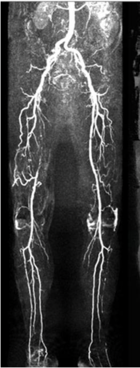
You are about to perform a lower extremity arterial duplex on a patient that recently had an MRA run-off exam. According to the attached image, what vessel will demonstrate reduced or absent flow in the right leg?
A. posterior tibial artery
B. superficial femoral artery
C. common femoral artery
D. anterior tibial artery
B. superficial femoral artery
Note the limited display of contrast in the superficial femoral artery of the right leg and the anterior tibial artery of the left leg. You should expect that Doppler evaluation of these vessels will demonstrate little to no flow.
When evaluating a pregnant patient for DVT, the patient should be placed in the _____ position
A. supine
B. right oblique or decubitus
C. Trendelenburg
D. left oblique or decubitus
D. left oblique or decubitus
The uterus can compress the IVC causing flow changes that lead to false results in the extremities. Placing the patient in a left oblique or decubitus position will reduce uterine pressure on the IVC.
Venography is usually performed with the patient in what position?
A. Supine with table horizontal
B. Supine with table at 60-degree reverse Trendelenburg
C. Lithotomy position
D. Supine with table at 60-degree Trendelenburg
B. Supine with table at 60-degree reverse Trendelenburg
Venography is performed with the patient in the semi-erect position to dilate the veins, making them more visible. When evaluating a patient for insufficiency, the semi-erect position helps to demonstrate the reflux that occurs when the patient is standing. When the patient lays on the table, the table can be adjusted to a 60-degree reverse Trendelenburg position to dilate the veins and increase hydrostatic pressure.