Practical- Cell identification + WBC/RBC Morphology + Haemogram Interpretation
 Call Kai
 Learn
Learn Practice Test
 Spaced Repetition
 Match
Match Flashcards
 Knowt Play
1/62

There's no tags or description
Looks like no tags are added yet.
Name | Mastery | Learn | Test | Matching | Spaced | Call with Kai | Chat |
|---|
No analytics yet
Send a link to your students to track their progress
63 Terms

Can you list 3 key differences between dog and cat erythrocytes?
1 - dog erythrocytes have a central area of pallor, whilst this is not generally visible.
2 - Cat erythrocytes are smaller in size compered to dog erythrocytes.
3 - Anisocytosis is usually present in cats, whilst dog erythrocytes are usually more uniform in size.

Identify the cells in this image
1 = Band neutrophil

Identify the cells in this image
2 = monocyte
3 = neutrophil

Identify the cells in this image
4 = eosinophil
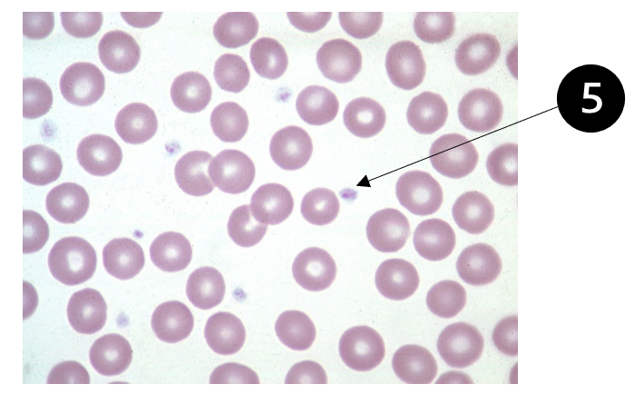
Identify the cells in this image
5 = platelets

Identify the cells in this image
6 - Lymphocyte - round nucleus. Little or no cytoplasm
And can see granular lymohocyte on the right?

Identify the cells in this image
7 - Neutrophil
8 - Eosinophil
9 - Lymphocyte

Identify the cells in this image
10 - Platelet
11 - Neutrophil
12 - Eosinophil

Identify the cells in this image
Stain precipitate

Identify the cells in this image and why this may occur
Hypochromasia - seen in iron deficiency anaemia
(Some polychromatic rbcs are also present on this slide)

Identify the cells in this image
Heinz bodies

Describe the RBC morphology in this smear
Red = Acanthocytes = Poikilocytosis
Target cells (codocytes) = Poikilocytosis
Blue = Elliptocytes = Poikilocytosis

Describe the RBC morphology in this smear
Microcytic Hypochromic erythrocyte = blue
normal erythrocyte = red

Describe the morphology of these neutrophils
Red circle = Left shift (band neutrophils)
Red arrow = Toxic changes (Döhle bodies, cytoplasmic basophilia)

Describe the morphology of these neutrophils
Marked neutrophilia
RED CIRCLES = STAIN PRECIPITATE
RED CIRCLE IN SECOND PICTURE = MONOCYTE
BLUE CIRCLE = TOXIC NEUTTROPHIL (Döhle bodies, cytoplasmic basophilia)

This is Microfiliaria, what layer of a blood smear would this be found in?
The feathered edge.

Describe this rabbit's blood smears
Significant anisocytosis
Poikilocytosis (acanthocytes = purple circle, echinocytes = light blue circle)
Polychromasia = dark blue circles
Heterophils with pink cytoplasmic granules = red circles

Mismatch is often seen when there are:
Left shift
Toxic changes
When is a mismatch like this common?
Analyser/smear mismatch: no evidence of neutropenia, lymphocytosis and monocytosis on the blood smear review

What dis?
degenerate neutrophil

What is the cause/type of neoplastic cell seen here?
Marked leucocytosis
Neoplastic cells -> Acute leukaemia
(Potentially lymphocytic)
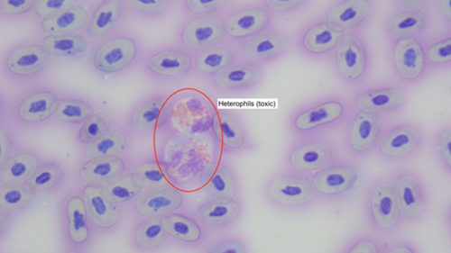
Describe this bearded dragon's WBC morphology
Heterophilia = red circle
Heterophil has toxic changes (reduced granularity, cytoplasmic basophilia)

Identify the cell formation in this image
Rouleaux
Describe what these rsults could be showing
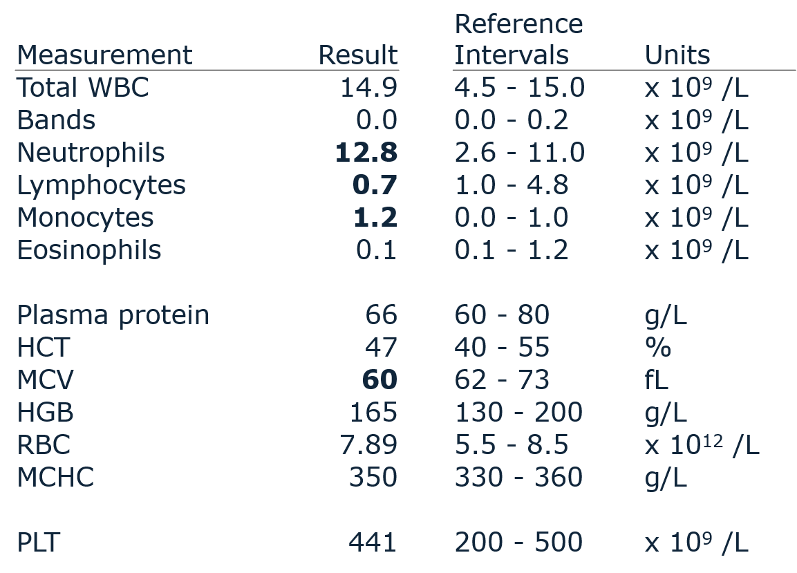
§There is a classical steroid response present as indicated by a mild mature neutrophilia, lymphopenia, and monocytosis. Lymphopenia is the most consistent finding.
§The steroid response is likely the most common leukocyte response. It is an indicator of major systemic illness or pain.
§A minor consideration is that the MCV is slightly decreased. There is no indication of anaemia. If inspection of RBC morphology and the RBC volume histogram are both normal, then the MCV is most likely normal for this individual.
Interpret these results

§There a moderate leukocytosis, likely an inflammatory neutrophilia with modest left shift in bands (1% bands would have been seen on the microscopy differential). The mature neutrophils would also indicate inflammation because they are greater than an approximate doubling of the upper reference limit.
§A superimposed steroid response is possible because the lymphocyte concentration is at the lower end of normal (a 1% change lower in the differential would result in lymphopenia). Note that this is probabilistic thinking.
§The mild monocytosis could be attributed to either inflammation and/or steroid release.
Summary: Combined inflammatory and steroid pattern, with inflammatory component being most clear.
Interpret these results

Blood smear examination confirmed platelet count (PLT)
§Lymphopenia indicates a steroid response, even though the expected neutrophilia is not present. The neutrophils may be increased within the reference interval. The expected monocytosis is variable and the absence of monocytosis does not rule out steroid release.
§
§The triad of anaemia, increased regeneration, and decreased protein is indicative of blood loss that is of at least a few days' duration as the marrow has had time to develop a regenerative response. The cause of blood loss is the thrombocytopaenia below. The dog should have visible petechial haemorrhages.
§
§There is marked thrombocytopaenia and this was confirmed by blood film examination. This is the cause of the blood loss. The magnitude of thrombocytopaenia in the face of adequate marrow function for WBC and RBC suggests that immune-mediated thrombocytopaenia is most likely.
Interpret these results

Blood was shipped to lab for analysis; ~48 hours old
Blood smear examination confirmed PLT
§The lymphocytosis is of a magnitude conclusive for lymphocytic leukaemia; it is also accompanied by blasts (undifferentiated blood cells). Cytometric analysis is indicated if contemplating treatment.
§There is non-regenerative anaemia with hypoproteinaemia. This is most likely due to blood loss given the marked thrombocytopaenia that is confirmed by blood film examination. But also the thrombocytopenia and non-regenerative anaemia could be due to BM suppression.
§Marrow failure due to lymphoproliferative infiltrate is highly probable; this would explain the failure to regenerate RBC, failure to produce platelets, and probable failure to produce neutrophils.
§Note the MCV and MCHC values: this can happen when blood ages in the tube. The RBCs will swell, giving a falsely high MCV and HCT. This will affect the calculation of MCHC (falsely low).

Interpret these results
Blood smear examination confirmed PLT
§There is marked panleukopaenia. Most notable is the severe neutropaenia. The absence of left shift and toxic change suggests the neutropaenia is due to a production failure.
§
§There is hyperproteinaemia. The most probable causes are either haemoconcentration or chronic antigenic stimulation leading to hyperglobulinaemia. The physical exam should address hydration state.
§
§There is borderline non-regenerative anaemia that could be somewhat more severe if the animal is haemoconcentrated.
§There is prominent thrombocytopaenia that is confirmed by blood film examination.
Interpret these results

Blood smear comments:
Toxic neutrophils present
Agglutination and many spherocytes are noted

§This is a relatively complex leukogram. There is neutrophilia with a prominent left shift and notation of toxic change; this is interpreted as an inflammatory pattern. The lymphopaenia is indicative of steroid release. There is rubricytosis as part of the regenerative response.
§
§There is severe anaemia that is prominently regenerative as indicated by reticulocytosis and rubricytosis. Because the protein is normal or high normal, haemolysis is most likely. Haemolysis is confirmed by finding a red cell haemolytic defect (agglutination) on the blood film. This pattern, in conjunction with many spherocytes, is characteristic of immune-mediated haemolytic anaemia.
§An inflammatory leukogram is usually present in this disorder.
Interpret these results

§Blood smear comments:
§Neutrophil toxic change present

§There is marked leukocytosis characterised by neutrophilia, a prominent left shift, and monocytosis. There is also neutrophil toxic change indicating accelerated marrow production of neutrophils.
§This collection of findings is interpreted as chronic inflammatory disease. A chronic inflammatory stimulus is required to establish a neutrophilia of this magnitude; the magnitude may be suggestive of a closed cavity process, such as pyometra.
§
§A minor consideration is that anaemia of inflammatory disease may occur in conjunction with this process. Even though the HCT is in the reference interval, it may be low for this individual as this breed of dog typically has a considerably higher HCT. This is an example of probabilistic consideration of the data.
Interpret these results

Blood smear comments:
Abnormal RBC morphology: keratocytes, fragments, hypochromic-appearing cells

§There is a moderate, mildly regenerative anaemia. In conjunction with the mildly decreased protein, this suggests bleeding.
§
§An important observation is moderate microcytosis. In conjunction with the blood smear, this is indicative of iron deficiency. This can be due to chronic or intermittent external blood loss.
§Thrombocytosis is a non-specific finding but can often be seen with iron deficiency blood loss.

Blood smear comments:
Marked neutrophil toxic change

Neutrophil concentration is within normal range, but there is a prominent left shift and marked toxic change. These suggest inflammation that is likely acute. Note that the balance between consumption and production of WBCs may result in an inflammatory pattern that does not create a leukocytosis; this is usually due to an acute process. A severe acute inflammation can result in neutropenia.
The lymphopenia indicates a concurrent stress (steroid) response.
The mild increase in haematocrit and protein suggests haemoconcentration or dehydration.
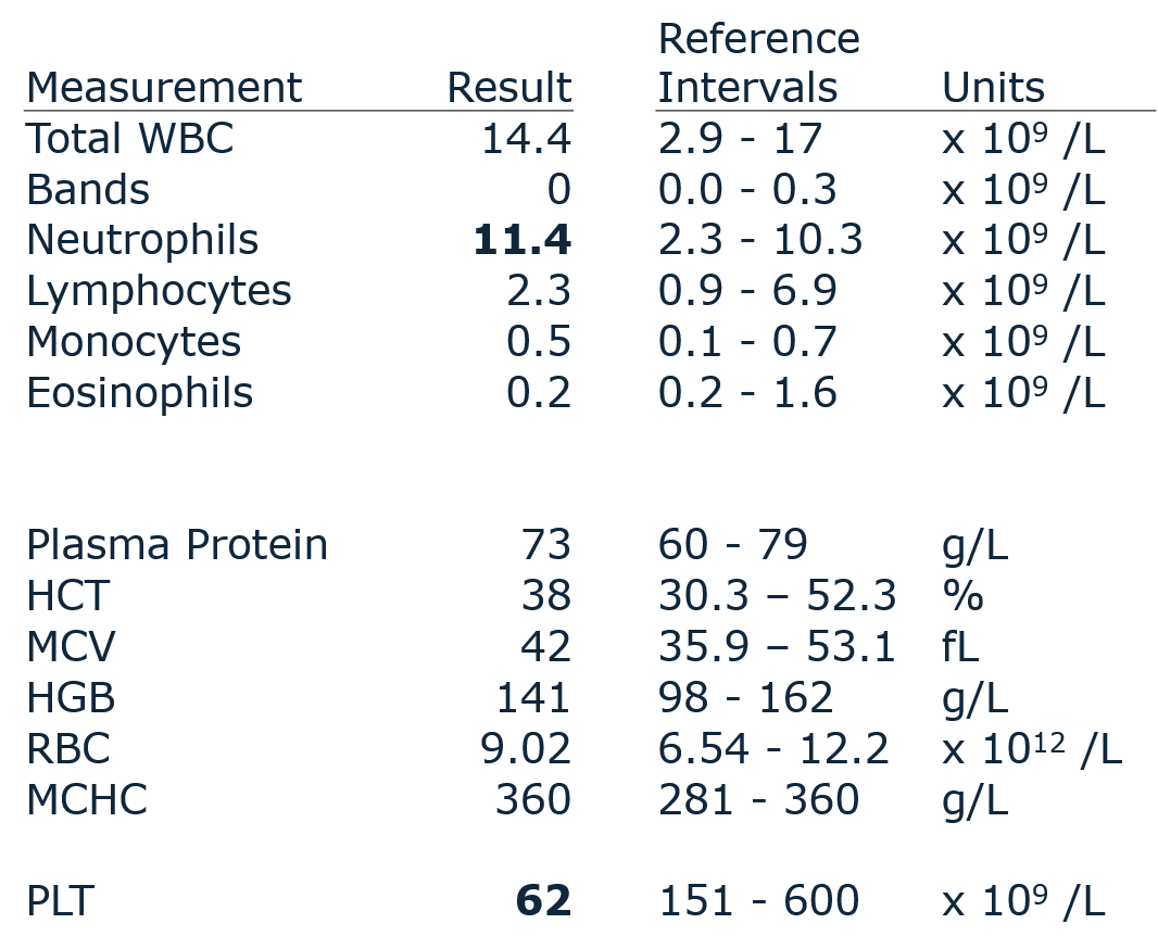
Blood smear comments:
Numerous large platelet clumps in feathered edge

There is a borderline mature neutrophilia. In the absence of any clinical disease referable to inflammation, this may be due to a physiologic leukocytosis associated with excitement. The very mild increase with the absence of left shift may suggest inflammation is less likely.
The main abnormality is a marked thrombocytopenia. This should prompt evaluation of the blood smear for platelet clumping which was confirmed in this case, but the monolayer should also be evaluated at high power for a minimum platelet count. Clumping is very common in the cat but also occurs in other species.

Blood film comments:
Polychromasia
The presence of a lymphopenia and monocytosis would support a stress (steroid) response.
Given the prominent rubricytosis (NRBCs) and polychromasia seen on the blood smear, a regenerative response would be expected. It is therefore a surprise that the animal is not anaemic but in fact has a mildly increased red cell mass.
A regenerative response in the face of a high normal or increased haematocrit suggests an oxygenation problem. Secondary polycythaemia is the likely explanation. This should be evaluated with a pulse oximeter or ideally arterial pO2 measurement. Polycythaemia vera is not expected to be associated with a regenerative response like this.

§There is a marked neutrophilia that can be interpreted as an inflammatory response. It is possible for variably chronic, well-compensated inflammatory responses to have a neutrophilia of this magnitude without a left shift.
§
§The magnitude of monocytosis would be consistent with a chronic inflammatory process.
§
§Note there is a borderline low haematocrit. This could be normal for this dog but also suggests the possibility of anaemia of inflammatory disease. A normal reticulocyte measurement (not available here) would be consistent with either of these possibilities.

The leukogram shows an inflammatory pattern, indicated by the degree of neutrophilia plus a left shift. The lymphopenia and eosinopenia suggest a superimposed stress (steroid) response.
The monocytosis could be attributed to either stress response or part of the inflammatory response. The relatively high magnitude favours inflammation as the more likely explanation.
There is prominent thrombocytosis which is difficult to explain. It can be associated with iron deficiency, but there is no indication of that in the erythron. A reactive thrombocytosis (due to inflammation) is usually mild. Thrombocytosis can occasionally be associated with neoplasia and therefore this may be a relatively rare paraneoplastic response.

Patient has presented collapsed
Case 14: 4yo FS mixed breed dog (cockapoo)
There is a mild leukocytosis due to a neutropenia. With a concurrent eosinophilia this could represent tissue demand for neutrophils in the presence of inflammation, but the primary abnormality here is the absence of a steroid (stress) response in a collapsed patient.
In a clinically unwell animal, failure to produce a stress response should prompt investigation for hypoadrenocorticism (Addison’s disease).
Most eosinophilias are mild; larger magnitudes (e.g. > 2 x10⁹L) may be seen in tissue-invading parasitism such as heartworm disease (Dirofilaria immitis), demonstrating the importance of travel history.
ferret haematology
Generally have higher HCT, HB, and RBC counts
White cell count generally quite low (3-4 x10⁹)
Ferrets appear to have only 1 blood group
describe ferret neutrophils
contain small, pale-red
granules
describe ferret eosinophils
granules rounded and bright red
rabbit haematology
Basophils are present quite commonly
Lymphocytes, eosinophils and monocytes similar to dog/cat
Typically the neutrophil to lymphocyte ratio is 1:1
(45%:45%)
The total white cell count rarely increases, instead the
neutrophil : lymphocyte ratio changes in response to stress or
disease
what is a common finding in rabbit blood
Anisocytosis and polychromasia
describe rabbit heterophils
have granular cytoplasm and may be mistaken for eosinophils
why is reptile haem usually manual
since automated techniques are made difficult due to nucleated RBCs
describe reptile heterophils
Predominant leukocyte
Equivalent to mammalian neutrophils
Intracytoplasmic fusiform granules
describe reptile azurophils
Unique to reptiles
Similar to monocytes, occur with
inflammation and granulomatous diseases
Red-purple cytoplasm
More common in snakes
why lymphopeania in reptiles
malnutrition, stress immunosuppression and poor husbandry
why lymphocytosis in reptiles
inflammation, parasitic and viral disease, wound healing, ecdysis
how can reptile eosinophils change
Eosinophils generally lower in summer
Eosinophilia → parasites and inflammation
why basophilia in reptiles
parasitic and viral disease
why monocytosis and azurophilia in reptiles
with inflammation and granulomatous diseases
how does haem cell size vary in birds
Cell size varies with species larger in ratites, smaller in passerine
what is normal in avian haem
Mild polychromasia and anisocytosis is normal
describe heterophils in birds
Function similar to mammalian neutrophil
Most abundant leukocyte in most avian species
Round cells with lobed basophilic nucleus and prominent eosinophilic needle-shaped, oval or round cytoplasmic granules
Cytoplasm is colourless
describe eosinophils in birds
Normally present in low numbers
Round cells, lobed basophilic nucleus
Cytoplasm stains clear blue
Granules brightly eosinophilic tend to be round
vs needle shaped
why heterophilia in birds
stress, infection
why heteropaenia in birds
acute or severe inflammation, sepsis, viruses
why raised eosinophils in birds
Eosinophil numbers may be elevated with tissue damage and parasitic diseases and possibly some viral infections
what birds are lymphocytic species
Amazon parrots and canaries → up top 70% lymphocytes
lymphocytosis in birds
excitement, leukaemia
why lymphopaenia in birds
chronic stress
what see in feathered edge
platelet clumps, big cells, big parasites
what see in body
rouleaux
agglutination
what see in monolayer
rbc density
wbc numbers
estimated differential