Outcome 5: Mitral Stenosis, MR, TV, Prosthetic Valves, and Corrective Procedures
 Call Kai
 Learn
 Practice Test
 Spaced Repetition
 Match
 Flashcards
 Knowt Play
1/141

Earn XP
Description and Tags
Mitral Stenosis, MR, TV, Prosthetic Valves, and Corrective Procedures
142 Terms

mitral valve structure
bicuspid valve
anterior MV leaflet (AMVL)
posterior MV leaflet (PMVL)
MV complex includes..
MV annulus
MV leaflets (commissures)
chordae tendinae
papillary muscles
opens in diastole
each MV leaflet has 3 scallops
A1-A3, P1-P3
the leaflet scallops are numbered from the lateral edge moving more medially on PSSAX view
scallop # can help discuss location of pathology
Which mitral valve scallops are typically visualized in the AP3 echocardiographic view?
A2, P2
mitral stenosis
blood flows into LV from LA is restricted
opening is smaller than normal from diff. mechanisms
in mitral or tricuspid stenosis, the respective atrium lacks the contractile strength to overcome the obstruction
» causing blood to back up and pool in the atrium
» leading to atrial dilation & inc. atrial pressure
clinical findings:
SOB
PHTN » can cause rt. HF
palpitations
atrial fibrillation
malar flush
red colour high on face/cheeks (due to high CO2 lvls)
not specific for mitral stenosis
fatigue
murmur of mitral stenosis
murmur:
low-pitched diastolic rumble with an opening ‘snap’
an added ‘tumour plop’ murmur w/ a mitral stenosis if due to lt. atrial myxoma
complications:
atrial fibrillation
LA thrombus
LA dilates as a consequence of stenosis » when chambers dilate, blood slows down & has a greater chance of clotting » can lead to development of LA thrombus
may see spontaneous echo contrast in LA
possible embolization of the thrombus
PHTN
left sided pathology causing right sided pathology
continued backup of blood into LA and pulm. veins increases pulm. vascular volume » raising pulm. pressures » causing PHTN
PHTN is a pressure overload for RV » can lead to rt. sided HF even tho mitral stenosis is a lt. sided pathology
atrial fibrillation
A condition where the left atrium dilates due to mitral stenosis, leading to irregular heart rhythms and potential complications.
left atrial thrombus
Blood pooling in the dilated left atrium can clot, potentially causing embolization to the brain or coronary arteries.
pulmonary hypertension (PHTN)
Increased pressure in pulmonary arteries due to blood backup from mitral stenosis, potentially leading to right-sided heart failure.
systemic embolization
Thrombi formed in the left atrium can travel to systemic circulation, causing strokes or other ischemic events.
right-sided heart failure
Resulting from pulmonary hypertension caused by mitral stenosis, leading to symptoms like peripheral edema and hepatic congestion.
spontaneous echo contrast
A phenomenon observed in the left atrium due to slow blood flow, indicating a risk for thrombus formation
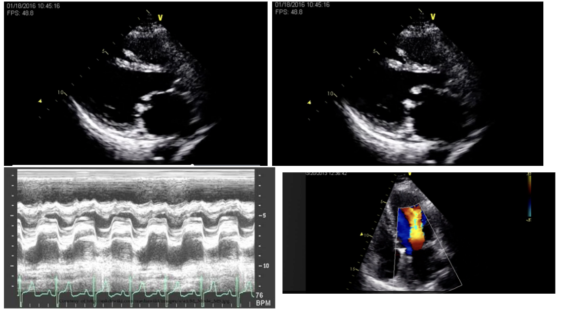
rheumatic heart disease - acquired MS
cause/etiology:
untreated strep throat infection
antibody complexes created during infection cause sclerotic and fibrotic changes to MV & damage the glomeruli in the kidneys
most common type of mitral stenosis; but has dec. in populations w/ access to antibiotic treatment
murmur:
low pitched diastolic rumble w/ opening snap
2D:
thickened and calcified leaflets of the affected valve(s)
calcified leaflets become tethered to e/o » restricting their movement & opening of the valve
subvalvular apparatus (chordinae tendinae) may also show calcific changes
anterior MV leaflet will have classic ‘hockey stick’ appearance and have doming leaflet movement upon opening
all valves can be affected
M-mode:
lines showing MV leaflet movement are thickened and have inc. echogenicity
occurs when valve is stenotic and calcified
leaflets move together in a box-like shape as the stenosis & tether of the MV leaflets worsen
colour:
accelerations and turbulence found at valve leaflets as the flow moves into LV
may or may not be MR
spectral:
inc. velocity thru MV due to calcification and stenosis
normal peak MV velo = ~1.2m/s
normal separate E and A waves fuse together & the stenotic waveform looks box-like


left atrial myxoma - acquired MS
cause/definition:
myxoma = benign tumour that can be found in all chambers of the heart, but are mostly found in the atria, esp. the LA
tumour is friable, so it needs to be surgically removed if found
how it causes stenosis:
if it grows large enough, it’ll fill LA and can fill opening of MV and obstruct blood flowing out
myxoma will move w/ blood flow
murmur:
‘tumour plop’ murmur from myxoma, followed by a low-pitched diastolic murmur of the MS
2D:
when found in atria, mass is attached by a stalk to the IAS
to confirm myxoma, echo images need to show its attachment to IAS
M-mode:
if it’s large enough to extend into opening of MV leaflets, it’ll be seen on MV M-mode
colour and spectral:
accelerated colour flow seen moving around myxoma in MV opening
CW used to measure high-velo flow if the stenosis created by the myxoma obstructing MV opening
waveform becomes more box-like as severity inc.
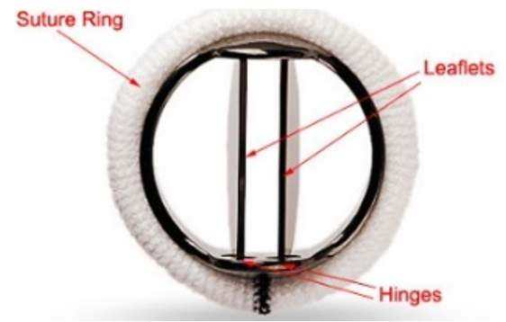
mechanical prosthetic mitral valves - acquired MS
2 types of prosthetic valves:
bioprosthetic and mechanical
all mechanical prosthetic valves will have some degree of stenosis
these valves have man-made leaflets that aren’t able to open as fully as a normal native leaflet » so the velo. thru these valves is higher than normal
all mechanical prosthetic replacement valves will have a range of flow velocities identified as normal for them
‘true’ stenosis of the prosthetic valves develops when the leaflets are blocked by pannus or vegetations, or bioprosthetic valve leaflets become calcified and resultant velo, thru valve is greater than normal established range

mitral annular calcification (MAC)
progressive, degenerative, chronic process in which calcifications is localized on MV annulus
usually occurs on posterior annulus first, as a bright echogenic mass
if anterior annulus is calcified too, rheumatic heart disease may be involved
many risk factors for development such as..
advanced age
HTN
renal disease
diabetes mellitus
marfan’s sydrome
HCM
severity best identified from PSSAX at MV
thickness of MAC is measured or estimated by looking at how much of the posterior MV annulus is covered w/ calcification
—
considered a cause of MS bcuz the calcification may extend onto leaflets » leading to restricted leaflet mobility & impaired opening
if MAC is severe enough to cause MS, accelerated colour flow and inc. velocities thru MV leaflets results
renal disease - acquired MS
multiple factors can cause sclerotic changes to MV, including..
uremia and chronic inflammation
usually the MV annulus and subvalvular apparatus are predominantly affected, rather than the leaflets

subvalvular mitral stenosis - congenital MS
cor triatriatum sinister (top image):
congenital malformation that has a membrane in the LA that blocks the flow from pulm. veins to MV
may be a single opening in the membrane to allow blood thru, or it may be fenestrated
opening is usually smaller than MV opening, so it’s considered a type of stenosis
echo images must show..
membrane on 2D, locations of colour doppler flow thru membrane, and measure highest flow velo. w/ CW to determine severity
may be assoc. w/ other congenital defects
ie. ASD, pulm. vein anomalies
cor triatriatum dexter = membrane in AR
supramitral ring (bottom image):
membrane that’s close to the MV and MV annulus
found w/ complex congenital heart disease malformations
may interfere w/ the opening of the valve leaflets

valvular (congenital) mitral stenosis
hypoplasia or atresia of MV
if there’s atresia, the valve hasn’t formed and there won’t be flow thru it
usually found in hypoplastic left heart syndrome (HLHS) and other complex heart malformations

parachute mitral valve - congenital MS
all the chordae of both leaflets of the MV are attached to one pap. muscle
can cause mitral stenosis and/or MR
diagnostic image will show the single pap. muscle
best image would be in PSSAX at Paps level
there may be smaller tissue that looks like a very small pap. muscle but there won’t be any chordae attached to it
a small posterior angulation of the probe in AP4 will also show solitary pap. muscle
colour and spectral for congenital malformations causing mitral stenosis
acc. colour flow thru area os stenosis
either thru MV leaflets or thru openings in a left atrial membrane
CW measures high-velocity flow caused by the stenosis
waveform becomes more box-like as severity inc.
2D imaging in mitral stenosis
for all types of stenosis:
LA dilation
LV size is small w/ normal wall thickness and systolic function
LV diastolic function will not be measurable w/ conventional parameters since MV inflow is abnormal due to stenosis
MV annular movement may also be abnormal if rheumatic or calcified MS is present w/ calcifications extending onto annulus

M-mode imaging in mitral stenosis
mitral valve:
look at rheumatic MV and MS due to myxoma
pulmonary valve:
PHTN will develop as severity inc.
absent A dip
‘flying W’ sign
colour and spectral doppler in mitral stenosis
colour:
turbulent flow across stenotic region
spectral:
E and A waves may fuse and inc. in velocity as severity inc.
box-like waveform w/ flatter slopes
evaluation focuses on the mean transmitral gradient rather than peak velocity or gradient
quantification of mitral stenosis severity
calculation of the mitral valve area (MVA) is the preferred assessment of MS severity
3 methods:
planimetry, PHT, continuity equation
a larger MVA indicated a wider valve opening that allows more blood flow, whereas smaller MVA restricts flow
MVA <2cm2 in considered stenotic
—
reference ranges for MVA:
mild MS » 1.5-2.0/2.5cm2
moderate MS » 1.0-1.5cm2
severe MS = <1.0cm2
(assuming a normal HR of 60-80bpm)

planimetry of MV area
can be calculated on 2D and 3D imaging

pressure ½ time (PHT) and deceleration time (DT) method
CW of MV inflow
calipers are placed along the slope of the E wave
E and A waves fuse as severity inc. and waveforms become box-like
decel. time is measured from this waveform & PHT is calculated from decel time.
PHT is the time it takes for the pressure to become one-half
PHT = 0.29(DT)
MVA (cm2) = 759/DT
MVA (cm2) = 220/PHT
a longer DT and PHT correspond to a more stenotic valve
factors that affect the accuracy of the PHT measurement
Ao. regurg.
MVA is overestimated and stenosis is underestimated
dec. LV compliance (LV diastolic dysfunction)
LVA is underestimated and stenosis is overestimated
ASD present (Lutembacher syndrome)
LVa is overestimated and stenosis is underestimated
atrial fibrillation
multiple waveforms need to be measured for better accuracy
‘2-step’ slope of E wave
sometimes the slope of E-wave dec. w/ 2 diff. slopes to follow
follow the mid-slope w/ PHT measurement
poster-percutaneous balloon mitral valvuloplasty (PBMV)
a corrective procedure for MS
first 1-3 days post-op, the PHT MVA calculations aren’t accurate
continuity equation for MV area
MVA = 0.785 (LVOT diam)2 x VTILVOT / VTIMV
—
required measurements and images needed:
LVOT diameter (zoomed, PSLAX)
LVOT VTI (PW, AP5, AP3)
mitral inflow VTI (CW, AP4 » avg. of 3-5 beats)
—
factors affecting accuracy:
Ao regug
MVA = overestimated, stenosis = underestimated
mitral regurg.
MVA = underestimated, stenosis = overestimated
atrial fibrillation
multiple waveforms need to be measured (5-10 beats)
accuracy of LVOT diameter measurement
heart rate (tachycardia)
less filling time of the ventricle w/ tachycardia » meaning smaller VTI of MV
MVA = overestimated, stenosis = underestimated
supportive findings of mitral stenosis severity
mean MV pressure gradient:
mean PG of MV inflow is a better indicator if severity than peak gradient across the mV
as the peak PG is reflective of LV dia. func. and LA compliance, rather than degree of stenosis
mean MV PG found on a VTI trace on CW waveform » same as the waveform used for measuring PHT
HR affects mean PG value
faster HR = larger mean gradient = greater stenosis reported
FACTORS AFFECTING..
flow dependence
high CO states and MR will inc. transmitral velocity and PGs » overestimating stenosis
dec. diastlic filling time (tachycardia) can inc. gradient
atrial fibrillation
REFERENCE RANGES..
mild MS = <5mmHg
moderate MS = 5-9mmHg
severe MS = >10mmHg
severity of PHTN:
RVSP= 4VTR2 + RAP
evidence of PHTN is also found when..
measuring RVOT acc. time on PW
flying W sign and absent a-dip on pulm. valve M-mode
REFERENCE RANGES..
mild MS = <30mmHg
moderate MS = 30-49mmHg
severe MS = >50mmHg
treatment of mitral stenosis
monitoring of the pt:
pt. will have a follow-up echocardiography testing ever 6 months or yearly, reviewing severity of stenosis to determine if surgical intervention is needed
balloon valvuloplasty:
balloon at the end of a catheter is advanced b/w the MV leaflets and is expanded
the calcification that tethers the leaflets together is broken & some mobility of the leaflets is regained
procedure may result in an unacceptable amt. of MR
prosthetic mitral valve replacement:
can be tissue or mechanical
further testing:
stress echocardiography may help identify pts. who require more immediate intervention
TEE and 3D echo
cardiac catheterization for LA pressures
mitral regurgitation (MR)
results from the MV leaflets not sealing together tightly or if there are perforations (‘holes’) in the valve leaflets that allow the blood to flow back into the atrium in systole
creates a LVVO
acute onset:
normal sized LA
LA doesn’t have time to dilate in response to the extra volume
pts. may rapidly develop pulm. edema and hypotension » making this a critical finding that requires prompt recognition & immediate communication w/ reading physician
in contrast, severe MR discovered in a stable pt. isn’t typically urgent
clinically, acute MR often present w/ a new murmur and sudden SOB
mechanisms..
endocarditis, ruptured pap. muscle, direct trauma, iatrogenic causes
chronic onset:
occurs slowly over time
LA dilates
over time & progression, the LV will dilate and develop a more spherical shape
LV systolic function may decline w/ chronic severe MR
pts. may experience general weakness, fatigue, and dec. exercise tolerance
backup of blood into the pulm. circulation causes PHTN when MR is severe
PHTN is a RVPO > will result in rt-sided heart pathology
MR can be a mechanical complication of a myocardial infarction » esp. if the damaged LV wall has a pap. muscle on it
mechanism..
structural defects of the leaflets, masses, annular dilatation, myocardial infarction
clinical findings with mitral regurgitation (MR)
chronic:
signs and symptoms generally become more noticeable as the severity increases
so mild/moderate MR ay be asympt. or cause few symptoms
severe MR leads to more obvious findings such as..
loud holosystolic murmur
chamber enlargement
possible HF symptoms
acute:
heart has no time to compensate, so even a moderate sudden leak can produce significant clinical symptoms like..
pulm. edema
hypotension
respiratory distress
characteristic MR murmur:
high-pitched, blowing, holosystolic murmur at the apex that then radiates to the axilla
acquired causes of mitral regurgitation (MR)
mitral annular dilation
myxomatous changes to the MV
MV prolapse
endocarditis (infective or non-infective)
flail MV leaflet
papillary fibroelastoma
rupture of the papillary muscle or chordae
systemic diseases
rheumatic heart disease and/or sclerosis
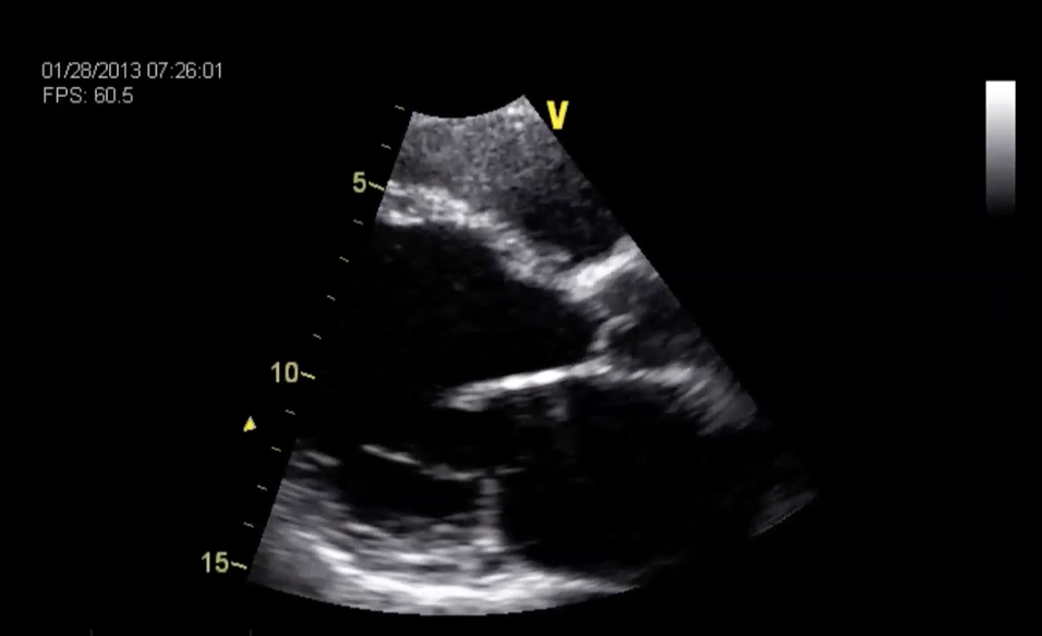
mitral annular dilation - acquired MR cause
a dilated LV will cause the MV annulus to dilate, and the leaflets are unable to coapt tightly
resulting in MR
results in a central MR jet » leaflets are equally pulled apart
examples of conditions that result in a dilated LV (and MV annulus) and MR:
post-myocardial infarction and wall damage
dilated cardiomyopathy
severe aortic regurgitation

myxomatous changes to the MV - acquired MR causes
leaflets are irregularly thickened w/ excessive valve tissue
leaflets w/ myxomatous degeneration may or may not progress further to a MV prolapse or flail leaflet
there’s excessive leaflet motion w/ this valve structure
MR is common w/ this type of valve structure, but may or may not be present
Barlow’s disease » a degenerative MV disease
the MV leaflets are myxomatous and prone to develop prolapse
the chordae are also thickened and elongated
age-related degenerative processes may also be a cause of myxomatous changes to the MV

mitral valve prolapse (MVP) - acquired MR causes
2D:
the ‘belly’ of the leaflet buckles or bows back into LA
tips of leaflets remain pointing towards the LV like they normally do, into the LV
lowest point of the ‘belly’ of the leaflet must be at least 2mm behind the visualized MR annular line
can only be diagnosed on PSLAX or AP3 views
M-mode:
will show MV prolapse w/ a ‘dipping down’ of the closure line in end-systole
colour:
colour can help determine if one or both leaflets are prolapsing from the direction of the colour jet
PMVL prolapse » jet directed anteriorly
AMVL prolapse » jet directed posteriorly
both MV leaflets » central jet
characteristic murmur of MV prolapse:
mid or late systolic click that can be augmented w/ the Valsalva
if there’s MR present along w/ prolapse, both the murmur of the prolapse and MR will be heard
—
pts. w/ MVP can be asymptomatic, or they can experience palpitations or chest pain
MVP found in pts. w/ connective tissue disorders (Marfan’s syndrome) presents w/ a long, redundant AMVL w/ prolapse
can be associated w/ secundum atrial septal defects
endocarditis (infective or non-infective) - acquired MR cause
characterized by the presence of vegetations on the leaflets
vegetations are a collection of platelets, fibrin, and inflammatory cells; they may contain microorganisms (infective) or may not (non-infective)
vegetations will always attach to the atrial surface of the atrioventricular valves (MV, TV) and the ventricular surface of the semilunar valves (AV, PV)
vegetations can cause regurg. of the affected valve bcuz they interfere w/ valve closure, or cause leaflet perforation or annular ring damage
vegetations can be very small and may be missed by transthoracic echo
a transesophageal echo is ordered if endocarditis is suspected and not found on TTE
TREATMENTS INCLUDE..
anticoagulants, antibiotics, or a valve replacement
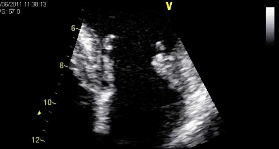
flail mitral valve leaflet - acquired MR cause
part of the mitral apparatus structure is weakened or compromised
ie. ruptured chordae can’t support the leaflet anymore, or a perforation/’hole’ in leaflet, etc.
leaflet tip of the flail leaflet will drop down into the LA and won’t remain pointing to LV
MR will always be present if there’s a flail MV leaflet
mechanisms..
infective endocarditis, trauma, ruptured pap. muscle
murmur:
doesn’t have a characteristic murmur like a prolapsing leaflet has; the murmur heard is just the resultant MR
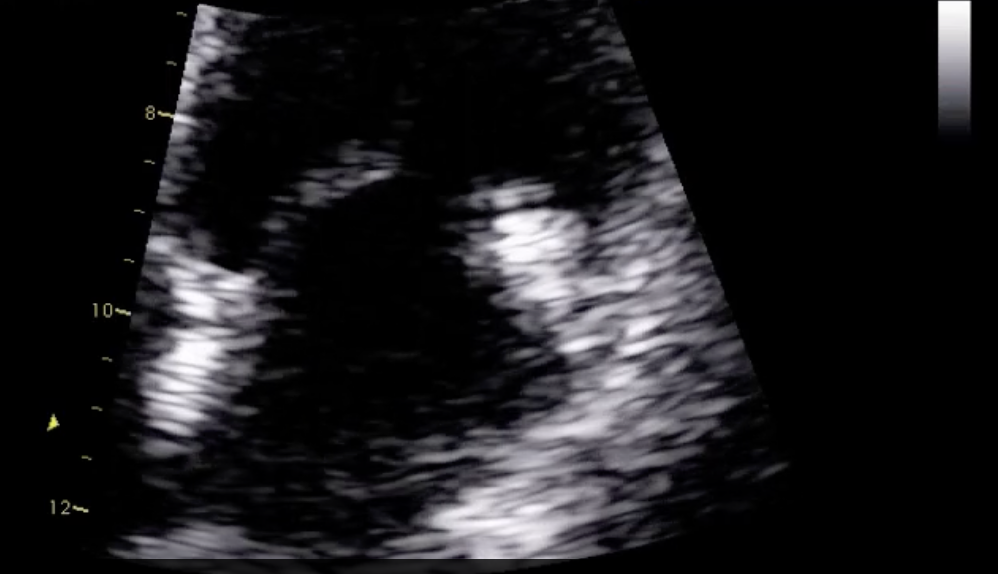
papillary fibroelastoma - acquired MR cause
usually found on AV leaflets but can also be a MV mass
pom-pom shaped mass attached w. a stalk (which can be difficult to see)

rupture of the papillary muscle or chordae - acquired MR cause
the pap. muscle breaks away from the LV wall
acute onset of MR
more severe MR if the pap. muscle ruptures « very few survive
‘mass’ at the end of the leaflet is the portion of the pap. muscle that has broken away from the LV wall
mechanisms..
infective endocarditis, trauma, myocardial infarction affecting the pap. muscle blood supply
systemic diseases - acquired MR cause
Marfan’s syndrome
MVP and +/- MR
presents w/ a long, redundant AMVL w/ prolapse
Ehler’s Danlos
MVP and +/- MR
Loey’s-Dietz
MVP and +/- MR
Ankylosing spondylitis
+/- MR
Rheumatoid arthritis
non-infective endocarditis nodules (+/- MR)
SLE (lupus)
non-infective endocarditis nodules (+/- MR)

rheumatic heart disease and/or sclerosis - acquired MR cause
calcification of the MV is usually caused by another disease process (rheumatic heart disease, renal disease)
calcification/sclerosis of the leaflets may prevent the leaflets from sealing tightly resulting in regurg.
calcification of the valve in rheumatic heart disease will cause stenosis of the leaflets, but can also cause the leaflets to seal poorly together and result in MR
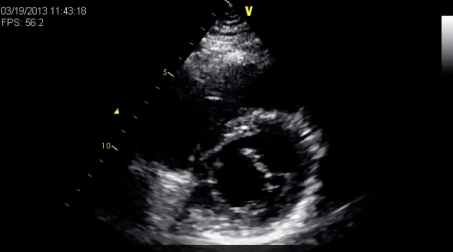
cleft mitral valve - congenital MR cause
there’ll always be MR w/ a cleft MV
MV leaflet has a break within it where it’s not continuous
it’s a component of a partial atrioventricular canal defect malformation
diagnostic image for anterior MV clefts is the PSSAX image of MV, but clefts in the posterior MV leaflet are difficult to see on TTE
on PSSAX, the AMVL should appear as a continuous line in a normal valve
w/ cleft valve, this continuity is disrupted

parachute mitral valve - congenital MR cause
this pathology can also have MR as well as or instead of mitral stenosis
PSSAX, at the paps is confirmation of the parachute MV only having a solitary pap. muscle
Which of the following statements about endocarditis and its relationship to mitral regurgitation are correct? Select all that apply.
Mitral regurgitation can result from leaflet perforation caused by endocarditis.
Vegetations attach to the atrial side of the mitral valve leaflets.
complications of mitral regurgitation (MR)
atrial fibrillation:
atrial dilation can lead to A. fib
the resulting ineffective atrial contraction and impaired emptying further enlarge the LA
left atrial thrombus/clot:
as chambers dilate, the risk of clot development inc. since the blood moves more slowly thru the larger chamber
potentially leading to complications like embolism or stroke
pulmonary congestion/hypertension:
the LA dilates to accommodate the regurgitant blood, but can only stretch so much
excess blood volume backs up in the pulm. circulation and can cause pulm. congestion and PHTN
treatment of mitral regurgitation (MR)
medical management (medications)
MV repair (native leaflets remain)
MV replacement (bioprosthetic or mechanical prosthetic valve)
MV clip/MV stitch
a clip to hold AMVL and PMVL tips tgt. of the native MV leaflets
annuloplasty (supportive ring placed in MV annulus)
echo findings of mitral regurgitation (MR) severity
2D:
investigate if there’s a structural reason for it
investigate cause
colour:
identifies the location of regurg. jet
determines if the regurg. is occurring along the coaptation lines of the leaflets (valvular MR), if it’s in the center of a leaflet due to perforation, or if it’s coming from the suture line of a prosthetic valve (paravalvular MR)
initial assessment for MR severity is determined by colour doppler clip
PISA radius (amt. of prox. flow), persistence of regurg. flow, and eccentricity of the jet

central MR colour jet
central MR colour jet
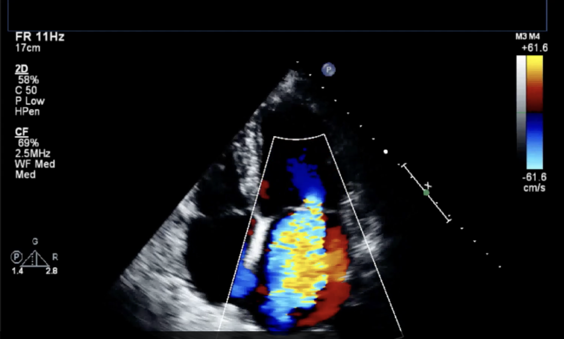

eccentric MR colour jet - anteriorly directed
eccentric MR colour jet - anteriorly directed


eccentric MR colour jet - posteriorly directed
eccentric MR colour jet - posteriorly directed


PISA, EROA, Regurgitant Volume - assessment of MR severity
proximal isovelocity surface area (PISA):
MR: after MV closure, LV pressure exceeds LA pressure » resulting in blood flowing back into the LA thru any area where the mitral leaflets fail to coapt properly
as blood approaches the regurg. orifice (opening b/w leaflets) on the ventricular side, its velo. increases, forming a flow convergence zone
this flow convergence zone can be visualized using color
flow convergence zone..
composed of multiple hemispheric shells of equal velocity (isovelocity surfaces)
convergence zone appears as a rounded dome of colour on the ventricular side of the valve, opp. direction of the regurg. jet » PISA dome or PISA ‘bubble’
height of this dome » known as the PISA radius » used to estimate severity
larger radius = more significant regurg.
reference ranges..
mild MR = <0.4cm
moderate MR = 0.4-0.89
severe MR = >/=0.9cm
effective regurgitant orifice area (EROA):
EROA (cm²) = 2 π r² VN ÷ VMR
reference ranges..
normal = <10mm2
severe = >35mm2
regurgitant volume (RV):
Regurgitant volume (RV) (mL) = EROA (cm²) x VTIMR (cm)


vena contracta - assessment of MR severity
vena contracta = narrowest part (the “waist”) of the regurg. jet
measurement done at the valve leaflets b/w prox. flow in the LV and the regurg. flow in LA
should be done w/ zoomed view of MV w/ colour
reference ranges:
mild MR = <0.3cm
moderate MR = 0.3-0.69cm
severe MR = >0.7cm

intensity and shape of MR waveform - assessment of MR severity
echogenicity/intensity of waveform:
inc. echogenicity » suggests more significant MR volume
intensity of the waveform sometimes may not reflect the true severity of the regurg. jet if the jet is eccentric and the sample line can’t align well w/ the flow
shape:
V-wave cut-off sign (or V-cut-off sign)
term used when the waveform of MR is V-shaped rather than parabolic shaped » suggests more severe MR
reference ranges:
mild MR = faint incomplete waveform
moderate MR = complete waveform w/ a parabolic shape
severe MR = densely echogenic waveform w/ a triangular shape
—
peak velocity DOES NOT reflect severity or amt. of MR
the peak MR PG reflects the pressure diff. b/w LV and LA during systole

flow reversal in the pulmonary veins - assessment of MR severity
PW doppler
w/ moderate MR, PW of pulm. veins will show blunting of systolic forward flow
if regurg. is severe, there will be flow reversal in systole
S wave on the pulm. vein can have a lower velocity than the D wave in young, super-normal individuals, but its presence in conjunction w/ significant MR supports its severity
pulmonary vein flow w/ MR:
mild MR = normal waveform
moderate MR = systolic blunting
severe MR = reversal in systole
regurgitant volume and regurgitant fraction - assessment of MR severity
regurgitant volume:
RVMR = 0.785(MV annulus diameter)2 x VTI(MV annulus) - 0.785(LVOT diameter)2 x VTI(LVOT)
regurgitant fraction:
RFMR (%) = RV(MV annulus) / SV(MV annulus) x 100
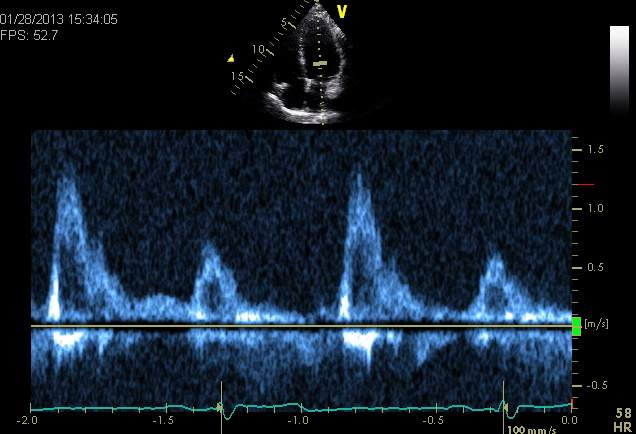
increased mitral inflow velocity - assessment of MR severity
w/ significant MR present, the inc. preload in the LA means there’s more blood available to go thru the orifice in diastole
results in an inc. in the velo. of the mitral inflow (E)
reference ranges:
mild MR = MV inflow velocities still normal
significant MR = inc. MV flow velocities (E >1.5m/s)
MR jet area trace - assessment of MR severity
area of the MR colour jet is traced and compared to the area trace of the LA
bigger area suggests larger jet
this is an old method of assessment of MR
no longer recommended by ASE guidelines
Which of the following is the equation used to determine the pressure gradient across a stenosis?
Bernoulli
What will torn chordae tendinae cause?
Mitral insufficiency
The posterior wall of the aorta is anatomically continuous with which of the following structures?
AMVL
Which of the following is the term to describe a dilated IVC that shows less than a 50% reduction in size with inspiration?
IVC plethora
The primary pacemaker in the heart is:
Sino-atrial node
Which valve is the most inferiorly located in the heart?
TV
A valve can be sclerotic and not be stenotic.
true
Which of the following would NOT be a possible cause of mitral annular calcification?
Scleroderma
Which of the following would be the most common symptoms of significant mitral stenosis?
SOB
Which of the following diseases is associated with mitral annular calcification? Choose all that are correct.
renal disease
HCM
HTN
What are the measurements needed for calculating the ERO area using PISA?
MR velocity, PISA radius, aliasing velocity
Which of the following would help a sonographer identify that there is severe mitral regurgitation present when the colour Doppler images are difficult to assess?
PW Doppler of the pulmonary veins
Which of the following is NOT a cause of an increased EPSS?
Aortic stenosis
The CW spectral Doppler of the MR is used to obtain measurements for the PISA calculation. Which of the following also uses measurements from the CW Doppler of the MR?
dP/dT

What does systolic bowing of the mitral valve leaflets suggest?
prolapse
Which of the following should only be measured using a CW spectral Doppler image of the mitral valve inflow? (may be multiple answers)
Maximum mean gradient mitral valve (mmHg)
Maximum mitral valve VTI trace (cm)
Mitral valve pressure half-time (ms)
Which of the following conditions has only one papillary muscle in the left ventricle for the atrioventricular valve chordae to attach to?
Parachute mitral valve
Mr.Smith's echo demonstrated an enlarged left atrium with systolic flow reversal into his pulmonary veins. What does Mr.Smith have?
Severe mitral regurgitation
The left ventricle size is enlarged when mitral stenosis is present.
false
What is Lutembacher syndrome?
Mitral stenosis + ASD

What is identified in this AP5 image with colour Doppler?
Mitral regurgitation and LVOT obstruction.
What is the most common etiology of mitral valve stenosis?
Rheumatic fever
A patient has bacterial endocarditis of the mitral valve. What disease process may this lead to?
Flail mitral valve leaflet
What is the cut-off value used to determine if the displacement of the mitral valve leaflet past the annular line fulfills the requirement to be diagnosed as a mitral valve prolapse?
>2mm
What is the most common etiology of mitral valve stenosis?
Rheumatic fever
Which of the following would support the value of moderate mitral regurgitation?
PISA radius = 0.6
Which of the following is a commonly seen arrhythmia with mitral stenosis?
Atrial fibrillation
All EXCEPT which of the following methods can be used to calculate the mitral valve area?
Vena contracta
When performing planimetry on the mitral valve orifice to calculate the valve area, the measurement is done following the outer leaflet surface.
false
Why is it important to determine the right ventricular systolic pressure in patients that have mitral stenosis?
The amount of pulmonary hypertension present in a patient helps determine the severity of mitral stenosis the patient has.
What images are needed to calculate the MVA using the continuity equation?
LVOT diameter
LVOT VTI
MV VTI
Explain the difference between mitral valve prolapse (MVP) and flail mitral valve. Explain the echo appearance of both and explain how to differentiate one from the other.
MV Prolapse – Only the middle of the leaflet bows into the left atrium. The MV leaflet tips remain pointed toward LV in systole.
Flail – MV leaflets and tips move/ point into LA in systole and LV in diastole
List the different methods to calculate the mitral valve area. List all that we have discussed in class for full marks.
Continuous area trace (planimetry)
PHT method
continuity equation
What is the purpose of a baseline study postoperative valve surgery? (List 2)
a. It can help identify the deterioration of the valve. Velocities will increase as the valve becomes impaired and you need to know what the 'normal' velocity was at the beginning, right after implantation.
b. It will also identify patients with patient-prosthesis mismatch. The velocities will be high right away. It isn't the deterioration of the valve, it is that the valve implanted into the patient is to small for them.
What is evaluated in a mitral valve score index?
When is it used?
Assess the Thickening + calcification of the leaflets, thickening of the subvalvular apparatus, leaflet mobility;
Used to assess patient's suitability for a Balloon valvuloplasty
Define the term dehiscence, as it means in echocardiography?
The prosthetic valve is seen 'rocking' (moving back and forth as the blood passes through it)
The stability of the valve is compromised

tricuspid valve: anatomy
atrioventricular valve
opens in diastole
opens before MV
velocities lower than MV due to the rt-sided pressures being lower than the lt. chambers
composed of..
3 leaflets
anterior TV leaflet (ATVL)
posterior TV leaflet (PTVL)
septal (medial) TV leaflet (STVL)

tricuspid stenosis (TS)
a restriction of the blood moving from the RA into the RV
RA dilates and blood backs up into the systemic veins (IVC, SVC) if it’s significant enough
extremely rare in adults
clinical findings:
hepatic congestion
ascites
leg edema
dilated jugular veins » Kussmaul’s sign
murmur:
mid-diastolic rumble (w/ a soft opening snap, for TS due to rheumatic heart disease)
can become louder w/ inspiration, amyl nitrate, or exercise
treatment:
depends on cause of stenosis
prosthetic valve replacement, excision of membrane or mass, and anticoagulants » possible treatments
causes of triscuspid stenosis
rheumatic heart disease
carcinoid heart disease
cor triatriatum dexter
large masses in the RA
rheumatic heart disease - cause of TS
uncommon for the TV to be affected
fibrosis and calcification occurs first on MV, then AV
calcification of TV leaflets, chordae, and annulus is due to damage from circulating immune complexes from a Streptococcus infection
may also result from TR, as well as the TS
may need a prosthetic valve replacement

carcinoid heart disease - cause of TS
a primary tumour located in the GI tract (appendix or ileum » most common), lung, biliary tract, pancreas, testis, or ovary, which secretes substances that are high in serotonin
circulating substances form plaque deposits on the TV and PV leaflets » resulting in fibrosis and retraction of the leaflets
valves affected may become stenotic and develop significant regurg. due to retraction of the leaflets
substances are inactivated in the pulm. circulation » so lt-sided heart valves are spares
they could be affected if there’s a rt-to-lt shunt present tho
may need prosthetic valve replacement
uncommon

cor triatriatum dexter - cause of TS
congenital malformation where there’s a membrane in the RA w/ an opening that restricts the blood from flowing to the TV and RV » causing tricuspid stenosis
membrane may have a single opening or be fenestrated
similar pathology to cor triatriatum sinister in the LA » causing mitral stenosis

large masses in the right atrium - causes of TS
the masses need to be large in order to obstruct the blood flowing out of the atrium into the ventricle
small tumours, vegetations, or thrombi don’t cause tricuspid stenosis
right atrial tumours:
myxoma in RA
extension of renal cell carcinoma
tricuspid valve vegetation:
endocarditis (infective) lesions on the TV that are large and obstructing the flow thru TV
right atrial thrombus