8 - Communication, Integration & Homeostasis
 Call Kai
 Learn
 Practice Test
 Spaced Repetition
 Match
 Flashcards
 Knowt Play
1/42

There's no tags or description
Looks like no tags are added yet.
43 Terms
Main Patterns of Physiological Communication (3)
Simple diffusion across small distances
Widespread distribution of molecules through the circulatory system
Rapid, specific delivery of messages by the nervous system
Main types of Physiological Communication (2)
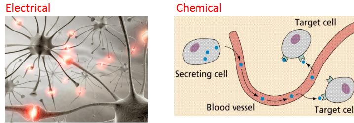
Electrical (causes change in cells’ membrane potential)
Chemical ( molecules secreted into the ECF)
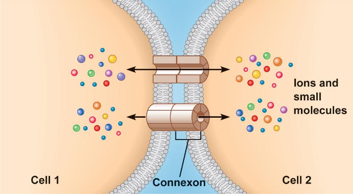
Gap Junctions (Cell-cell communication)
Direct cytoplasmic transfer of electrical/chemical signals between adjacent cells
Protein channel creates bridge between adjacent cells (forms from the union of connexins on two adjacent cells - membrane-spanning proteins)
Allows transfer of amino acids, ATP, etc.
Electrical signals can pass directly cell to cell
Channels can open or close
Multiple open channels can allow several cells to act together as one multi-nucleated cell (know as syncytium)
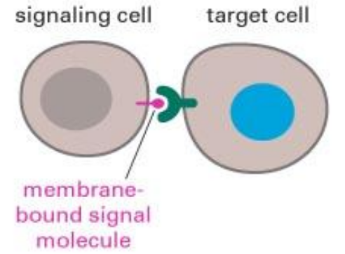
Contact Dependent Signals (cell-to-cell communication)
Surface molecules on one cell’s membrane binds to the surface molecules on another cell’s membrane
Cell adhesion molecules (CAMs) act as receptors in cell-cell signalling
Linked to the cytoskeleton or intracellular enzymes
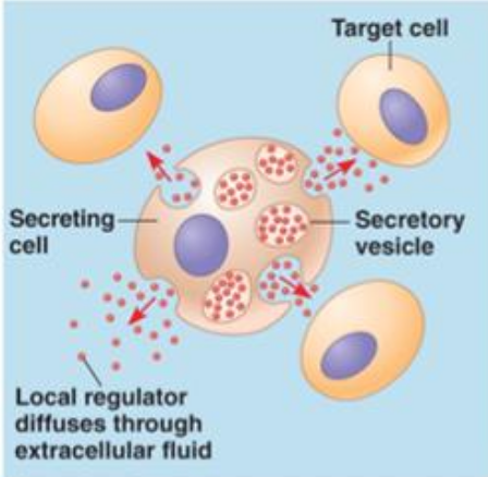
Secretory signals (cell-to-cell communication)
Cell releases chemicals that diffuse through ECF and act on those close by (paracrine signalling) or on cell that released them (autocrine)
NOT HORMONES, just chemicals being secreted to neighbouring cells
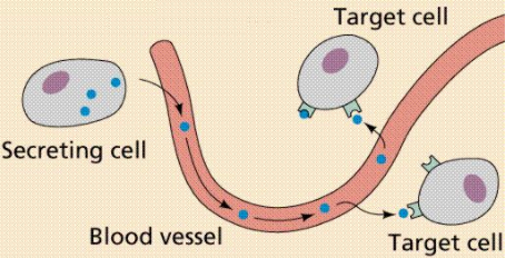
Long Distance Communication (cell-to-cell communication)
Involves nerves and/or blood for transportation
Hormones: chemical signals that are secreted into the blood & distributed all over the body via circulation (blood vessels)
Neurocrine System: the nervous system uses a combination of chemical signals & electrical signals to communicate over long distances.
An electrical signal travels along a nerve cell until it reaches the very end of the cell, where it is translated into a chemical signal secreted by the neuron
Cytokines
Associated primarily w/ immune responses, such as inflammation, but also control cell development & cell differentation.
In development and differentiation, cytokines usually function as autocrine or paracrine signals
In stress & inflammation, some cytokines may act on relatively distant targets & may be transported through the circulation just as hormones are
Cytokines vs Hormones
Cytokines:
any nucleated cell can secrete cytokines at some point in its life span
made on demand
Intracellular signal pathways usually different
Hormones:
produced by specialized endocrine glands/epithelial cells
made in advanced & stored in the endocrine cell until needed
Receptor Proteins
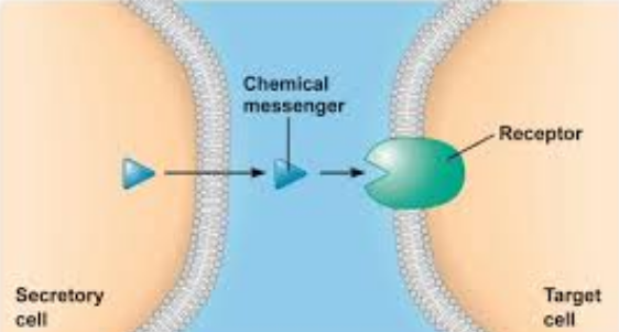
A cell can respond to a particular chemical signal only if the cell has the appropriate receptor protein to bind that signal
If a target cell has the receptor for a signal molecule, binding of the signal molecule to the receptor protein initiates a response
Signal Pathway Steps (5)
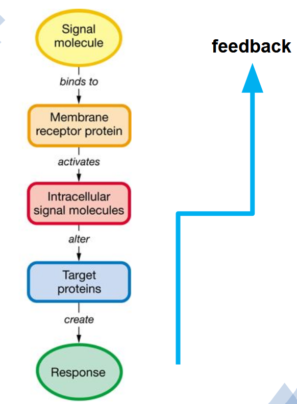
Signal molecule (ligand) binds to a protein receptor
Activation of receptor by ligand
Receptor activates one or more intracellular signal molecules
last molecule in the pathway initiates synthesis of target proteins or modifies existing target protein for response
Negative feedback
Target cell receptor proteins may be found in the nucleus, the cytosol, or on the cell membrane as integral proteins
Lipophobic Signal (signal receptor)
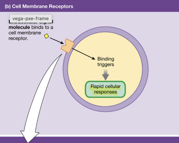
Bind to the surface membrane receptors and produce effects through a second-messenger system/cascade
Hates lipids, likes H2O (doesn’t want to bypass the lipid bilayer)
Last messenger of cascade acts on intracellular targets to create response
Response can include changes to/creation of:
an enzyme
muscle contraction
cytoskeletal movement
change in protein synthesis
alteration of membrane transport
Rapid cellular responses
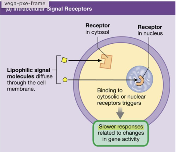
Lipophilic Signal (signal receptor)
Ligand diffuses through lipid membrane to bind with intracellular receptors
Likes lipids (wants to bypass the lipid bilayer
Binds to receptor in the cell or nucleus
Activates a specific gene that contains code for synthesizing a particular enzyme
Transcription to mRNA, which leaves nucleus & enters cytoplasm
Binds with ribosomes to direct synthesis of new proteins (& ultimately physiological response)
Slower responses related to changes in gene activity
4 Major Lipophobic Signal Receptor Types
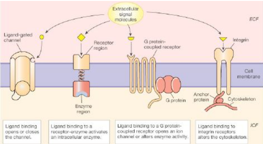
Receptor channels: chemically gated channels that open when the receptor binds
Receptor enzymes
G protein coupled receptors
Integrin Receptors
Last 3: signal is passed across membrane to initiate intracellular response (like a telephone call)
Receptor Channels (Lipophobic receptor types)
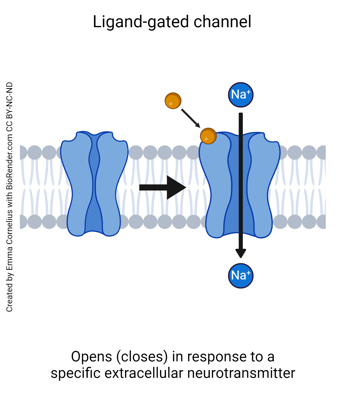
The activation of receptor-channels initiates the most rapid intracellular responses of a receptors
When an extracellular ligand binds to the receptor-channel protein, a channel gate opens or closes, altering the cell’s permeability to an ion
Increasing or decreasing ion permeability rapidly changes the cell’s membrane potential, creating an electrical signal that alters voltage-sensitive proteins
Receptor Enzymes (Lipophobic receptor types)
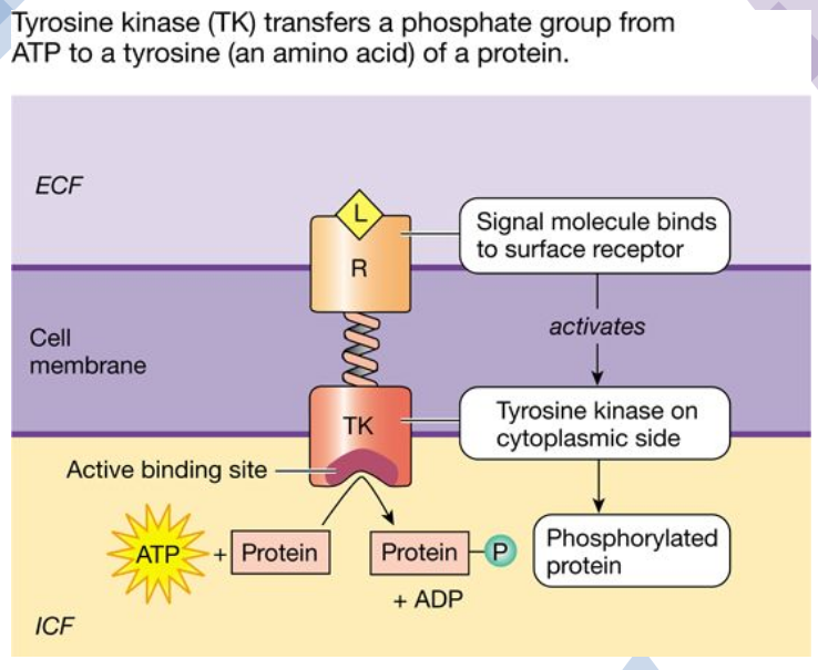
Catalytic receptors that have 2 regions:
A receptor region on the extracellular side of the cell membrane
An enzyme region on the cytoplasmic side
Ligand binding to the receptor activates the intracellular enzyme
Catalytic receptor enzymes include protein kinases
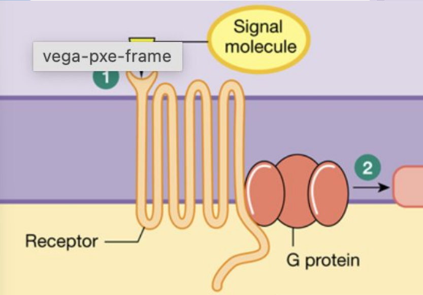
G-Protein Coupled Receptors (Lipophobic receptor types)
Crosses the phospholipid bilayer seven times
The cytoplasmic tail of the receptor protein is linked to a three-part membrane transducer molecule known as a G protein
Inactive G proteins are bound to guanosine diphosphate (GDP)
Exchanging the GDP for guanosine triphosphate (GTP) activates the G protein
When G proteins are activated, they either
Open an ion channel in the membrane or
Alter enzyme activity on the cytoplasmic side of the membrane
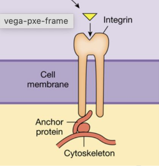
Integrin Receptors (Lipophobic receptor types)
On the extracellular side of the membrane, integrins bind either to proteins of the extracellular matrix or to ligands such as antibodies & molecules involved in blood clotting
Inside the cell, integrins attach to the cytoskeleton via anchor proteins
Ligand binding to the receptor causes integrins to activate intracellular enzymes or alter the organization of the cytoskeleton
Cell adhesion, activate immune cells, & cell movement
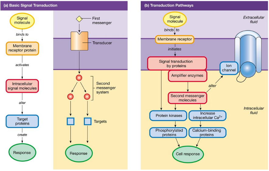
Signal Transduction
The process by which an extracellular signal molecule activates a membrane receptor that in turn alters intracellular molecules to create a response
The extracellular signal molecule is the first messenger, & the intracellular molecules form a second messenger system
An extracellular signal molecule ( the first messenger) binds to & activates a membrane receptor
The activated membrane receptor turns on its associated proteins and starts an intracellular cascade of second messengers
The last second messenger in the cascade acts on intracellular targets to create a response
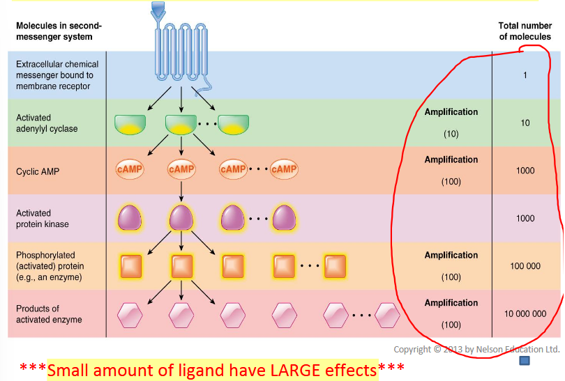
Second Messenger system amplifies the response; T or F?
True
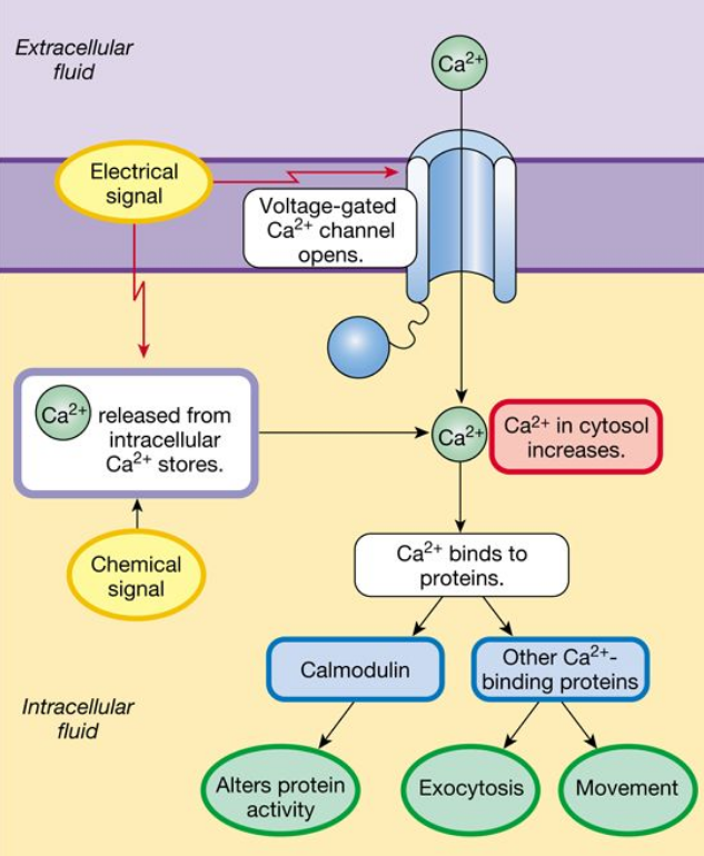
Calcium (Ca2+) (Key signal molecules)
Can enter the cell through a wide variety of channels, mostly stored in endoplasmic reticulum (ER)
Can also be released from intracellular compartments by second messengers
Functions:
Alter enzyme/transporter activity, channel gating
Movement of contractile proteins (cytoskeleton)
Trigger exocytosis of secretory vesicle
Initiate development of embryo when egg is fertilized
Muscle contraction
Soluble Gases (Key signal molecules)
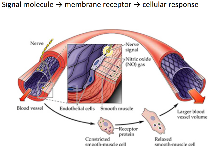
Short-acting paracrine/autocrine signal molecules that act close to where they are produced
Includes Nitric Oxide (NO), carbon monoxide (CO), & hydrogen sulfide (HS)
Nitric Oxide (Key signal molecules)
Produced by endothelial cells lining blood vessels
Causes smooth muscle cells to relax & blood vessels to dilate
Also acts as neurotransmitters and neuromodulator in the brain
Carbon monoxide (CO)
Activates guanylyl cyclase and cGMP, but may also work independently to exert its effects
Carbon monoxide targets smooth muscle & neural tissue
Hydrogen Sulfide (H2S)
Acts in the cardiovascular system to relax blood vessels
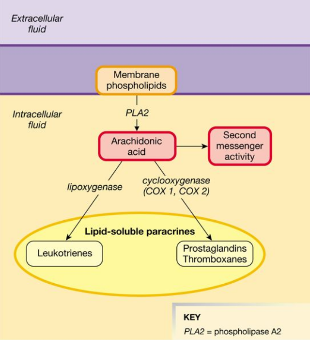
Eicosandoids
lipid-derived paracrine signals that play important roles in many physiological processes
Derived from arachidonic acid, a 20-carbond fatty acid
Act on their target cells using G protein-coupled receptors
The synthesis process for eicosanoids is a network called the arachidonic acid cascade
Properties of Receptors
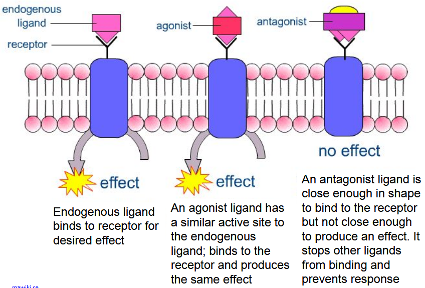
Receptors behave like all other proteins:
Specificity - certain ligands can only bind to specific receptors
Competition - ligands w/ similar structure can bind to the same receptor
Saturation - a point is reached where all receptors are bound
Receptor Agonists & Antagonist
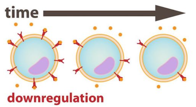
Down-regulation
Target cell decreases in receptor #
Removal from membrane (endocytosis) or receptor desensitization
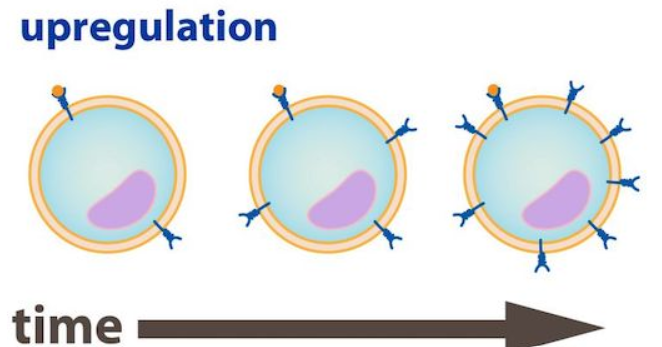
Up-regulation
Target cell inserts more receptors into the membrane
Allows cells to carry responsiveness to signal molecules
Stopping the Signal
Ligand can be degraded by enzymes in extra-cellular space
Chemical messengers can be taken up into neighbouring cells
Endocytosis of receptor-ligand complex (receptor returned by exocytosis)
Bottom line: process initiated by signal can be stopped when necessary
In more complicated reflex control pathways, info must be transmitted throughout the body using chemical signals or a combination of electrical & chemical signalling; T or F?
True
Regulated Variables & Control Systems
The nervous system has a role in preserving the “fitness” of the internal environment
Some systems of the body are under tonic control
Some systems of the body are under antagonistic control
One chemical signal can have different effects in different tissues
Reflex Pathways
Usually involves chemical & electrical signals (nervous & endocrine control)
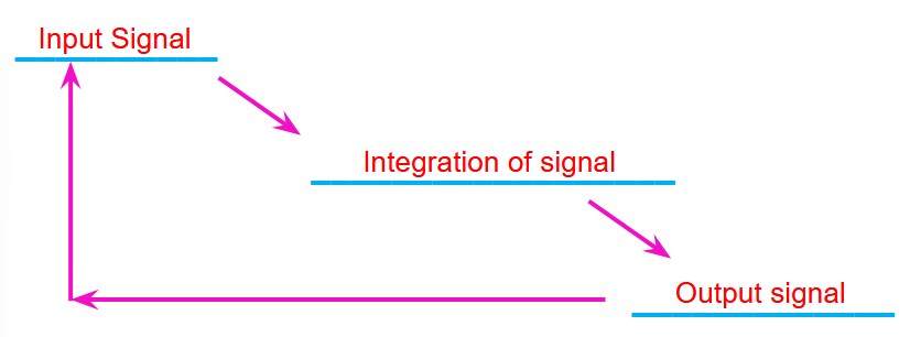
Response loop has 3 main components
Input signal
Integration of Signal
Output signal
1) Stimulus (Steps in a Reflex Pathway)
Starts the reaction
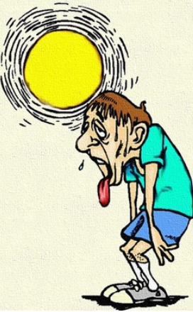
Can be changes in temp, oxygen, blood pressure, etc. (any regulated variable)
E.g. body temp control on a hot day
2) Sensory Receptors (Steps in a Reflex Pathway)
Located throughout the body sense change in stimulus
Central receptors - located in or closely related to the brain (eyes, ears, nose, tongue, central, chemo-, osmo-, & thermoreceptors)
Peripheral Receptors - found elsewhere in the body (joints, skin, blood vessels, etc.), for pH, osmolarity, temp, pressure, etc.
E.g. Increase in body temp sensed by thermoreceptors
3) Activated Sensor (Steps in a Reflex Pathway)
sends input signal to integrating centre (usually CNS)
E.g. thermoreceptors send message to hypothalamus
4) Integration centre (Steps in a Reflex Pathway)
compares input signal to desired setpoint
E.g. hypothalamus assesses if body temp is >37 degrees
5) Output signal (Steps in a Reflex Pathway)
If signal is out of range, integrating centre initiates output signal
E.g. Hypothalamus sends message to skin, muscle & sweat glands
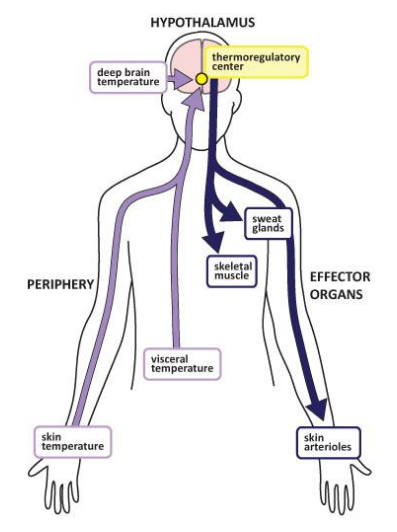
6) Output signal to target (Steps in a Reflex Pathway)
Output signal travels to the target (effector)
7) Response (Steps in a Reflex Pathway)
Effector carries out response to bring variable back to normal
Responses can be cellular or systemic
E.g. Arterioles dilate, muscles relax, sweating starts, body temp decreases
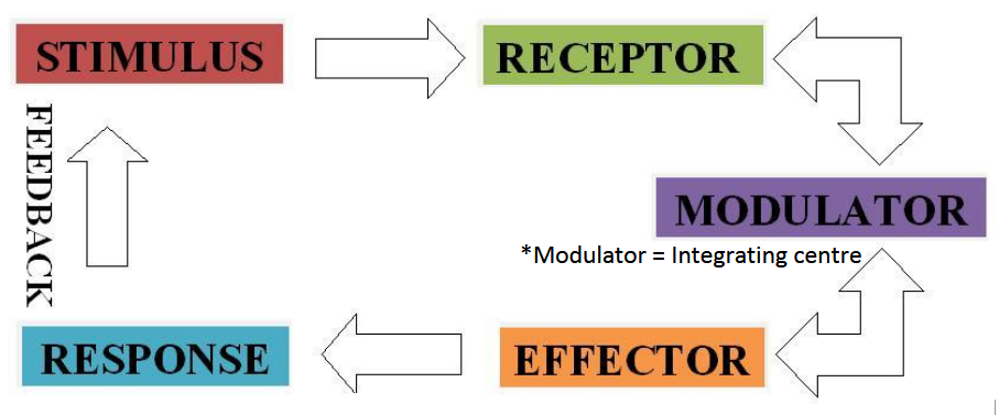
Summarized Steps in a Reflex Pathway
Stimulus occurs
Sensory receptors sense change
Activated sensor sends input signal to integrating centre (afferent signal)
Integrating centre compares input signal to desired set-point
These 4 steps are always active - always looking for small changes & sending it to the integrating centre to sense if the change is within homeostasis
If signal is out of range, integrating centre sends output signal (efferent signal)
Output signal travels to the target (effector)
Target carries out response to bring variable back to normal
Nervous System vs. Endocrine System
Reflex control can be done by one system or both
Nervous & endocrine systems often overlap (neuroendocrine)
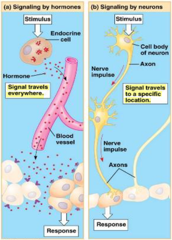
Specificity
Nervous system:
Each neuron has a specific target cell
Endocrine system:
Chemical messengers reach many cells through the blood (moves around the whole body)
Receptors determine response
Several tissues can respond to the same hormone
Nature of the signal
Nervous system:
Electrical (neurons) & chemical (neurotransmitters)
Endocrine system:
Purely chemical (hormones) & larger
Speed:
Nervous system:
Very fast (electrical signals up to 120m/sec)
Endocrine system:
Slower
Requires distribution through circulatory system and diffusion from capillaries to receptors
Duration of Action
Nervous system:
Relatively short
Neurotransmitters rapidly removed from vicinity of action
Sustained response requires multiple neural signals
Endocrine system:
Last longer
Controls “longer” processes like metabolism and reproduction
Coding for Stimulus Intensity
Nervous system:
Signal strength is constant
Intensity of response depends on signal frequency
There’s always a space between responses (gives the cell a rest)
Endocrine system
Stimulus intensity is proportional to hormone release
Simple vs Complex Control Pathways
Simple Control Pathway
Generally have one integrating centre
Direct response to a single stimulus
Can be either neural or hormonal
Complex control
Involves more than one integrating centre
Can involve both neural and hormonal (endocrine control)
Typically symbolized by the brain