2 - 10 29 Spinal Cord & PNS - PHYL 301
 Call Kai
 Learn
 Practice Test
 Spaced Repetition
 Match
 Flashcards
 Knowt Play
1/68

There's no tags or description
Looks like no tags are added yet.
69 Terms
Peripheral Nervous System (PNS)
Branches off the CNS to the rest of the body
Spinal nerves
Cranial nerves
Central Nervous System (CNS)
Brain
Spinal Cord
Spina Bifida
Birth defect
Folate (vitamin B 9 ) deficiency increases risk of spina bifida
Caused by incomplete neural tube closure
Location & severity varies
Which primary and secondary brain vesicles give rise to the Cerebrum?
Prosehow do i phrancephalon (forebrain) → Telencephalon → Cerebrum
Which primary and secondary brain vesicles give rise to the Eye cup, Thalamus, hypothalamus, and epithalamus?
Prosencephalon (forebrain) → Diencephalon → Eye cup, Thalamus, hypothalamus, and epithalamus
Which primary and secondary brain vesicles give rise to the Midbrain?
Mesencephalon (Midbrain) → Mesencephalon → Midbrain
Which primary and secondary brain vesicles give rise to the Pons and Cerebellum?
Rhombencephalon (Hindbrain) → Mesencephalon → Pons and Cerebellum
Which primary and secondary brain vesicles give rise to the Medulla Oblongata?
Rhombencephalon (Hindbrain) → Myelencephalon → Medulla

How How does the neural plate become the neural tube?does the neural plate become the neural tube?
Part of the ectoderm thickens to form the neural plate (neuroectoderm).
The center of the plate dips inward to form the neural groove, while the edges rise to form the neural folds.
The neural folds converge (move toward each other) and fuse, forming the neural tube beneath the surface ectoderm. Cells at the edges of the folds separate to form the neural crest.
The neural tube head end develops into the brain and the rest develops into the spinal cord.
Meninges
Specialized membranes wrapping entire CNS
Provide protection, physical stability, and shock absorption
Spinal meninges continuous with cranial meninges
Meningeal layers
Dura mater: Tough, fibrous outermost layer
Arachnoid mater: Middle delicate meshwork layer
Pia mater: Soft, innermost layer; adheres to CNS tissue
Dura mater
Tough, fibrous outermost layer

Arachnoid mater
Middle delicate meshwork layer

Pia mater
Soft, innermost layer; adheres to CNS tissue

What is the dura mater, and what type of tissue is it made of?
Tough, fibrous outermost layer of the meninges.
made of dense irregular connective tissue.
How does the dura mater help stabilize the spinal cord?
Its cranial and sacral attachments help stabilize the spinal cord lengthwise within the vertebral canal
Where is the spinal epidural space?
Space between the dura mater and lining of the vertebral bones
What forms the coccygeal ligament?
The dura mater blends with the filum terminale, forming the ligament that anchors to the coccyx
Where is the arachnoid mater?
Middle meningeal layer, between the dura mater and pia mater
Arachnoid trabeculae and where do they extend from?
Thin, web-like strands that extend from the arachnoid mater to the pia mater
Where is the subarachnoid space, and what flows through it?
Between arachnoid (middle) and pia (inner)mater
Cerebrospinal fluid flows within subarachnoid space
Subdural space
Potential space between the dura (outermost) mater and arachnoid mater
Not found in healthy living people, but may appear in disease or in cadavers
What is the Pia Mater and what does it contain?
Deepest meningeal layer
Contains blood vessels in this layer
Firmly bound to brain and spinal cord tissue
Denticulate ligaments:
extensions of pia mater attach it and arachnoid mater to the dura mater
Meningitis
-itis” = inflammation
meningitis - acute inflammation of the meninges
CSF should be sterile
Many possible causes of infection
fungi
parasites
viruses
bacteria (e.g., Neisseria meningitidis)
Gray Matter
Mostly consists of neuronal cell bodies (somas) and glial cells
Surrounds central canal (inner butterfly)
Forms horns
grAy = mAss = cell bodies
White matter
Consists of axons (myelinated and unmyelinated)
Axons organized into tracts or columns (outer)
Generally superficial to gray matter
white outline, axons WX
What is a nucleus in the central nervous system?
Cluster of neuron cell bodies in the CNS (brain or spinal cord)
What is a ganglion?
cluster of neuron cell bodies in the PNS
The dorsal root ganglion is one example.
What is a tract?
Bundle of axons within the CNS
What is a nerve?
Bundle of axons in the PNS, wrapped in connective tissue
It can carry sensory signals, motor signals, or both
What does the posterior (dorsal) root carry?
Somatic Sensory (afferent) and viseral sensory nuclei signals into the spinal cord.
top
What does the Lateral horns (intermediate horns) carry?
Visceral motor unit nuclei
Middle
What does the anterior (ventral) root carry?
Somatic Motor (efferent) signals out of the spinal cord.
Bottom

Where is the dorsal root ganglion, and what does it contain?
Swelling on the dorsal root near the intervertebral foramen. It contains sensory neuron cell bodies
What are the three connective tissue layers of a spinal nerve?
Endoneurium wraps each axon.
Perineurium wraps each fascicle (bundle of axons).
Epineurium wraps the whole nerve
How does a typical spinal nerve form, and why is it mixed?
he dorsal and ventral roots join near the intervertebral foramen. The resulting spinal nerve carries both sensory and motor axons.
What are the two main branches of a spinal nerve, and what do they supply?
The dorsal ramus supplies the deep back muscles and skin of the back. The ventral ramus supplies the front and sides of the trunk and the limbs. Both rami are mixed.
Peripheral neuropathy
Condition resulting from peripheral nerve damage
Many potential causes
Trauma, infection, Vitamin deficency, alchol use, type 2 diabetes
Peripheral neuropathy - Sensory Symptoms
Numbess, Tingling, Pain
False signals, feeling sensations singals sent to wrong part
Sharp, throbbing, freezing, or burning pain
Peripheral neuropathy - Motor Symptoms
Muscle Weakness, paralysis
Lack of coordination & falling
Peripheral neuropathy - Treatment
Trauma
Medications, physical therapy, surgery, etc.
infection
Medications (e.g. antibiotics)
Vitamin deficency
Diet & supplements
alchol use
Limit or discontinue alcohol
type 2 diabetes
Weight, diet, & lifestyle management
Leprosy (Hansen's disease)
Caused by Mycobacterium leprae bacteria
Symptoms can take years to appear
Can cause peripheral neuropathy
Loss of sensation
Paralysis of hands and feet
Prof. Alice Augusta Ball
University of Hawaiʻi’s first female and African American chemistry professor
1915 – Developed early injectable treatment for leprosy from chaulmoogra tree
Leprosy now curable with antibiotics
The 31 pairs of spinal nerves
8 cervical nerves
12 thoracic nerves
5 lumbar nerves
5 sacral nerves
1 coccygeal nerve
Main difference of cervical spinal nerves?
8 Cervical spinal nerves

Smallest to largest of Peripheral Nerve Connective Layers
Endometrium → Perineurium → Epineurium

Endoneurium
Smallest, innermost
Loose Connective Tissue
Surrounds each axon (nerve fiber)

Perineurium
Surrounds each Fascicle (fascicles), bundle of axons
Connective Tissue

Epineurium
Most outermost layer
Irregular connective tissue
Surrounding entire spinal nerve; continuous with dura mater
What are the main branches of a spinal nerve, how do they connect to the sympathetic nervous system?
Main branches: The dorsal (posterior) ramus and ventral (anterior) ramus. Both carry sensory and motor fibers.
Sympathetic connections: White and gray rami communicantes connect spinal nerves to the sympathetic trunk. T1–L2 have both. Gray rami are present at all spinal levels.
Dermatome
An area of skin supplied by sensory fibers from a single spinal nerve root
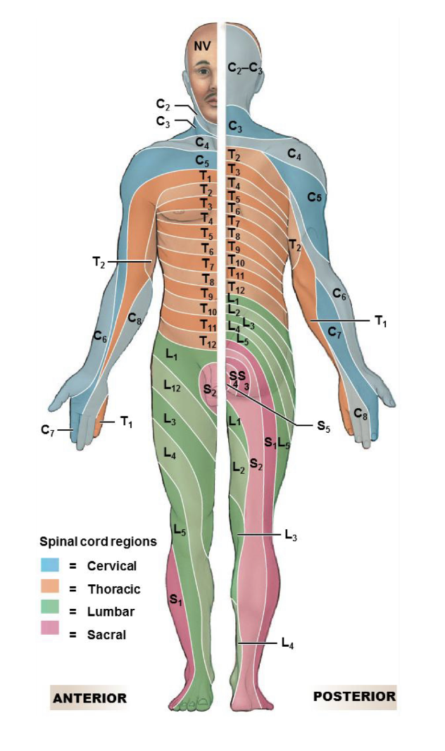
When neuron cell bodies are grouped together what is it called?
CNS: Nucleus
PNS: Ganglion (like dorsal root ganglion)
When axons are grouped together what is it called?
CNS: tract (carries signals in CNS)
PNS: Nerves (carries sensory, motor, or both signals)
Structure of nerve: Smallest to largest
Nerve fiber → fascicle (bundle of fibers) → whole peripheral nerve
wrapped in: endoneurium → perineurium → epineurium
Innervation
Distribution or supply of nerves to a body region
Which direction do signals travel in sensory innervation?
Receptors in the tissue → CNS
Sensory signals are afferent, meaning they travel toward the CNS
Which direction do signals travel in motor innervation?
CNS → muscles or glands
Motor signals are efferent, meaning they travel away from the CNS
Somatosensation Tests
Tested along dermatomes
Used in assessing spinal cord or brain damage
Unequal/reduced/absent sensation may indicate nerve or CNS damage
Shingles
Varicella-zoster virus (“chicken pox”)
Reactivates
Painful rashes & blisters
Affected area may feel painful, burning, tingling, numb sensations (or a combination!)

Shingles – Cause
Viral latency
Viruses lie dormant in nerves
Can reappear later in life
Avg. age ~50+
1 in 3 lifetime chance
the virus can remain dormant in sensory ganglia, such as a dorsal root ganglion
Shingles – Prevention
Symptoms last ~2 to 4 weeks
Antiviral drugs help with symptoms
Vaccinations reduce chance of shingles
Nerve Plexus
Interwoven network of nerves (plexus = “to braid)
Cervical Plexus
Ventral rami of C1 to C4 spinal nerves
Some C5 spinal nerve fibers
Cutaneous and muscular branches
Cutaneous branch innervates head, neck, & chest
Lesser occipital nerve (Cervical Plexus)
Innervates skin of neck and posterior head
Supraclavicular nerve (Cervical Plexus)
innervates skin of neck and shoulder
Great auricular nerve (Cervical Plexus)
innervates skin of neck and posterior ear
Transverse cervical nerve (Cervical Plexus)
innervates anterior triangle of the neck
Ansa cervicalis nerve
innervates five extrinsic laryngeal muscles
U shaped
Phrenic nerve
innervates diaphragm
Breathing