Clinical Diagnosis Quiz 4: Renal/GU
 Call Kai
 Learn
 Practice Test
 Spaced Repetition
 Match
 Flashcards
 Knowt Play
1/188

There's no tags or description
Looks like no tags are added yet.
189 Terms
Pre-renal
____-renal AKI is caused by problems with blood flow into the kidneys.
Post-renal
____-renal AKI is caused by problems with urine flow out of the kidneys (BPH, kidney stones).
Intra-renal
____-renal AKI is caused by problems with the functioning of the glomeruli and tubules of the kidney.
Hypovolemia
The most common cause of pre-renal AKI is ____.
pre-renal, hypovolemia
Even if a patient has a normal blood pressure, processes such as diarrhea, over-diuresis, hemorrhage, and excess sweating can cause ____-renal AKI due to _____.
CHF, cirrhosis
What are two classic causes of pre-renal AKI due to decreased effective circulating volume?
pre-renal AKI, hypotension
Sepsis, cardiogenic shock, anaphylaxis, and anesthesia are causes of ____-renal AKI due to _____.
Relative hypotension
***Body gets used to operating at a very high blood pressure. When the BP is decreased to 120/80, the body responds as though it is hypotensive.
_____ hypotension is a cause of pre-renal AKI that occurs when a patient with severe chronic hypertension (200s SBP) has their blood pressure lowered to a normal range.
pre-renal
An aortic dissection that obstructs blood flow through a renal artery would cause ____-renal AKI due to changes in renal hemodynamics.
Pre-renal
***I know this is weird because it sounds more intra-renal than pre-renal. Technically, "pre-renal" conditions are conditions that cause decreased blood flow/hydrostatic pressure to the nephron without actually damaging the kidney parenchyma. Efferent arteriole dilation causes blood that enters the glomerulus to exit very quickly, leading to decreased hydrostatic pressure within the glomerulus. If it deals with afferent/efferent arteriole, think pre-renal. If the glomeruli or tubules are actually being damaged, think intra-renal.
ACEi can cause ____-renal AKI by dilating the efferent arteriole, therefore reducing the amount of blood and hydrostatic pressure in the glomerulus.
Pre-renal
***Decreased delivery of blood to the glomerulus --> technically pre-renal, not intra-renal. If the condition involves problems with filtration or tubular function, that would be intra-renal. Afferent/efferent arteriole problems = pre-renal
NSAIDs can cause ____-renal AKI by constricting the afferent arteriole,.
Decreased effective circulating volume
A heart failure patient with a BP of 120/80 presents with pre-renal AKI. What is the most likely cause of the AKI?
pre-renal, hypotension
A patient with 2 days of diarrhea presents with a BP of 90/55 and AKI. His AKI is most likely ___-renal caused by ____.
Abnormal skin turgor
***But the likelihood ratio is still only 3.5, so it's not super great. Labs are your best bet at determining if someone is actually hypovolemic
Which physical exam finding of hypovolemia is the strongest indicator that a patient is hypovolemic:
-dry axilla
-dry mucous membranes
-longitudinal furrows of the tongue
-sunken eyes
-abnormal skin turgor
Subclavian
Abnormal skin turgor is best assessed by pinching the skin in the ____ region.
1. Hemoglobin
2. Albumin
3. Calcium (a significant amount is bound to albumin)
***Other solutes (Na, Cl, K) may change a little in response to hypovolemia, but they have the ability to move in and out of cells and in and out of urine easily, so their serum concentrations remain fairly steady with changes in plasma volume. Hemoglobin and albumin are too big to traverse cell membranes/glomerulus. Calcium is bound to albumin, so it behaves like albumin.
What are the three solutes in blood that are most affected by plasma volume.
Higher than normal.
***Lower plasma volume, but constant amount of Ca bound to albumin --> higher calcium concentration in blood
A patient presents with severe diarrhea and hypovolemia. Would we expect his serum calcium level to be higher or lower than normal?
Hemodiluation from the 3L of IV fluids
***Hypocalcemia (or lowered albumin levels)--> clue that it is hemodilation
A patient presents to the ER with Hgb of 11 from a GI bleed. His anemia is not severe enough to give a blood transfusion. He is given 3L of normal saline because he is hypovolemic. A repeat Hgb drops to 9 and his calcium drops from 10 to 8. What do you think is the primary reason for his Hgb drop?
High
***Kidneys are able to increase their reabsorption of urea in order to retain water in circulation. The body cannot reabsorb creatinine, so the BUN will increase more than the creatinine.
Will pre-renal AKI present with a low or high BUN:Creatinine ratio?
When glucose is over 200, the PCT cannot reabsorb all of the glucose in urine, so it remains in urine and draws water into the urine, leading to diuresis.
Why does severe hyperglycemia lead to hypovolemia?
Bicarbonate
***This is the contraction alkalosis seen in sweating, vomiting, and diuresis.
When the body loses large amounts of chloride, the kidneys retain the anion ____.
1.018
A patient who is hypovolemic will have a urine specific gravity that more than _____.
IV fluids
What is the main treatment for hypovolemia?
BUN:creatinine > 20:1
Urine Na < 20
fena<1
Prerenal AKI
BUN/Cr: >20:1
Urine Na⁺: 500 mOsm/kg
Urine specific gravity: >1.020
Urine sediment: Hyaline casts
Mechanism: ↓ Renal perfusion → kidneys retain Na⁺ and water
Response: Improves with IV fluids
A clue that a patient may be hypovolemic is that their BUN:creatinine ratio is greater than ____ and that their urine sodium is less than ____.
Cryoglobulinemia
Condition | Symptoms / associations |
|---|---|
Membranoproliferative glomerulonephritis (MPGN) | Hematuria, proteinuria, edema, hypertension, ↓ kidney function |
Mixed cryoglobulinemia | Palpable purpura (especially legs), arthralgias, weakness, Raynaud phenomenon |
Vasculitis | Palpable purpura, skin ulcers, peripheral neuropathy, arthralgias |
Hepatitis | Fatigue, nausea, RUQ discomfort, jaundice (often asymptomatic initially) |
High-yield associations
HCV → mixed cryoglobulinemia → MPGN.
Cryoglobulins precipitate in cold temperatures → small-vessel vasculitis.
Classic triad: Purpura + arthralgia + weakness.
MPGN: hematuria + proteinuria + hypertension.
Low C4 is characteristic of mixed cryoglobulinemia.
HCV can also cause B-cell non-Hodgkin lymphoma.
What vasculitis that can cause glomerulonephritis is strongly associated with hepatitis?
Henoch-Schonlein Purpura
Henoch-Schönlein Purpura (HSP) – IgA vasculitis
Small-vessel vasculitis caused by IgA immune complex deposition, commonly following an upper respiratory infection. Most common in children.
Feature | Symptoms / associations |
|---|---|
Skin | Palpable purpura, especially buttocks and lower extremities |
Joints | Arthralgia, arthritis (knees, ankles) |
GI | Colicky abdominal pain, nausea, vomiting, GI bleeding |
Kidneys | Hematuria, proteinuria, IgA nephropathy |
Other | Scrotal edema/pain in boys |
Classic tetrad – memorize
Palpable purpura (without thrombocytopenia)
Arthralgia
Abdominal pain
Renal involvement (hematuria)
A patient presents with a maculopapular rash, joint pain, and UA with RBCs, red cell casts, and 2+ protein after a GI illness. What vasculitis would you most suspect as a cause?

SLE (systemic lupus erythematous)
SLE causes immune complex deposition (type III hypersensitivity) in the kidneys → complement activation → inflammation and renal damage.3. Classic lupus nephritis associations
Finding | Association |
|---|---|
Anti-dsDNA | Associated with lupus nephritis and disease activity |
Anti-Smith | Highly specific for SLE |
↓ C3 and ↓ C4 | Immune complex consumption |
Wire-loop lesions | Class IV diffuse proliferative |
Spike-and-dome | Class V membranous |
Full-house immunofluorescence | IgG, IgA, IgM, C3, C1q |
Proteinuria + hematuria | Renal involvement |
Feature | Class IV: Diffuse proliferative | Class V: Membranous |
|---|---|---|
Main pathology | Endocapillary proliferation and inflammation | Subepithelial immune complex deposition |
Immune deposits | Subendothelial | Subepithelial |
Light microscopy | Wire-loop lesions | Thickened capillary walls |
Special histology | Wire-loop appearance | Spike-and-dome appearance (silver stain) |
Deposit location | Between endothelium and GBM | Between podocytes and GBM |
Clinical presentation | Nephritic syndrome | Nephrotic syndrome |
Symptoms | Hematuria, hypertension, oliguria, proteinuria | Edema, frothy urine, heavy proteinuria |
Complement | ↓ C3, ↓ C4 | May be decreased |
Severity | Often aggressive | Often presents with nephrotic syndrome |
Classic association | Severe lupus nephritis | Nephrotic lupus nephritis |
Treatment | Steroids + mycophenolate or cyclophosphamide | Supportive care; immunosuppression if significant proteinuria |
A 32 yo female presents with a facial rash, joint pain, and UA with RBCs, red cell casts, and 2+ protein. What do you suspect is the underlying disorder causing her glomerulonephritis?

Granulomatosis with polyangitis (Wegeners)
Granulomatosis with polyangiitis (GPA) – Wegener's
Necrotizing granulomatous vasculitis of small- and medium-sized vessels. Associated with c-ANCA (anti-PR3).
Feature | Findings |
|---|---|
Pathology | Necrotizing granulomatous inflammation + vasculitis |
Vessels affected | Small- and medium-sized vessels |
Antibody | c-ANCA (anti-PR3) |
Upper respiratory | Chronic sinusitis, nasal ulcers, epistaxis, saddle-nose deformity |
Pulmonary | Cough, hemoptysis, dyspnea, pulmonary nodules, cavitary lesions |
Renal | Rapidly progressive glomerulonephritis (RPGN), hematuria, proteinuria |
Clinical presentation | Upper airway + lung + kidney involvement |
Kidney biopsy | Pauci-immune necrotizing crescentic GN |
Complement | Usually normal |
Granulomas | Necrotizing granulomas, especially respiratory tract |
Diagnosis | c-ANCA/PR3 + clinical findings + tissue biopsy |
Treatment | Glucocorticoids + rituximab or cyclophosphamide for severe disease |
Classic association | Saddle-nose deformity + hemoptysis + hematuria |
Feature | Anti-GBM disease (Goodpasture) | GPA (Wegener) |
|---|---|---|
Cause | Autoantibodies against α3 chain of type IV collagen | ANCA-associated autoimmune vasculitis |
Antibody | Anti-GBM IgG | c-ANCA (anti-PR3) |
Pathology | Antibody-mediated basement membrane injury | Necrotizing granulomatous vasculitis |
Immunofluorescence | Linear IgG deposition | Pauci-immune (minimal immune deposits) |
Lungs | Alveolar hemorrhage → hemoptysis, dyspnea | Pulmonary nodules, cavitation, hemoptysis |
Kidneys | RPGN → hematuria, proteinuria, renal failure | RPGN → hematuria, proteinuria |
Histology | Crescentic GN | Crescentic GN |
Complement | Usually normal | Usually normal |
Classic association | Pulmonary hemorrhage + renal failure | Sinusitis + pulmonary nodules + renal disease |
Treatment | Plasmapheresis + glucocorticoids + cyclophosphamide | Glucocorticoids + rituximab or cyclophosphamide |
A patient presents with chronic sinusitis, hemoptysis, and UA with RBCs, red cell casts, and 2+ protein. What do you suspect is the underlying vasculitis causing glomerulonephritis?
Intra-renal
AKI caused by IgA nephropathy would be an example of ____-renal AKI.
Hemolytic uremic syndrome
***In HUS, thrombi containing platelets form and lodge in the arterioles and glomeruli --> renal failure.
Classic triad:
Microangiopathic hemolytic anemia (MAHA)
Thrombocytopenia
Acute kidney injury (AKI)
Cause
Most commonly Shiga toxin-producing E. coli (EHEC O157:H7).
Often follows bloody diarrhea after contaminated food (undercooked beef).
Shiga toxin damages vascular endothelium, especially in renal microvasculature.
Causes platelet-rich microthrombi → RBC fragmentation and renal ischemia.
Labs
↓ Hemoglobin
↑ LDH
↑ Indirect bilirubin
↓ Haptoglobin
Schistocytes on peripheral smear
↓ Platelets
↑ BUN and creatinine
Normal PT/PTT (helps distinguish from DIC)
Clinical presentation
Bloody diarrhea (often preceding HUS)
Pallor and fatigue
Decreased urine output
Hematuria
Edema
Hypertension
Treatment
Supportive care and IV fluids (carefully managed).
Correct electrolyte abnormalities.
Dialysis if severe AKI.
RBC transfusion if indicated.
Avoid antimotility agents and generally avoid antibiotics in suspected STEC infection because they may increase toxin release.
A child presents severe diarrhea and abdominal pain a few days after eating a chipotle burrito. He is found to have thrombocytopenia and acute renal failure. PTT and PT are normal. What condition do you suspect?
DIC
***TTP and HUS produce thrombi made of platelets --> clotting factors are not used up to the same extent that they are in DIC
What thrombotic microangiopathy has increased PTT and PT:
- hemolytic uremic syndrome
- thrombotic thrombocytopenic purpura
- Disseminated intravascular coagulation
Proteinuria
***Hematuria in intra-renal AKI is usually due to glomerular problems
Which is more likely to be a feature of AKI due to tubular injury: hematuria or proteinuria?
Muddy brown cast, Acute tubular necrosis
What is this? What do you think is the cause of this patient's renal failure?

Acute tubular necrosis
The presence of more than 10 muddy brown casts per high-power field on a microscopic UA is 100% specific for _____.
No
Is hematuria a common clinical feature of pre-renal AKI?
Acute interstitial nephritis
The presence of eosinophils in urine is highly suggestive of ____.
Sarcoidosis and lymphoma
What are two conditions that can cause AKI by infiltrating the renal interstitium?
Neurogenic bladder
***Spinal cord injury --> loss of innervation to the bladder --> bladder cannot empty so urine backs up into the kidneys --> post-renal AKI
_____ is a cause of post-renal AKI that is frequently seen in patients with spinal cord injuries.
Ureter
A retroperitoneal hematoma can cause post-renal AKI by compressing the ____.
BPH
What is the most common cause of urinary tract obstruction in men?
Stricture
Repeated trauma from catheterization --> fibrosis of the urethra --> urethral stricture
Repeated trauma to the urethra from urinary catheterization can lead to the formation of a urethral ____.
Bleeding
***High urea levels interfere with platelet and clotting factor functioning --> very prone to bleeding
Would severe uremia (BUN > 100) cause a patient to be more prone to blood clots or bleeding?
Rhabdomyolysis causing acute tubular necrosis
***Muscles breakdown and release myoglobin --> filters freely through the glomerulus and damages proximal tubular cells
An elderly woman falls in her home and is lying on the ground immobile for 17 hours. When she is found and brought to the hospital, she is found to be in acute renal failure and has a CK of 100,000. What is the cause of her renal failure?
Dietary intake of beets, blackberryies, and red food coloring
A patient presents to the ER because she is concerned that she is peeing blood. Urine dipstick is negative for heme and microscopy is negative for RBCs. Further questioning reveals that she ate a beet salad, blackberry pie, and half of a red velvet cake for lunch. What do you think is the cause of her red urine?
Nitrofurantoin
***Rifampin also causes red urine, but this is not typically used for UTIs
A patient presents to the ER with concerns for hematuria. UA is negative for heme or RBCs. She was recently treated for a UTI with an antibiotic. What antibiotic mostly likely caused her red urine?
Rifampin
A patient presents to clinic with red urine. UA is negative for heme or RBCs. He is currently on medications for latent TB. What antibiotic do you think is causing his red urine?
Phenytoin
What is an antiepileptic that can cause heme-negative red urine?
1. Rhabdomyolysis (dipstick is actually detecting myoglobin)
2. Hemolytic anemia
What are two conditions that can cause a patient to have blood on urine dipstick but no RBCs on microscopic exam?
Macroscopic hematuria (20-25% have bladder cancer compared with 3.7% for microscopic)
Which is a stronger indicator that a patient has bladder cancer: macroscopic hematuria or microscopic hematuria?
3 RBCs
A microscopic exam that shows less than ____ RBCs per high power field is considered to be normal.
***HPF = what is in your field of vision when you are looking into a microscopic at a high magnification
40
Malignancy is extremely rare in patients under the age of ___ with microscopic hematuria.
Pursue additional testing to rule out malignancy.
***UTIs are common and can cause hematuria, but it is also likely that the hematuria is caused by malignancy and the UTI is just an incidental finding. This patient is high risk and malignancy needs to be ruled out.
A 80 yo male presents with gross hematuria and is found to have a UTI. Should you just treat the UTI or should you pursue additional testing to rule out malignancy?
Glomerular source
***Dysmorphic RBCs, red cell casts, worsening htn, proteinuria, and rising creatinine are all features of dysfunction within the glomerulus/kidney. These are not features of problems with organs distal to the kidney
A patient presents with microscopic hematuria, dysmorphic RBCs, red cell casts, and worsening hypertension. Do you think the hematuria is caused by a glomerular source or by a problem with the ureters/bladder?
Dysmorphic RBCs
Most likely coming from the glomerulus
What are these called? Where are they most likely coming from?
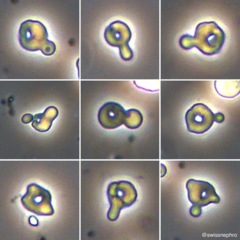
Red cell casts, glomerulus
What are these called? These indicate that there is a problem with the ____.

IgA nephropathy
What is the most common cause of glomerulonephritis worldwide?
Aggressive blood pressure control
What is the most important aspect of preventing progression of renal disease in a patient with IgA nephropathy?
Trauma catheterization
The most common cause of hematuria coming from the urethra is ____.
Urine culture
***UA is also important, but it won't tell you organism or what antibiotics it is sensitive to
A _____ is the most important test in determining if a patient has a UTI.
1. Obtain a urine culture to assess for UTI.
2. Obtain UA to assess for glomerular sources (red cell casts, dysmorphic RBCs, Cr, urine protein)
3. Refer to urology for cystoscopy or multiphasic CT urography to rule out malignancy
What are the 3 steps in evaluating hematuria?
To assess if the UTI was the cause of the hematuria.
***If repeat UA is negative for blood --> UTI probably caused the hematuria
If repeat UA is still positive for blood --> something else is causing the hematuria
If a patient has hematuria and is found to have a UTI, why do you need to repeat a UA after finishing antibiotics?
Glomerulus, renal biopsy
If a patient with hematuria is found to have an elevated creatinine and a UA with protein, red cell casts, and dysmorphic RBCs, you should suspect that the hematuria is caused by a problem with the _____ and you should consider ordering a _____.
White males
What demographic is 3-4 times more likely to develop bladder cancer than the rest of the population?
40
Bladder cancer is very unlikely under the age of ____.
Bladder cancer
Industrial chemical/toxin exposures account for 20% of what urologic cancers?
10-20 years
Bladder cancers tend to appear ____ years after an industrial chemical/toxin exposure.
Aniline dyes
What compound found in wood stains and clothing dyes is strongly associated with bladder cancer?
Aromatic amines
What compounds found in commercial hair dyes, tobacco smoke, diesel exhaust, and manufacturing plants are associated with bladder cancer?
Bladder cancer.
Coal, arsenic, aromatic amines, and aniline dyes are strongly associated with ____ cancer.
Diesel exhaust (contains aromatic amines)
Bus drivers are high risk for developing bladder cancer due to their exposure to ____.
Bladder cancer
Miners, bus drivers, rubber workers, and hair dressers are all at an increased risk for developing ____ cancer due to prolonged exposure to industrial chemicals.
Renal cell carcinoma
***Usually not palpable until in a very late stage
What condition classically presents with a triad of hematuria, flank pain, and a palpable abdominal mass?
As an incidental finding on abdominal imaging.
***Symptoms of hematuria, flank pain, and an abdominal mass do not present until a very late stage
How is early stage renal cell carcinoma usually identified?
Cystoscopy
What procedure is used to visualize the inside of the bladder and collect biopsy samples?
Multiphasic CT urography
What imaging study is used to detect renal cell carcinoma, renal stones, and other GU tumors?
Yes - patients over 40 are at a high risk for bladder cancer. Cystoscopy should be performed in cases of hematuria even if glomerular disease is present, especially if risk factors are present.
A 50 year old patient is found to have hematuria that is suspected to be from IgA nephropathy. Would you still recommend obtaining a cystoscopy to assess for malignancy?
No. Small stones within the renal pelvis are unlikely to cause hematuria. They can sit there for years and not cause any clinical symptoms. He should be evaluated for another source such as malignancy.
A patient presenting with hematuria is found to have a 1 mm stone in the left renal pelvis. Do you think this is the cause of his hematuria?
IgA nephropathy
A patient presents with hematuria after a URI. Found to have red cell casts, protein on UA, and hypertension. What is the most likely cause of his presentation?
thin basement membrane disease
A patient presents with hematuria and notes that multiple family members have hematuria but none have developed CKD. What do you think is the most likely source of his glomerular disease?
Alport syndrome
Alport syndrome is caused by a mutation in type IV collagen (COL4A3, COL4A4, or COL4A5).
Most commonly X-linked dominant → COL4A5 mutation.
Defective type IV collagen damages the glomerular basement membrane (GBM).
GBM becomes irregular, with splitting and thickening → impaired filtration.
Classic triad
Hematuria (often the earliest finding)
Sensorineural hearing loss
Ocular abnormalities (anterior lenticonus, retinal abnormalities)
High-yield pathology
UCLA Health (@UCLAHealth) on X
Electron microscopy: basket-weave appearance of GBM.
Progressive CKD → eventual renal failure.
Can have proteinuria as disease progresses.
A patient presents with hematuria. He has a family history of hematuria with progressive renal disease and sensorineural hearing loss. What do you think is the most likely cause of his glomerular disease?
Yes - lisinopril is used to for strict blood pressure control and for it's renal protective effects. Blood pressure control is the most important aspect of preventing CKD progression in patients with IgA nephropathy.
A patient has IgA nephropathy and a blood pressure of 120/80. Is it appropriate to start lisinopril even though his blood pressure is normal.
Smoking
Although all of these are risk factors, smoking is more significant than low-level, non-occupational exposures to hair dye or diesel exhaust
A patient is found to have bladder cancer. She has a 40 year pack history, routinely gets her hair dyed, and notes that her husband drives a diesel pickup truck. What was the most significant risk factor that predisposed her to developing bladder cancer?
Absolutely not - this is reserved for patients who have complaints about the area
Do you perform a digital rectal exam on all male patients during the yearly physical?
Yes
A patient reports erectile dysfunction. Should you perform a digital rectal exam?
Yes - you need to evaluate the prostate
A patient reports difficulty voiding. Do you perform a digital rectal exam?
Top (pointy apex is at the bottom)
Is the base of the prostate at the top of bottom of the prostate gland?

Epididymis
Which is more likely to be tender to palpation (when palpated with normal force): testicles or epididymis
Induration, infection
Peyronie disease – abnormal fibrous hardening (plaque formation) of tissue within the penis.
It is often associated with penile curvature during erection, pain, and erectile dysfunction.
Abnormal hardening of tissue within the penis is called _____. This is often an indication of _____.
Base to tip
When palpating the penis, do you go base to tip or tip to base?
Crepitus
***Often a sign of gangrene
The presence of air bubbles in penile tissue that makes it feel as though there are "rice crispies" under the skin is described as _____.
Hypospadius (or epispadius if it occurs on the ventral aspect of the penis, but hypospadius is much more common)
The presence of a urethral opening in an abnormal part of the penis is called ____.
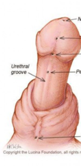
Phimosis (tight fibrous ring of the foreskin). Do not attempt to retract (if anything let the patient do it)
What is this called? Should you attempt to retract the foreskin?

Paraphimosis - the patient had a tight phimotic ring that was pulled down during catheterization, but nobody pulled the foreskin back over the head, so it got stuck around the shaft of the penis
This is seen in a patient that we recently hospitalized and required catheterization. What is this called and what caused it?

Balanitis (infection of the head of the penis)
Feature | Findings |
|---|---|
Symptoms | Redness, swelling, itching, pain, tenderness |
Other findings | Discharge, foul odor, dysuria |
Common cause | Candida albicans |
Risk factors | Diabetes mellitus, poor hygiene, uncircumcised status, phimosis |
Other causes | Bacterial infections, irritants, soaps, STIs |
Diagnosis | Usually clinical; swab/culture if indicated |
Treatment | Topical antifungal (clotrimazole) for Candida; antibiotics if bacterial; avoid irritants and improve hygiene |
What is this called?

Head of the penis
Balanitis is infection of the ____.
Fournier's gangere
_____ is a condition caused by an infection from gas-producing bacteria under the fascia of the penis and testicles.

Crepitus
A patient has Fournier's gangrene from a gas-producing bacteria. What physical exam finding would you expect based on the presence of gas in the tissue underneath the fascia?
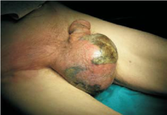
Peyronie's disease - caused by a plaque on the posterior aspect of the penis that causes it to curve abnormally
Correct! Peyronie disease is caused by fibrous plaque formation in the tunica albuginea of the penis.
Cause: Fibrosis/scarring of the tunica albuginea.
Location: Usually dorsal (upper) aspect of the penis.
Effect: Reduced elasticity on the affected side → penis curves toward the plaque during erection.
Symptoms: Penile curvature, painful erections, palpable hard plaque, possible erectile dysfunction.
Association: Can develop after repetitive penile microtrauma.
What is this called?

Peyronie's disease
_____ occurs when there is a plaque that develops on the posterior aspect of the penis between the corporal bodies, causing the penis to curve abnormally.
Penile cancer
Malignancy of the penile squamous epithelium, commonly involving the glans or foreskin.
Feature | Findings |
|---|---|
Histology | Squamous cell carcinoma |
Major risk factor | HPV infection (especially types 16 and 18) |
Other risk factors | Uncircumcised status, poor hygiene, phimosis, smoking |
Protective factor | Neonatal circumcision |
Presentation | Painless penile lesion, ulcer, mass, or nonhealing sore |
Location | Glans penis and foreskin |
Appearance | Exophytic, fungating mass or ulcerative lesion |
Spread | Initially to inguinal lymph nodes |
Diagnosis | Biopsy |
Treatment | Surgical excision, glansectomy, or penectomy depending on stage; lymph node evaluation |
What is this?
