Cardiovascular System: Anatomy, Circulation, and Blood Vessels
 Call Kai
 Learn
 Practice Test
 Spaced Repetition
 Match
 Flashcards
 Knowt Play
1/58

Earn XP
Description and Tags
Comprehensive vocabulary flashcards covering the anatomical structures, circulatory routes, coronary vasculature, and blood vessel histology of the cardiovascular system.
59 Terms
Mediastinum
The central compartment of the thoracic cavity located between the lungs that houses the heart within the pericardial cavity, positioned anterior to the vertebral bodies and posterior to the sternum and costal cartilages.
Pericardium (Pericardial Sac)
A tough membranous sac surrounding the heart that separates it from surrounding mediastinal structures and contains an outer fibrous layer and an inner serous layer.
Fibrous Pericardium
The tough, outer layer of the pericardial sac composed of dense connective tissue that protects the heart and attaches to the base of the great vessels.

Serous Pericardium
The inner double-layered membrane of the pericardium consisting of a parietal layer fused to the fibrous pericardium and a visceral layer (epicardium) fused directly to cardiac muscle.
Pericardial Cavity
The fluid-filled space situated between the parietal pericardium and visceral pericardium containing lubricating pericardial fluid to reduce friction.
Epicardium
The outermost, superficial layer of the heart wall, identical to the visceral layer of the serous pericardium.

Myocardium
The middle, thickest layer of the heart wall composed of cardiac muscle arranged in figure-of-8 bands around the ventricles; it is thickest in the left ventricle to generate higher pumping pressure.
Endocardium
The innermost layer of the heart composed of endothelium (simple squamous epithelium) that lines the chambers, forms heart valves, and is continuous with the endothelial lining of blood vessels.
Apex of the Heart
The inferior, pointed tip of the heart positioned between the 4th and 5th ribs, directed slightly to the left of the sternum.
Base of the Heart
The superior surface of the heart where the great vessels—superior vena cava, inferior vena cava, aorta, and pulmonary trunk—are attached.
Auricle
A leaf-like, superficial extension or flap located on the external surface of each atrium.
Coronary Sulcus
A groove on the external surface of the heart marking the boundary between the atria and ventricles, housing major coronary blood vessels and fat.
Anterior Interventricular Sulcus
A groove located on the anterior surface of the heart between the right and left ventricles that contains coronary vessels and fat.
Posterior Interventricular Sulcus
A groove situated on the posterior surface of the heart demarcating the division between the ventricles, containing coronary blood vessels and adipose tissue.
Interventricular Septum
A thick muscular extension of the myocardium dividing the left and right ventricles.

Skeleton of the Heart (Cardiac Skeleton)
A framework of dense connective tissue with openings and fibrous rings that encircle and support the four heart valves.
Trabeculae Carneae
Muscular ridges of cardiac muscle lined with endocardium located along the internal walls of the ventricles.
Chordae Tendineae
Tendinous cords composed of collagen and elastin fibers that anchor the cusps of atrioventricular valves to papillary muscles.
Papillary Muscles
Muscular projections from the ventricular myocardium that contract during ventricular systole to pull on chordae tendineae and keep the atrioventricular valves closed.
Tricuspid Valve (Right Atrioventricular Valve)
A three-cusped endothelial valve located between the right atrium and right ventricle that prevents blood backflow into the right atrium during ventricular contraction.
Bicuspid Valve (Mitral Valve)
A two-cusped endothelial valve located between the left atrium and left ventricle that prevents the backflow of oxygenated blood into the left atrium during ventricular systole.
Pulmonary Semilunar Valve
A valve composed of three pocket-shaped endothelial cusps located at the base of the pulmonary trunk that lacks chordae tendineae and prevents backflow into the right ventricle during ventricular diastole.
Aortic Semilunar Valve
A valve composed of three pocket-shaped flaps at the base of the aorta that closes as blood fills its cusps during ventricular diastole, preventing backflow into the left ventricle.
Atrial Systole
The phase of the cardiac cycle when atria contract, forcing blood through open atrioventricular valves into the ventricles while semilunar valves remain closed.
Ventricular Systole
The contraction of the ventricles that pushes blood upward, closing the atrioventricular valves (producing the first heart sound) and forcing open the semilunar valves to eject blood into the pulmonary trunk and aorta.
Ventricular Diastole
The relaxation phase of the ventricles where pressure drops, causing backflowing blood in the pulmonary trunk and aorta to catch and close the pocket-shaped semilunar valves (producing the second heart sound).
Pulmonary Circuit
The blood pathway that routes carbon-dioxide-rich deoxygenated blood from the right ventricle through pulmonary arteries to alveolar capillaries for gas exchange and returns oxygenated blood via pulmonary veins to the left atrium.
Systemic Circuit
The circulatory route that distributes oxygen-rich blood from the left ventricle through the aorta and systemic arteries to body tissues and returns deoxygenated blood via the venae cavae to the right atrium.
Superior Vena Cava
A large vein draining deoxygenated systemic blood from body structures situated superior to the diaphragm—including the head, neck, upper limbs, and thoracic cavity—directly into the right atrium.
Inferior Vena Cava
A large systemic vein that collects deoxygenated blood from regions inferior to the diaphragm, including the lower limbs and abdominopelvic cavity, and delivers it to the right atrium.
Pulmonary Trunk
The major arterial vessel exiting the right ventricle that bifurcates into the right and left pulmonary arteries carrying deoxygenated blood to the lungs.
Pulmonary Veins
Four vessels (two from each lung) that transport oxygenated blood from pulmonary capillaries back to the left atrium of the heart.
Coronary Circulation
The specialized blood supply to the myocardium that delivers blood when the heart muscle is relaxed and stops during contraction, receiving its highest perfusion in the left ventricle.
Left Coronary Artery (LCA)
An arterial branch arising from the base of the ascending aorta that gives rise to the anterior interventricular artery and the circumflex artery, supplying the left atrium, left ventricle, and interventricular septum.
Anterior Interventricular Artery (Left Anterior Descending / LAD)
A large branch of the left coronary artery traveling down the anterior interventricular sulcus to supply the anterior ventricular walls and interventricular septum.
Circumflex Artery
A branch of the left coronary artery that travels along the coronary sulcus to the left side of the heart, supplying the left atrium and posterior left ventricle.
Right Coronary Artery (RCA)
An arterial branch originating at the base of the ascending aorta that courses along the coronary sulcus, supplying the right atrium, portions of both ventricles, and giving rise to the marginal and posterior interventricular arteries.

Coronary Sinus
A large venous vessel lying in the atrioventricular (coronary) sulcus on the posterior heart surface that collects venous blood from the cardiac veins and empties directly into the right atrium.
Great Cardiac Vein
A cardiac vein ascending through the anterior interventricular sulcus that drains blood from the myocardium into the coronary sinus.
Middle Cardiac Vein
A cardiac vein located in the posterior interventricular sulcus paralleling the posterior interventricular artery that drains blood into the coronary sinus.
Small Cardiac Vein
A venous channel running parallel to the right coronary artery along the inferior margin of the heart that drains into the coronary sinus.
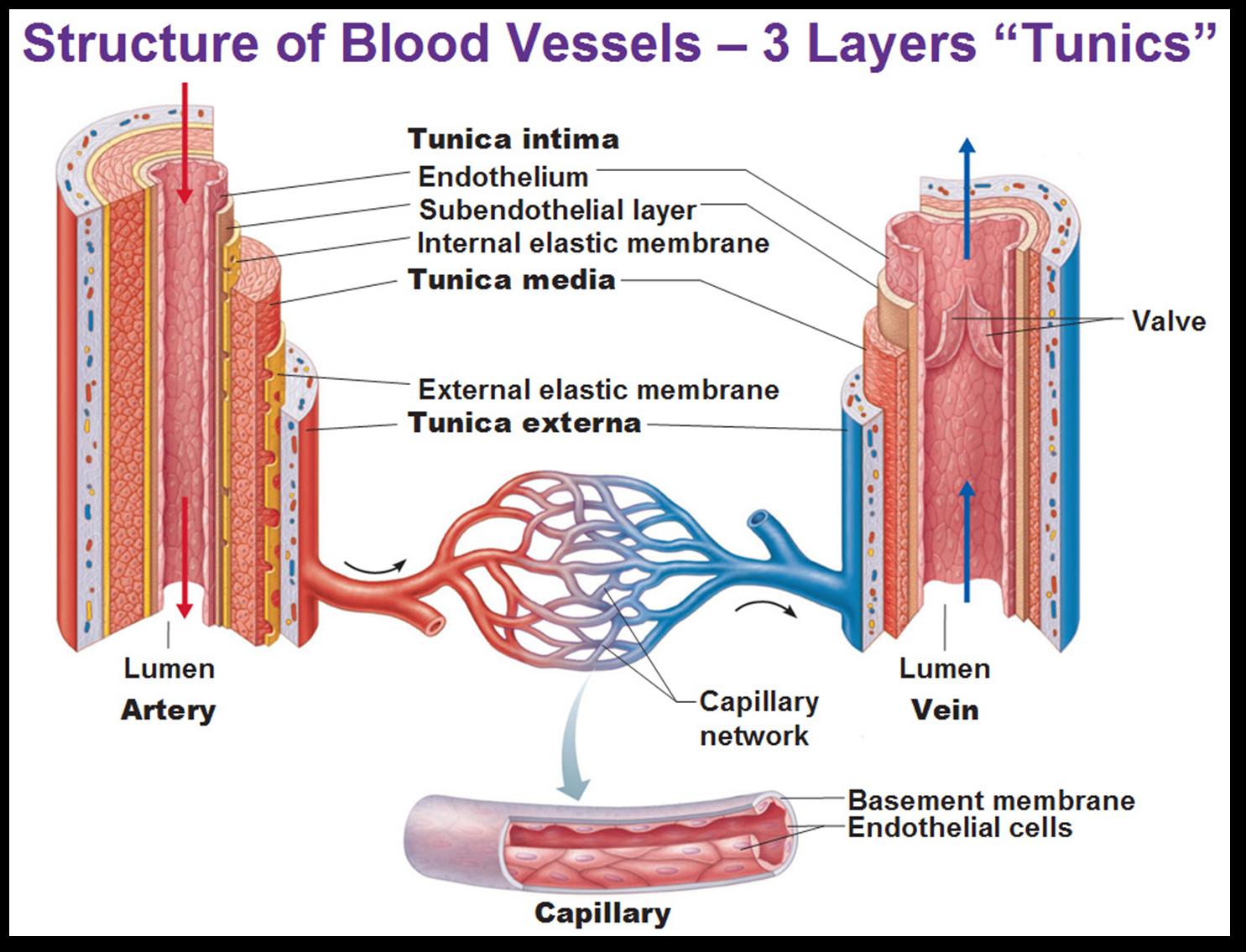
Tunica Intima
The innermost tunic of blood vessels, composed of endothelium and a basement membrane; in larger arteries it contains an internal elastic membrane, and in veins its endothelial folds form one-way valves.
Tunica Media
The middle and usually thickest vascular layer in arteries, composed of smooth muscle and elastic fibers that regulate vascular diameter via vasoconstriction and vasodilation.
Tunica Externa
The outermost layer of blood vessels made predominantly of collagenous connective tissue fibers; it is the thickest coat in veins and contains the vasa vasorum and nervi vasorum.
Vasoconstriction
The contraction of vascular smooth muscle in the tunica media that narrows the vessel lumen and increases blood pressure, regulated by sympathetic neural tone.
Vasodilation
The relaxation of smooth muscle within the tunica media resulting in an expanded vessel lumen, increased blood flow, and reduced blood pressure.
Elastic Arteries
Large conducting vessels situated near the heart whose abundant elastic fibers allow them to expand under high ejection pressure and recoil to maintain continuous blood pressure throughout the arterial tree.
Muscular Arteries
Medium-sized distributing arteries that contain a thick tunica media of smooth muscle and play a primary role in vasoconstriction and directing blood flow.
Arterioles
Small resistance vessels leading into capillary networks that possess a thin smooth muscle layer and cause the greatest decrease in blood pressure.
Continuous Capillaries
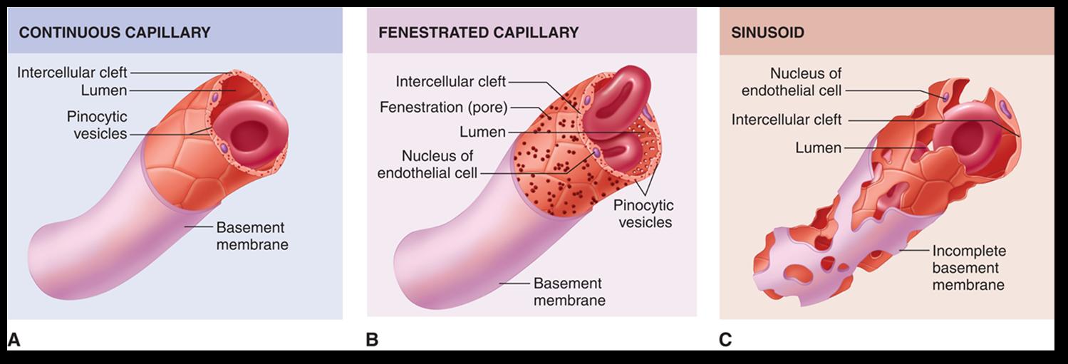
Capillaries featuring an uninterrupted endothelial lining secured by tight junctions with small intercellular clefts for fluid, gas, and small molecule exchange; found in skeletal muscle, lungs, connective tissues, and modified in the blood-brain barrier.
Fenestrated Capillaries
Capillaries characterized by small pores (fenestrations) in the endothelial cell membranes in addition to intercellular clefts, specialized for high filtration or absorption in kidneys, the small intestine, and endocrine glands.
Sinusoids
Leaky, large-lumen capillaries with extensive clefts, fenestrations, and an incomplete or absent basement membrane that permit large plasma proteins and formed elements to pass; located in bone marrow, the liver, spleen, and lymph nodes.
Venules
Microscopic venous vessels formed by converging capillaries that merge together to create veins.
Capacitance Vessels
A functional term for veins referring to their thin walls, high distensibility, and capacity to accommodate large volumes of blood under low pressure as reservoirs.
Venous Reserve
The approximately 21% of total blood volume residing within the venous networks of the liver, bone marrow, and spleen that can be mobilized to circulation via venoconstriction.
Skeletal Muscle Pump
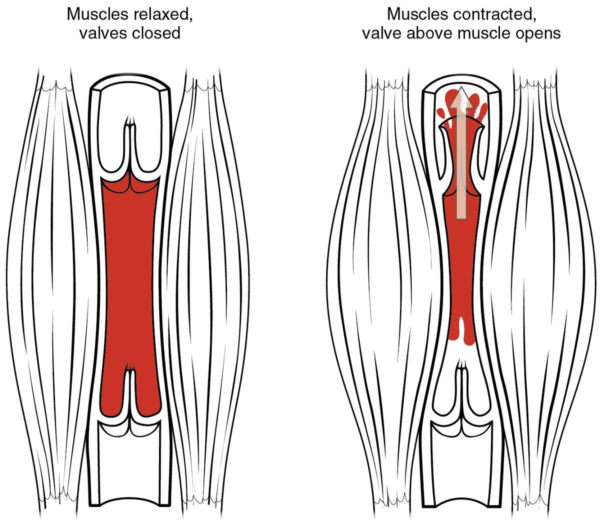
A mechanism of venous return where skeletal muscle contractions compress deep veins, elevating intravenous pressure and driving blood past upper one-way valves toward the heart while lower valves prevent retrograde flow.
Respiratory Pump
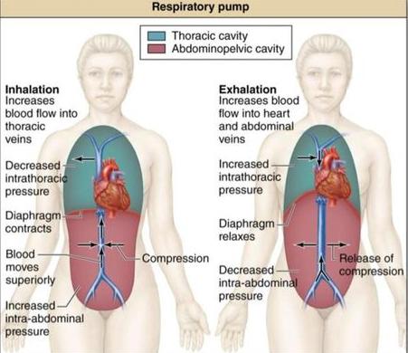
A physiological mechanism aiding venous return: during inhalation, diaphragmatic descent increases abdominal pressure while lowering intrathoracic pressure to draw venous blood into the thoracic cavity; exhalation elevates thoracic pressure to propel blood into the right atrium.
Brachiocephalic Trunk
The first and largest branch arising from the arch of the aorta that divides into the right common carotid artery and the right subclavian artery.
Celiac Trunk
An anterior visceral branch of the abdominal aorta that bifurcates into the left gastric, splenic, and common hepatic arteries.