Anatomy Exam 4
 Call Kai
 Learn
 Practice Test
 Spaced Repetition
 Match
 Flashcards
 Knowt Play
1/330

There's no tags or description
Looks like no tags are added yet.
331 Terms
Central nervous system (CNS)
The brain and spinal cord.
Four major regions of the brain
Cerebrum, diencephalon, brainstem, and cerebellum.
Cerebrum
Two hemispheres; five lobes per hemisphere.
Outer surface of the brain
The outer surface is folded.
Gyri
Ridges on the brain surface.
Sulci
Depressions between ridges; fissures are deep sulci.
Gray matter
Made of neuron cell bodies, dendrites, and unmyelinated axons.

name all the pieces of this image
Cerebral cortex
Gray matter surface of the cerebrum.
Cerebral nuclei
Regions of gray matter (clusters of cell bodies) found deep in the cerebrum.

name the parts of this
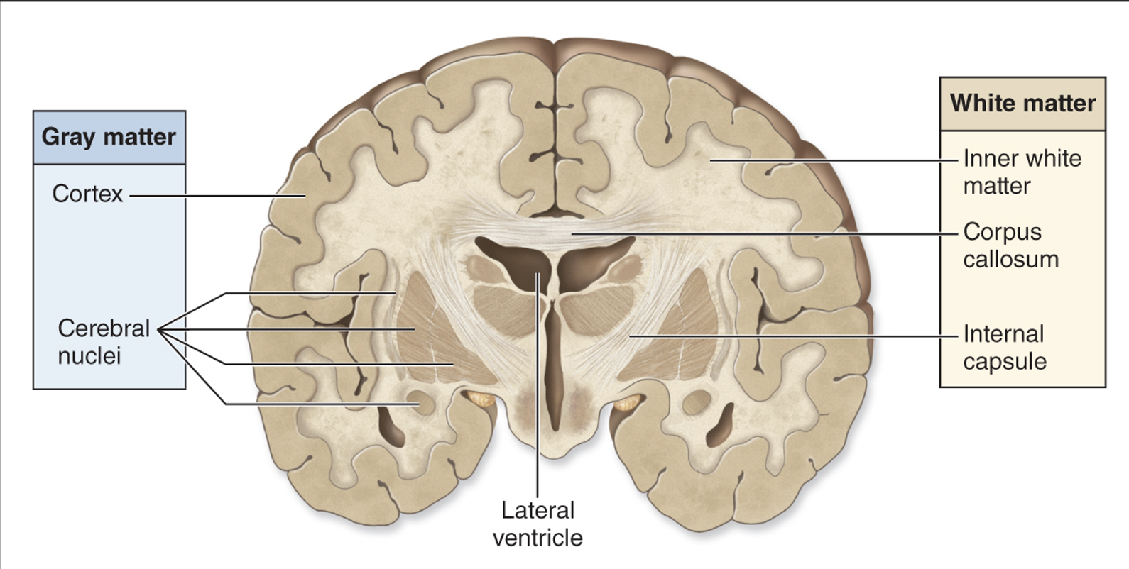
White matter
Consists of myelinated axons.

name all the parts of this image

what is this
medulla oblongata
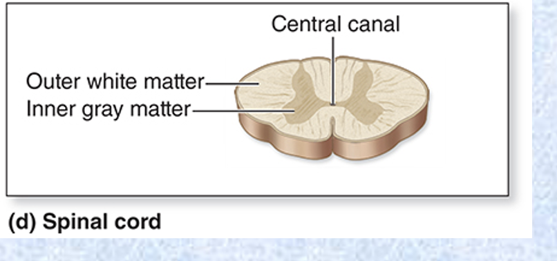
what is this
spinal cord
Cranial meninges
Three connective tissue layers that protect and support soft tissue of the brain, enclose and protect blood vessels supplying the brain, and help contain and circulate cerebrospinal fluid.
Cranial meninges: layers from deep to superficial
Pia mater, arachnoid mater (arachnoid membrane), dura mater.
Pia mater
Innermost of the meninges; adheres to brain surface. Thin layer of areolar connective tissue.
Arachnoid mater
Lies external to pia mater and deep to dura mater. Made of a web of collagen and elastic fibers.
Arachnoid trabeculae
Extend to pia mater through the subarachnoid space.
Subarachnoid space
Contains cerebrospinal fluid (CSF).
Subdural space
A potential space that can fill with blood if a vein is ruptured (subdural hematoma).
Dura mater
Tough, outer membrane made of dense irregular connective tissue in two layers.
Dura mater: two layers
Meningeal layer (deeper layer of dura) and periosteal layer (more superficial layer of dura).
Periosteal layer of dura mater
Forms the periosteum on the internal surface of cranial bones.
Dural venous sinuses
The dura layers are usually fused, but in some areas they separate to form dural venous sinuses that drain blood from the brain.

name the parts of this

Epidural space of the cranium
A potential space between dura and skull; contains arteries and veins.
Ventricles, and what are they lined with?
Cavities within the brain that contain cerebrospinal fluid; lined with ependymal cells; connect with each other and with the spinal cord's central canal.
Four ventricles
Two lateral ventricles, the third ventricle, and the fourth ventricle.
Lateral ventricles
Large cavities in the cerebrum, separated by a medial partition called the septum pellucidum.
Septum pellucidum
Medial partition separating the two lateral ventricles.
Third ventricle
Narrow space in the middle of the diencephalon.
Interventricular foramen
Connects each lateral ventricle to the third ventricle.
Fourth ventricle
Sickle-shaped space between the pons and cerebellum.
Cerebral aqueduct
Connects the third ventricle to the fourth ventricle.
Fourth ventricle, what does it connect to?
Narrows before merging with the central canal of the spinal cord; opens to the subarachnoid space via median and lateral apertures.

understand the parts of this
Cerebrospinal fluid (CSF)
Clear, colorless liquid surrounding the CNS; circulates in the ventricles and subarachnoid space.
Functions of CSF
Provides buoyancy and reduces the brain's apparent weight by 95%; protects the CNS by providing a liquid cushion; keeps the CNS environment stable; helps transport nutrients and wastes; protects against chemical fluctuations.
Choroid plexus
Specialized tissue in each ventricle where CSF is formed.
What is the choroid plexus made of?
A layer of ependymal cells and blood capillaries.
How CSF is formed
Blood plasma is filtered through capillaries and modified by ependymal cells.
T/F: CSF is continuously formed and reabsorbed.
true
CSF flow pathway
Choroid plexus of ventricles → lateral ventricles → third ventricle → fourth ventricle → through apertures into the subarachnoid space and down into the central canal of the spinal cord.
CSF reabsorption
Excess CSF flows into arachnoid villi and drains into dural venous sinuses.
Cerebrum
Two large hemispheres on the superior aspect of the brain; origin of all complex intellectual functions.
Functions of the cerebrum
Intelligence and reasoning; thought, memory, and judgment; voluntary motor control; special sense interpretation (e.g., visual and auditory).
Cerebral hemispheres
The cerebrum is composed of left and right hemispheres.
Longitudinal fissure
Deep cleft separating the cerebral hemispheres.
Corpus callosum
Largest white matter tract providing a connection between the cerebral hemispheres.
Cerebral hemisphere connections
At a few locations, white matter tracts connect the hemispheres.
Crossed connections with the body
Connections with the body are generally crossed. The left hemisphere receives sensory signals from the right side of the body and sends motor signals to the right side of the body.
Five lobes of each cerebral hemisphere
Frontal, parietal, temporal, occipital, and insular.
Four lobes named for cranial bones
Frontal, parietal, temporal, and occipital.
Insular lobe
Not visible at the surface.
Frontal lobe
Anterior part of the cerebrum; posterior border is the deep central sulcus.
Precentral gyrus
Controls voluntary movement.
Frontal lobe functions
Motor control, concentration, verbal communication, decision making, planning, and personality.
Parietal lobe
Serves general sensory functions, such as evaluating the shape and texture of objects.
Temporal lobe
Located inferior to the lateral sulcus; functions include hearing and smell.
Occipital lobe
Functions in vision and visual memories.
Insula functions
Small lobe observed by pulling away the temporal lobe; functions in memory and sense of taste.
Motor areas of the cerebrum
Housed within the frontal lobes.
Primary motor cortex
Located in the precentral gyrus; also called the somatic motor area; controls skeletal muscle activity on the opposite side of the body.
Contralateral motor projections
Motor areas project to the opposite side within the brainstem or spinal cord.
Motor homunculus
The controlled body regions map as a motor homunculus. Distorted proportions reflect the amount of cortex dedicated to each part.
Why hands are large on the motor homunculus
A large area of the brain controls their precise movements.
Motor speech area
Located in the left frontal lobe in most people; controls movements for vocalization.
Frontal eye field
On the superior surface of the middle frontal gyrus; regulates eye movements needed for reading and binocular vision.
Premotor cortex
Also called the somatic motor association area; located anterior to the primary motor cortex; coordinates learned, skilled activities.
Primary somatosensory cortex
Located in the postcentral gyrus of the parietal lobes.
Primary somatosensory cortex receives
Somatic sensory information from proprioceptors and touch, pressure, pain, and temperature receptors.
Sensory homunculus
Areas of the body sending input can be mapped as a sensory homunculus. Distorted proportions reflect the amount of sensory information collected from that region.
Large regions on the sensory homunculus
Lips, fingers, and genital regions.
Somatosensory association area
Immediately posterior to the postcentral gyrus in the parietal lobe; integrates touch information, allowing us to identify objects by feel.
Primary visual cortex
Located within the occipital lobe.
Visual association area
Surrounds the primary visual cortex; integrates color, form, and memory to allow us to identify things we see, such as faces.
Primary auditory cortex
Located within the temporal lobe.
Auditory association area
Located in the temporal lobe; interprets sounds and stores and retrieves memories of sounds.
Primary olfactory cortex
Located within the temporal lobe; provides conscious awareness of smells.
Primary gustatory cortex
Located within the insula; involved in processing taste information.
Functional brain regions
Integrate information from multiple association areas.
Prefrontal cortex
Located rostral to the premotor cortex in the frontal lobe; involved in complex thought, judgment, personality, planning, and deciding; still developing in adolescence.
Wernicke area
Typically located in the left hemisphere (temporal lobe); involved in language comprehension.
Gnostic area (common integrative area)
Integrates information from a variety of sensory association areas. Sights, smells, and sounds converge, and the brain becomes aware of the situation (e.g., lunchtime).
Central white matter
Lies deep to the gray cerebral cortex and is composed of myelinated axons grouped into tracts.
Association tracts
Connect regions of the cerebral cortex within the same hemisphere.
Arcuate fibers
Short association tracts connecting neighboring gyri.
Longitudinal fasciculi
Longer association tracts connecting gyri in different lobes.
Commissural tracts
Commissures connect regions in different hemispheres; include the corpus callosum, anterior commissure, and posterior commissure.
Projection tracts
Link the cerebral cortex to inferior brain regions and the spinal cord.
Corticospinal tracts
Carry motor signals from the cerebral cortex to the spinal cord.
Cerebral lateralization
Hemisphere specialization; the two sides of the cerebrum exhibit differences in higher-order functions.
Categorical hemisphere
Usually the left hemisphere; specialized for language abilities and functions in categorization and analysis; contains Wernicke area and the motor speech area.
Representational hemisphere
Usually the right hemisphere; concerned with visuospatial relationships, imagination, and comparison of senses.
Communication between hemispheres
The two hemispheres communicate through the corpus callosum and other commissures.
Development of lateralization
Develops in early childhood and is seen prior to 5–6 years of age.
Lateralization and handedness
Lateralization is correlated with handedness. In right-handers, the left hemisphere is almost always categorical and speech-dominant. Left-handed individuals may have either hemisphere be categorical.
Diencephalon
Includes the epithalamus, thalamus, and hypothalamus.