Chapter 13 - Central Nervous System
 Call Kai
 Learn
 Practice Test
 Spaced Repetition
 Match
 Flashcards
 Knowt Play
1/18

There's no tags or description
Looks like no tags are added yet.
19 Terms
Describe the embryonic brain development


Label the ventricles

What are the four regions of the brain?
Cerebrum, brain stem, cerebellum, diencephelon
Describe the distribution of gray and white matter in the brain and spinal cord
In the brain you can find gray matter surrounding the brain in the cortex and adjacent to the ventricles. White matter makes up the inner surface. In the spinal cord white matter is on the outside and gray matter is on the inside .


Describe the medulla oblongata. What cranial nerves attach?
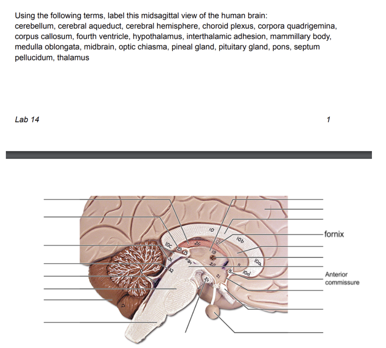
Inferior cerebellar peduncle connects the brainstem to the cerebellum. Four pairs of cranial nerves attach (XIII, IX, X, XII)
Describe the pons. What cranial nerves attach
A bridge between the midbrain and medulla oblongata. Three pairs of cranial nerves attach (V, VI, VII) Sends axons to the cerebellum through the middle cerebellar peduncle
Describe the midbrain
Lies between diencephalon and pons. Cerebral peduncles located on the ventral surface of the brain. Superior cerebellar peduncles connect midbrain to cerebellum.
Describe the cerebellum
Located dorsal to pons and medulla. Helps maintain equilibrium, smooths and coordinates body movements. Consists of two hemispheres connected by the vermis. Surface is folded into ridges called folia. Hemispheres each subdivided into the anterior lobe, posterior lobe, and flocculonodular lobe
What are the three divisions of the diencephalon and what are their functions?
The three divisions are the thalamus, hypothalamus, and epithalamus. The thalamus acts as a relay station for incoming sensory messages. Every part of the brain communicating with the cerebral cortex relays signals through the thalamus. The hypothalamus lies between the eoptic chiasm and mamillary bodies. It is the main visceral control center of the body. The epithalamus includes the pineal gland which secretes melatonin.
What are the fissures of the cerebrum?
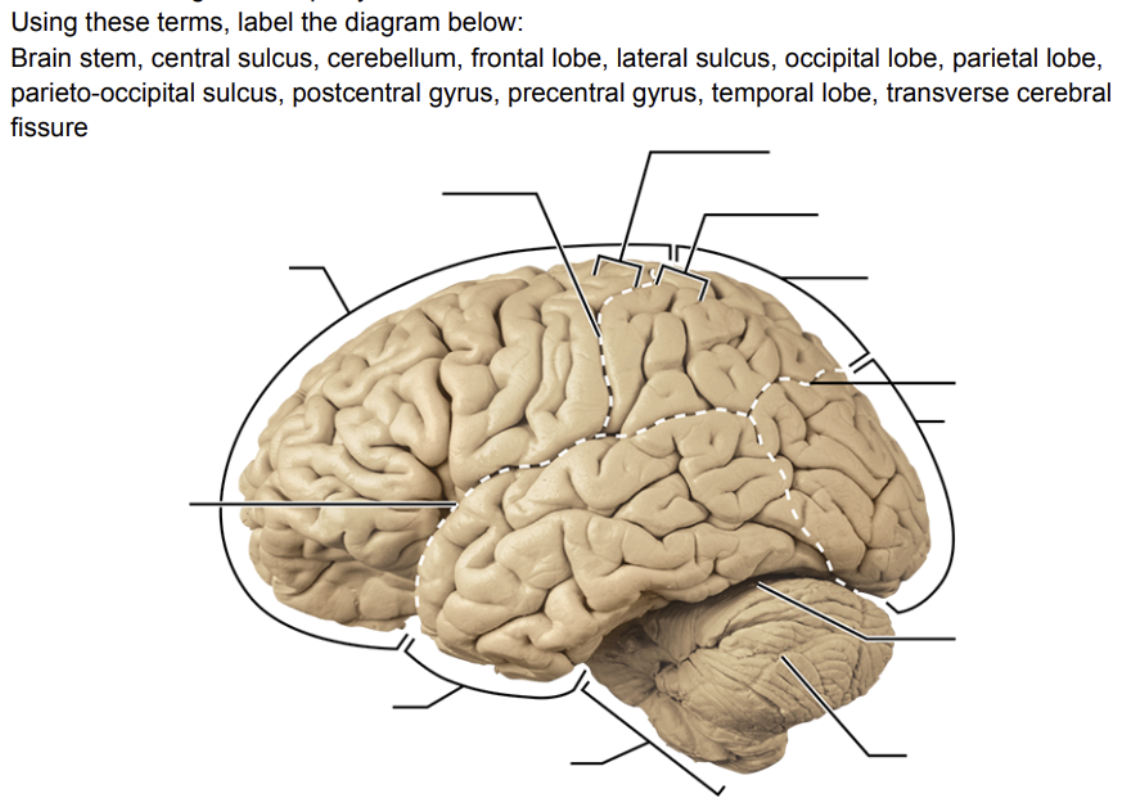
Transverse fissure (separates cerebrum and cerebellum) and the longitudinal fissure (separates cerebral hemispheres)
What are gyri and sulci?
Gyri are twisted ridges between sulci. Sulci are grooves on the surface of the cerebral hemispheres.
What does the cerebral cortex do?
It is the home of our conscious mind and enables us to be aware of ourselves and our sensations. Initiates and controls voluntary movements. Communicate, remember, and understand.
What are the cerebral cortex areas and what do they do?
There is a sensory area for each of the major senses each of which has an association area that processes sensory information, Multimodal association areas receive and integrate input from multiple regions of the cerebral cortex. Motor areas plan and initiate voluntary motor movements.
Describe the sensory areas
Primary somatosensory cortex is located along the postcentral gyrus. Primary visual cortex is on the medial part of the occipiaal lobe. Primary auditory cortex is located along the superior edge of the temporal lobe. Vestibular cortex is located in the posterior part of the insula and is responsible for conscious awareness of sense of balance. Gustatory cortex is located in the insula. Olfactory cortex lies on the medial aspect of the cerebrum.
Describe the motor areas
Primary motor cortex is located in the precentral gyrus. Speaking is only found in the left cerebral cortex and is controlled by two areas. Broca’s area is responsible for the direct primary motor area to stimulate the appropriate muscles for speaking and writing. Wernicke’s area helps us understand both the written and spoken word and sends the information to the Broca’s area.
Describe the meninges (dura mater, arachnoid mater, and pia mater)
The dura mater is the strongest of the meninges and is composed of two layers: periosteal layer and meningeal layer.
The arachnoid mater is located deep to the dura mater. Subarachnoid space contains CSF. Arachnoid villi project through the dura mater and allow csf to pass into the dural blood sinuses.
Pia mater is delicate connective tissue and clings tightly to the surface of the brain.
What is cerebrospinal fluid? How is it formed
Fills the hollow cavities of the brain and spinal cord. Provides a liquid cushion for the spinal cord and brain. It nourishes the brain and spinal cord, removes wastes, and carries chemical signals between parts of the CNS. It is formed in choroid plexuses in the brain ventricles.