Potassium
 Call Kai
 Learn
Learn Practice Test
 Spaced Repetition
 Match
Match Flashcards
 Knowt Play
1/50

There's no tags or description
Looks like no tags are added yet.
Name | Mastery | Learn | Test | Matching | Spaced | Call with Kai | Chat |
|---|
No analytics yet
Send a link to your students to track their progress
51 Terms
Why intracellular K+ is high
Na/K‑ATPase pumps 3 Na+ out, 2 K+ in → maintains −90 mV resting potential.

How do we regulate high circulating levels of K+? Why?
We shift K+ into the cells until they can be excreted by the kidneys or GI system. High levels of K+ can lead to arrythmias

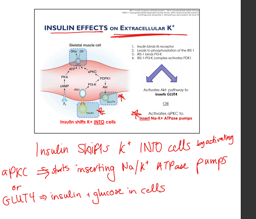
Effect of insulin on K+
Activates PI3K → aPKC → inserts Na/K‑ATPase + activates Akt which inserts GLUT4→ shifts K+ INTO cells.
Effect of β2‑agonists on K+
↑ cAMP/PKA → ↑ Na/K‑ATPase → shifts K+ INTO cells.
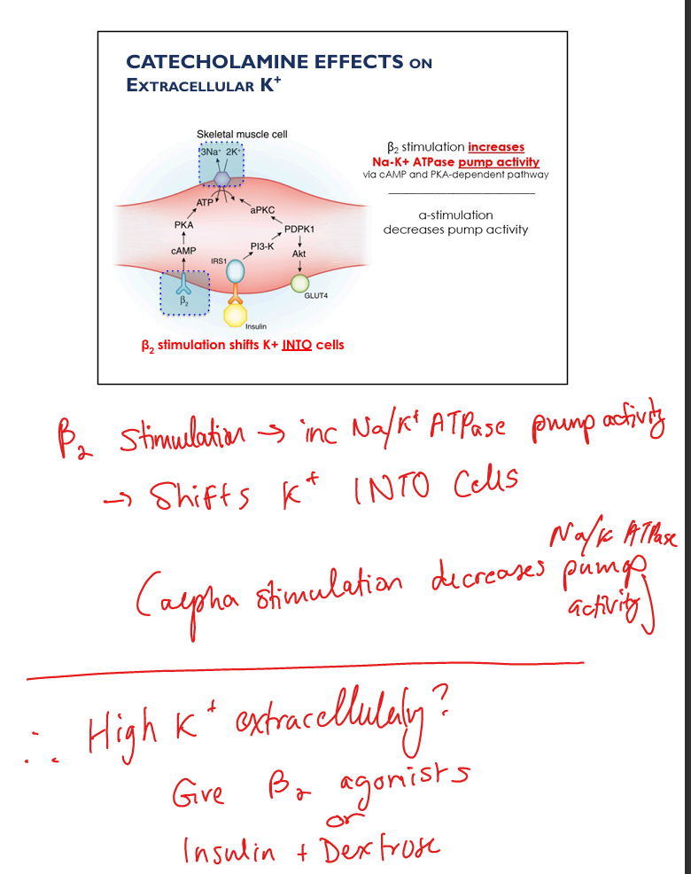
Effect of α‑agonists on K+
Decreases Na/K‑ATPase → K+ OUT of cells.

Effect of mineral (inorganic) acidosis on K+
↓ extracellular pH → ↓ Na/H exchanger1/Na/HCO3 exchanger → ↓ intracellular Na → ↓ Na/K‑ATPase → K+ OUT of cells.

Effect of organic acidosis on K+
OA− enters cell via MCT → ↑ intracellular Na → maintains Na/K‑ATPase → NO significant change in serum K+.

Effect of hyperosmolarity on K+
Water leaves cells → solvent drag pulls K+ OUT → hyperkalemia.
Effect of exercise on K+
K+ released from contracting muscle → transient ↑ serum K+.

Effect of cell lysis on K+
Massive K+ release → hyperkalemia (TLS, rhabdo, hemolysis).

Effect of digitalis on K+
Inhibits Na/K‑ATPase → K+ OUT → hyperkalemia.

Renal K+ handling (overview)
PT reabsorbs 65–70%, TAL reabsorbs 20%, DT/CCD secrete K+, α‑intercalated reabsorb K+ in hypokalemia.

PT K+ reabsorption mechanism
Passive paracellular via solvent drag + lumen‑positive voltage.

TAL K+ handling
NKCC2 reabsorbs K+, ROMK recycles K+ to lumen → creates lumen‑positive voltage → paracellular K+ reabsorption.

Effect of loop diuretics on K+
Inhibit NKCC2 → collapse lumen‑positive gradient → ↓ K+ reabsorption → hypokalemia.

Early DCT K+ handling
NaCl cotransporter (NCC) → ↑ intracellular Cl− → activates K‑Cl cotransporter → K+ secretion.

Effect of thiazides on K+
Inhibit NCC → ↑ Na delivery to CCD → ↑ K+ secretion → hypokalemia.

Collecting Duct principal cell K+ secretion
ENaC reabsorbs Na+ → lumen becomes negative → ROMK/Maxi‑K secrete K+.

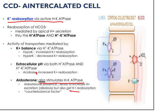
α‑intercalated cell K+ handling
H‑K ATPase reabsorbs K+ (↑ in hypokalemia, ↓ in hyperkalemia).
Major regulators of renal K+ secretion
Distal Na delivery, tubular flow rate, aldosterone, extracellular K+.

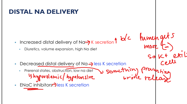
Effect of distal Na delivery
↑ Na → ↑ ENaC activity → ↑ lumen negativity → ↑ K+ secretion.
Effect of tubular flow rate on K+
High flow washes K+ away → ↑ gradient → ↑ K+ secretion.

Effect of aldosterone
↑ ENaC, ↑ Na/K‑ATPase, ↑ ROMK → ↑ K+ secretion.

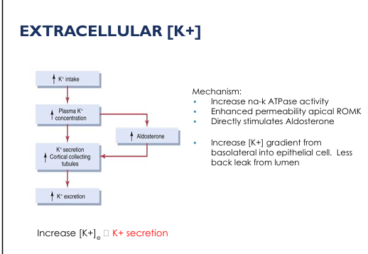
Effect of extracellular K+
Directly stimulates aldosterone + ↑ Na/K‑ATPase → ↑ K+ secretion.

Causes of hypokalemia (overview)
Poor intake, shifts into cells, renal losses, GI losses, skin losses, pseudohypokalemia.

Hypokalemia: intracellular shifts
Insulin, β‑agonists, alkalosis.

Hypokalemia: renal losses
Thiazides, loops, amphotericin B, cisplatin, hyperaldosteronism, Bartter, Gitelman, Liddle, RTA I/II.

Hypokalemia: GI losses
Diarrhea, vomiting, NG suction, laxatives.

Hypokalemia: pseudohypokalemia
Extreme leukocytosis (AML).

Hypokalemia clinical manifestations
Weakness, cramps, rhabdo, ileus, respiratory failure, arrhythmias.

Hypokalemia EKG findings
Flattened T waves, ST depression, prominent U waves, prolonged QU interval.


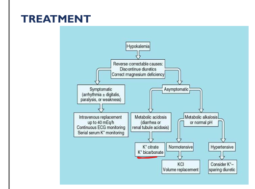
Hypokalemia treatment (general)
Correct Mg first, stop diuretics, give KCl, treat underlying cause.
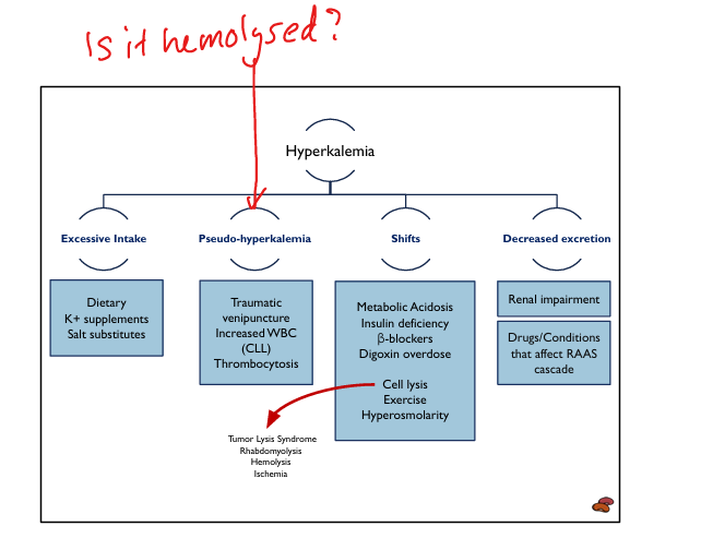
Causes of hyperkalemia (overview)
Excess intake, shifts out of cells, decreased excretion, pseudohyperkalemia.

Hyperkalemia: intracellular shifts
Acidosis (mineral), insulin deficiency, β‑blockers, digoxin toxicity, hyperosmolarity, exercise, cell lysis.
Hyperkalemia: decreased excretion
CKD, AKI, hypoaldosteronism, ACEi, ARBs, NSAIDs, K‑sparing diuretics, TMP‑SMX, cyclosporine, tacrolimus.

Hyperkalemia: pseudohyperkalemia
Hemolysis(bc lysed cell releases K), thrombocytosis, leukocytosis (CLL).

Hyperkalemia clinical manifestations
Muscle weakness, paralysis, arrhythmias, cardiac arrest.

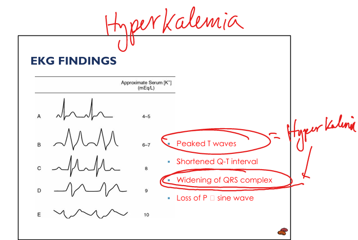
Hyperkalemia EKG findings
Peaked T waves → prolonged PR → loss of P → wide QRS → sine wave → asystole.

Hyperkalemia emergency stabilization
IV calcium gluconate or CaCl2 → stabilizes cardiac membrane.

Hyperkalemia redistribution therapies (what do you do if you C A BIG K)
IV insulin + dextrose, β2‑agonists, sodium bicarbonate (if acidotic).=>shift K into cell (CABIGK= Calcium gluconate, Albuterol(b2 agonist), Bicarb, Insulin, Glucose, Kayexalate(K+ binder))

Hyperkalemia elimination therapies
Loop diuretics, K‑binders (SPS, patiromer, SZC), dialysis.

Drugs causing hyperkalemia
ACEi, ARBs, spironolactone, eplerenone, amiloride, triamterene, TMP‑SMX, NSAIDs, heparin, cyclosporine, tacrolimus, β‑blockers, digoxin.

Why Mg deficiency worsens hypokalemia
Mg inhibits ROMK→allows for excess K+ to leak out into the urine→cant replenish and maintain internal K+

Why diarrhea causes low urinary K+
GI loss → low serum K+ → low filtered load → low urinary K+.
Expected K+ changes in acid‑base disorders
Mineral acidosis → ↑ K+
Expected K+ changes with insulin therapy
Insulin shifts K+ INTO cells → ↓ serum K+.
Expected K+ changes with β‑blockers
Block β2 → ↓ Na/K‑ATPase → ↑ serum K+.
Expected K+ changes with digoxin
Inhibits Na/K‑ATPase → ↑ serum K+.
Expected K+ changes with aldosterone
↑ K+ secretion → ↓ serum K+.
Expected K+ changes with hypoaldosteronism
↓ K+ secretion → ↑ serum K+.