Abdominal Viscera & Peritoneum
 Call Kai
 Learn
 Practice Test
 Spaced Repetition
 Match
 Flashcards
 Knowt Play
1/80

There's no tags or description
Looks like no tags are added yet.
81 Terms
In the development of the gut tube, what does the septum transversum become
the ventral mesentry
Define mesentery
A membranous fold attaching an organ to the body wall
Role of mesenteries and ligaments in the abdomen
provide pathways for vessels, nerves, and lymphatics to travel back and forth between abdominal organs
Location of ventral mesentery
Only found between the foregut and the upper portion of the duodenum
Ventral mesentery is pulled towards which side of the body
Ventral mesentery is pulled towards the right side of the body
What organs are associated with the ventral mesentery
liver
lesser curvature of the stomach
Location of the dorsal mesentery
Extends continuously from the foregut to the end of the hindgut
Dorsal mesentery is pulled towards which side of the body
Dorsal mesentery is pulled towards the left side of the body
What is the Dorsal mesentery associated with (what wall of the abdomen)
The posterior abdominal wall
How does the greater omentum form
The dorsal mesogastrium (attached to the stomach) grows and expands downward, forming a double-layered fold
Double layers of the peritoneum fuse together to form a large apron-like peritoneal fold hanging from the stomach - the greater omentum
What is the lesser omentum
A smaller peritoneal fold connecting the stomach to the liver.
When the anterior abdominal wall is reflected to expose the contents of the abdominal cavity, everything that can be seen is covered in what
peritoneum (thin, serous membrane)
What is the space between the parietal & visceral layers of peritoneum
the peritoneal cavity
What structures/otherwise are in the peritoneal cavity
No structures
Fluid occupies this space to allow movement and mobility of certain organs
The peritoneal cavity can be divided into 2. What are the 2 parts called
Greater sac – main compartment
Lesser sac – hidden from view
Where does the greater sac of the peritoneal cavity extend from & to
Extends from the Diaphragm to Pelvis
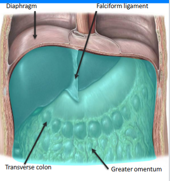
Where is the lesser sac of the peritoneal cavity / omental bursa
Posterior to → Stomach & greater omentum
Anterior to → Parietal peritoneum & pancreas
(yellow = lesser sac)
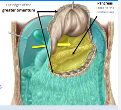
Name of opening between the greater & lesser sacs
The omental foramen / Epiploic foramen / Foramen of Winslow
(provides a direct communication between the greater & lesser sacs)
What is the sexual dimorphism with the peritoneal cavity
Males → Peritoneal cavity is completely closed
Females →There is an opening in the peritoneum through the uterine tubes (fallopian tubes & oviducts)
What parts of the abdominal viscera have a relationship to the peritoneum
All of it
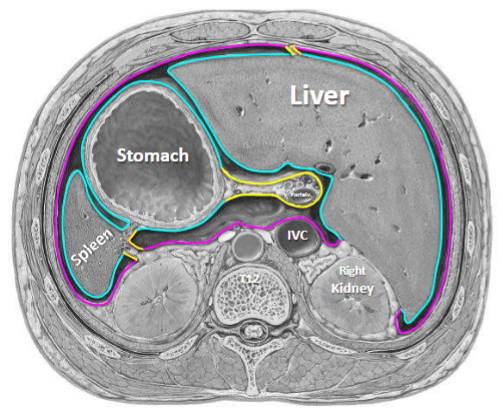
Where is the parietal layer of peritoneum
Covers and lines the internal walls of the abdomen
Pink part of diagram
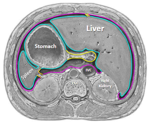
Where is the Visceral layer of peritoneum
Covers and invests abdominal organs that protrude into the peritoneal cavity
Blue part of diagram
In which layer of peritoneum is pain well localised. In which is it more generalised
Why is that the case?
Parietal = well localised (very sensitive) - Served by the same neurovasculature as the adjacent wall/structure
Visceral = generalised (referred) - Served by the same neurovasculature as the organ it is covering
What are some structures that are formed from peritoneum
Formed from more than one layer of peritoneum:
greater omentum
lesser omentum,
mesentery
peritoneal ligament
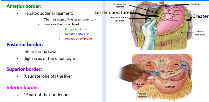
Boundaries of the Omental Foramen
Which of these is not a structure that is associated with, or derived from, peritoneum:
Fibrous pericardium
Mesentery
Omenta (greater & Lesser)
Peritoneal ligaments
Peritoneal recesses
Peritoneal fossae
Fibrous pericardium - this is to do with the heart
Role of mesentery
provide pathways for vessels, nerves, and lymphatics to pass between abdominal organs
helps to provide a continuous connection between the visceral and parietal peritoneum
connect organs to the posterior abdominal wall
What is contained within mesentery
Contains fat as well as the arteries, veins, lymphatic vessels, and nerves that supply the abdominal organs
Mesentery are named according to what
the organs they connect
Mesoesophagus (embryonic)
Mesogastrium (embryonic)
The “Mesentery” proper (of small bowel)
Mesoappendix
Transverse mesocolon
Sigmoid mesocolon
How many layers of peritoneum make up the lesser omentum & greater omentum
Lesser = 2 layered peritoneal fold
Greater = 4 layered peritoneal fold
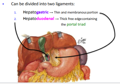
What does the lesser omentum connect
Connects the liver to the LESSER curvature of the stomach and the first part of the duodenum
The lesser omentum can be divided into 2 ligaments - what are they called and what is their structure
What does the greater omentum connect
Connects transverse colon to GREATER curvature of stomach
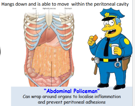
What does the greater omentum do
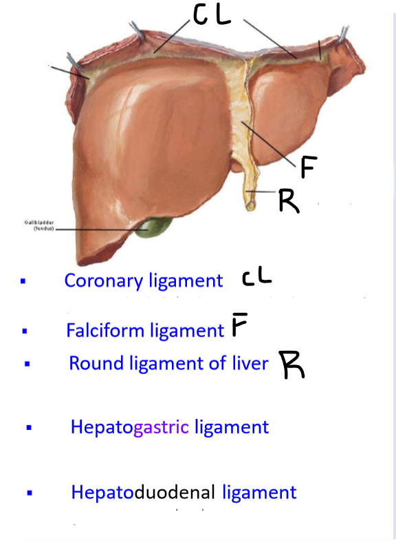
name the peritoneal ligaments of the liver
name the peritoneal ligaments of the stomach
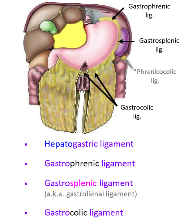
name the peritoneal ligaments of the spleen
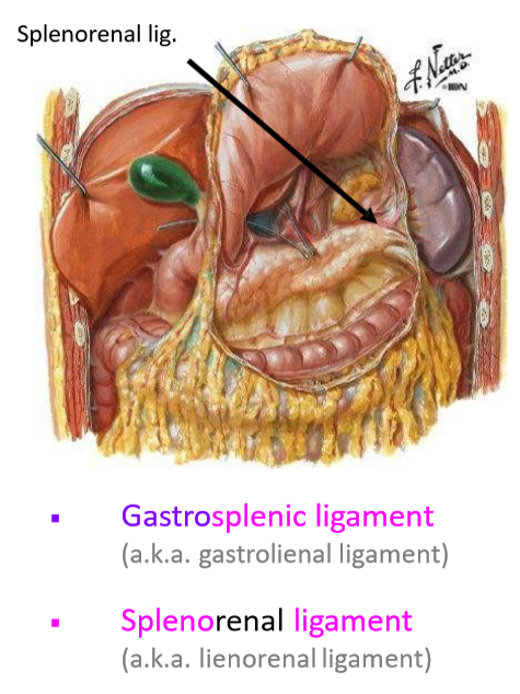
Ligaments can connect organs to what
Another organ or the abdominal wall
Name the peritoneal recesses
Paracolic gutters
Subphrenic recesses
Subhepatic space
Hepatorenal recess
Location of each of the peritoneal recesses
Paracolic gutters: between the abdominal wall and the lateral aspect of the ascending or descending colon
Subphrenic recesses: Located below the diaphragm
Subhepatic space: Immediately inferior to the liver
Hepatorenal recess: Extension of the subhepatic space between the visceral surface of the liver and the right kidney
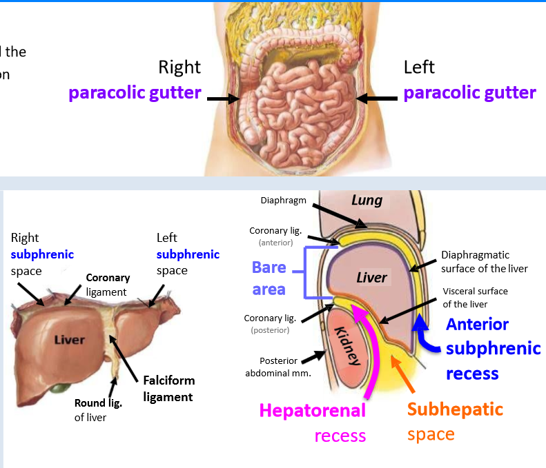
Function of Paracolic gutters
Allows communication between the supracolic and infracolic regions of the greater sac
Important for peritoneal fluid flow
Function of Subphrenic recesses
Potential site for fluid collection
Function of Hepatorenal recess
Potential site for fluid collection
Which recess is split into right & left.
What is it split by
Subphrenic recesses
Separated into left and right by the falciform ligament
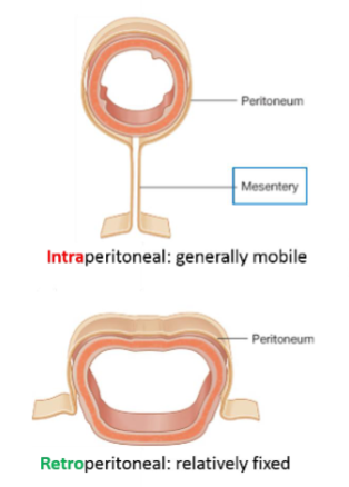
Abdominal organs can be intraperitoneal or retroperitoneal. What does this mean
Intraperitoneal organs
almost entirely wrapped in visceral peritoneum
usually suspended by mesentery in the abdominal cavity
highly mobile
Retroperitoneal organs
lie behind the peritoneal cavity with only part of its surface covered by peritoneum.
generally immobile
Which of these organs is intraperitoneal / retroperitoneal
Stomach
Duodenum
Small Intestine
Transverse colon
Rectum
Liver
Pancreas
Aorta
intraperitoneal | retroperitoneal
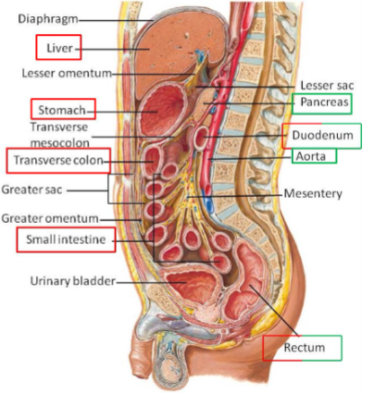
Name the intraperitoneal organs
• Stomach
• Liver
• Spleen
• Tail of the pancreas
• Duodenum: 1st part
• Jejunum
• Ileum
• Cecum & appendix
• Transverse colon
• Sigmoid colon
What defines something as primary or secondary retroperitoneal
1° - Primary retroperitoneal structures are retroperitoneal from the start of their development.
2° - Secondary retroperitoneal structures were once suspended within the abdominal cavity by mesentery but migrated posteriorly to end up behind the peritoneum.
Name the primary & secondary retroperitoneal structures of the abdomen
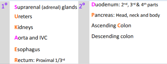
What are infraperitoneal organs
& example
Defined as being beneath the peritoneal cavity
Only part of its surface is covered by peritoneum
E.g. rectum (distal 2/3rds) & urinary bladder
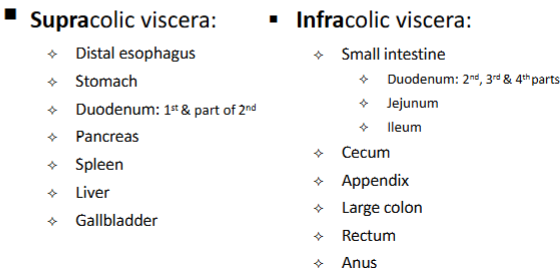
What defines the boundary between supra & infracolic abdominal compartments
the transverse mesocolon
Where is defined as supracolic
Visceral structures below the diaphragm and above the transverse mesocolon
Label these as either supra/infracolic viscera:
Pancreas
Appendix
Liver
Distal esophagus
Rectum
Gallbladder
Large colon
Duodenum
Cecum
Stomach
Spleen
Anus
Small intestine
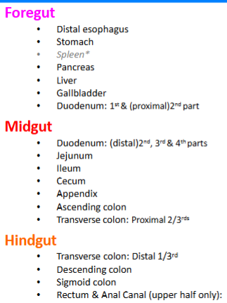
Split these into foregut, midgut & hindgut:
Duodenum
Jejunum
Stomach
Pancreas
Appendix
Sigmoid colon
Cecum
Distal esophagus
Gallbladder
Rectum & Anal Canal
Transverse colon
Ascending colon
Spleen*
Ileum
Liver
Descending colon
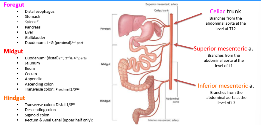
Arterial supply to foregut
Celiac trunk: Branches from the abdominal aorta at the level of T12
Arterial supply to midgut
Superior mesenteric artery: Branches from the abdominal aorta at the level of L1
Arterial supply to hindgut
Inferior mesenteric artery: Branches from the abdominal aorta at the level of L3
What is peritonitis
Infection can occur if gas, faecal matter or bacteria enter the peritoneal cavity which would result in inflammation of the peritoneum
Can be caused by exudate (fluid/pus that seeped out from an inflamed organ/vessel) , ascitic fluid & paracentesis (surgical puncture of peritoneal cavity)
What are Peritoneal adhesions & examples
fibrous bands that form abnormal connections, e.g.:
▪ Visceral peritoneum attaching to an adjacent organ
▪ Visceral peritoneum attaching to the parietal peritoneum
How do Peritoneal adhesions form
Form as a result of damage (acquired through injury, infection or surgery) to the peritoneal surface when sticky fibrin appears in order to assist with the healing process
Problems & complications with Peritoneal adhesions
Adhesions limit the normal movement of viscera and could lead to complications:
intestinal obstruction (a.k.a. volvulus)
Chronic pain
What is a laparotomy
Surgical incision into abdominal cavity prior to major surgery
What is an adhesiotomy
Surgical separation of adhesions
What is an omental bursa herniation
Part of GI tract (usually a loop of the small intestine) can pass through the omental foramen (Epiploic foramen of Winslow) and become twisted and strangulated inside the lesser sac
Is an omental bursa herniation common/rare
rare
What is treatment for an omental bursa herniation
Surgery is required to manually reposition and free the trapped intestinal tract
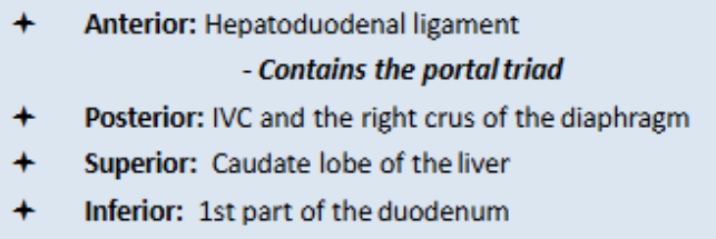
Boundaries of the omental (epiploic) foramen
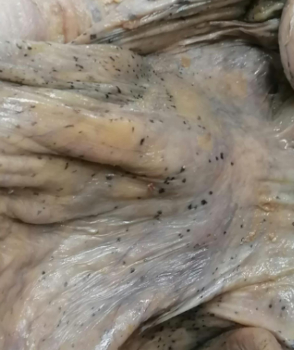
what are these
Dark macular spots found on peritoneal layers (visceral & parietal)
From intraperitoneal spillage & spread of India ink from preoperative endoscopic tattooing
At what point is an embryo called a foetus
At week 9
Where does the foregut extend from
From the mouth to the 1st part of the duodenum
Is the peritoneum attached to all of the abdominal wall (anterior & posterior)
It is attached to the complete posterior wall & some of the front
2 layers of peritoneum come together to form what 3 types of structures
a ligament, mesentery, or omentum
largest continuous compartment in the body
Peritoneum
What is the Pringle manoeuvre
a surgical technique used to control bleeding during liver surgery, particularly when there is bleeding from the liver or its vascular structures.
It involves temporarily clamping the hepatoduodenal ligament, which contains the portal vein, hepatic artery, and common bile duct. By clamping this ligament, blood flow to the liver is reduced, which helps control bleeding from the liver, especially during procedures like liver resections or trauma surgery.
The maneuver is often used to determine whether the source of bleeding is from the liver itself or from other areas, such as the surrounding blood vessels. If bleeding stops when the Pringle maneuver is applied, the bleeding is likely from the liver's blood supply. If bleeding continues, the source may be outside of the liver (e.g., from the retroperitoneum).
Some organs are connected to the abdominal wall by a mesentery and can move within the abdominal cavity. Name some of these organs
Small intestine
Transverse colon
Sigmoid colon
Appendix
The gastrocolic ligament connects the stomach to what
The transverse colon
The Falciform ligament connects the liver to what
anterior abdominal wall and the diaphragm
Which ligament is important in hypertension
The coronary ligament
Which organs have mesentery and which don’t (which groups of organs)
Intraperitoneal organs have mesentery
Retroperitoneal organs don’t have mesentery
The embryonic aorta has how many unpaired branches
Name them
The embryonic aorta has 3 unpaired branches:
the celiac trunk
the superior mesenteric artery
the inferior mesenteric artery
The appendix is associated with what dermatone
T10