Unit 1: Cells (copy)
 Call Kai
 Learn
 Practice Test
 Spaced Repetition
 Match
 Flashcards
 Knowt Play
1/79

Earn XP
Description and Tags
80 Terms
Cells
Basic structural unit of all living organisms
Cell Theory
Cells are SMALLEST unit of life
Organisms are made of ≥1 cell
Cells arise from pre-existing cells
Structures common to cells in all living organisms + Reasons
Cell Membrane
Phospholipids spontaneously arrange into a bilayer
Encloses cell + Protects contents from environment
Controls interactions between cell contents and exterior (Signalling & transport)
Cytoplasm
Mainly water matrix called cytosol (Contains all ingredients i.e. carbon compounds, ions + Provides medium for metabolic reactions); Responsible for giving cell its shape
Ribosomes (?)
DNA
Able to form large molecules which carries and transfers genetic material between organisms; Controls production of enzyme and proteins within organism
Prokaryotic Cell Structure (HINT: 10 features)
Cell wall: Rigid outer layer of peptidoglycan
Function: Maintains shape and prevents lysisPlasma membrane: Semi-permeable; Selective barrier surrounding cell
Naked loop of DNA: Bacterial chromosome; Circular
70S ribosomes: RNA + Protein Complex
Function: Polypeptide synthesisCytoplasm: Internal fluid
Plasmid: Autonomous circular DNA molecules; Transferrable between bacteria (Horizontal gene transfer)
Nucleoid Region: Where DNA is located in cytoplasm; Non-compartamentalized
Function: Cell control and reproductionSlime capsule: Thick polysaccharide layer
Function: Protection against desiccation and phagocytosisFlagella: Long, slender projections; Contains motor protein
Function: MovementPili: Hair-like extensions
Function: Attachment pili = Adhesion to surfaces; Sex pili = Bacterial conjugation
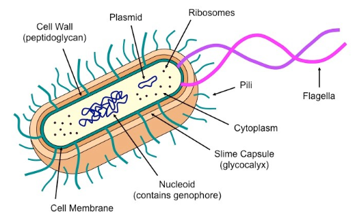
Types of Prokaryotic Cell Structure (+ Relevant Examples)
Gram-positive eubacteria
Thick peptidoglycan layer
No outer lipid membrane
—> Retains crystal violet stainExample: Bacillus, Staphylococcus
Gram-negative
Phytoplasma
Mycoplasma
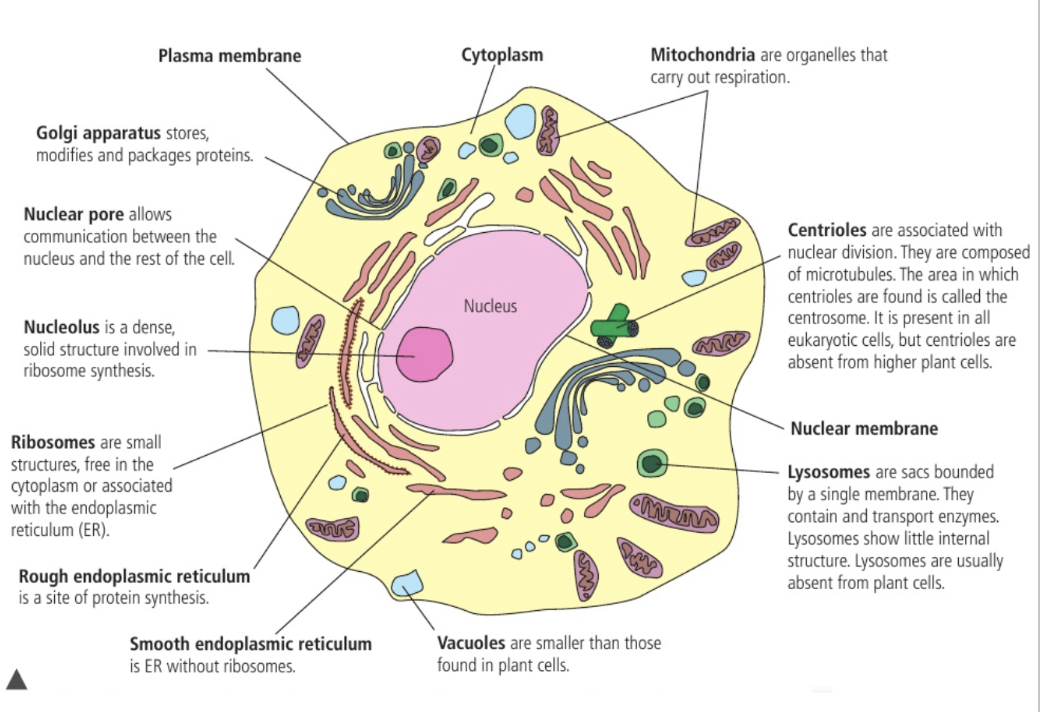
Common Eukaryotic Cell Structures (HINT: 4)
Plasma Membrane
Cytoplasm
80S Ribosomes
Bound = Protein synthesis for transport
Free-moving = Protein synthesis for cell
Nucleus
DNA bound to histones; Contained in a double membrane, nuclear envelope with nuclear pores → Controls movement of molecules between nucleus and cytoplasm
Organelles in Animal / Plant Cells (HINT: 11, 4 special)
Nucleus
Mitochondria
Has its own genome; Reproduce by fission; Double membrane-bound
Makes energy, stores Ca, Controls cell growth and deathRough Endoplasmic Reticulum (RER)
Surface is covered in ribosomes
Synthesise proteins for use outside of cell
Smooth Endoplasmic Reticulum (SER)
Synthesises lipid and hormones
Golgi apparatus
Processing + Secretion of protein; Package proteins and lipids into vesicles for delivery out of cell
Transport vesicles are received on cis side
Modified as it moves through cisternae + Attachment of signalling chemical which directs it to destination
Packaged and transported on trans side
Vesicles
Small sacs that transports molecules into cell by fusing with membrane
Vacuoles
Small and numerous
Large space; If central area = Central vacuole
Store food, toxins, metabolic waste, water + Uptake of water → Rigidity, Provide nutrition
Lysosomes
Sacs bound by single membrane
Contain and transport enzymes
Cytoskeleton
System of protein fibres (microtubules and microfilaments)
Hold organelles in place; Maintain structure and shape of cell
Centrioles
At centrosome
Assembles microtubules for structure & movement + Nuclear division
Chloroplasts
A plastid (Organelle for manufacturing / storing energy)
Converts light to chemical energy via photosynthesis
Kingdoms of Eukaryotes + Differences
Protista
Uni OR Multicellular; No specialised tissue
Fungi
Chitin cell wall; Nutrition via heterotrophic absorption (Obtain by absorption from environment; Broken down externally)
Plantae
Cellulose cell wall; Autotrophic nutrition (Photosynthesis)
Animalia
No cell wall; Heterotrophic ingestion for nutrients
Compare & Contrast Prokaryotic and Eukaryotic Cells (Min. 3 for Each)
PROKARYOTES
Simple structure
Small size
Peptidoglycan cell wall
70S Ribosomes
Unicellular
No membrane bound organelles
BOTH
DNA
Ribosomes
Cell membrane
Cytoplasm
EUKARYOTES
Complex structure
Prominent nucleus
Large size
Chitin / Cellulose cell wall (some)
Uni or Multicellular
80S Ribosomes
Membrane bound organelles
Processes of Life in Unicellular Organisms (HINT: MR H N GERM) + In Paramecium
Homeostasis: Maintenance of constant internal environment
Maintains internal conditions e.g. expel water by contractile vacuolesMetabolism: Chemical reactions within organism
Produce enzymes to catalyse chemical reactions in cytoplasmNutrition: Ability to acquire energy to sustain life
Feeds on smaller organisms by endocytosis (Ingestion and digestion in vesicles)Movement: Ability to move
Thousands of cilia beatExcretion: Ability to release unnecessary / harmful materials into environment
Expel metabolic waste (CO2) from respiration by diffusionGrowth: Development
Increase in size and dry mass by accumulating organic matter and minerals from foodResponse to stimuli: Include environmental adaptations
Reverses direction of movement upon contact with solid objectReproduction: Ability to produce offspring
Asexual reproduction via mitosis or sexual reproduction via meiosis and gametes
Differences in Eukaryotic Cell Structures (Animals, fungi, and plants)
Comment on: Presence and composition of cell walls, differences in size and function of vacuoles, presence of chloroplasts and other plastids, and presence of centrioles and centrosomes, basal bodies, cilia, flagella, and budding scar
Presence / Composition of Cell Wall
Fungi: Chitin
Animal: N/A
Plant: CelluloseSize and Function of Vacuoles
Fungi: Small and numerous; Degradation of molecules
Animal: Small and numerous; Store carbohydrates as glycogen
Plant: Large; Store carbohydrates as starch; Water uptake
NOTE: Contractile vacuoles in unicellular eukaryotesPlastids
Fungi: Mitochondria
Animal: Mitochondria
Plant: Mitochondria, ChloroplastsCentrioles
Fungi: Centrosomes ONLY
Animal: Yes + Centrosomes
Plant: Centrosomes ONLYCilia
Fungi: Some
Animal: Some
Plant: N/AFlagella
Fungi: Some
Animal: Some
Plant: SomeBasal Bodies
Fungi: N/A
Animal: Yes
Plant: N/ABudding Scar
Fungi: Yes
Animal: N/A
Plant: N/A
Atypical Cell Structures in Eukaryotes (Number of nuclei)
Aseptate fungal hyphae
Multi-nucleated, long tube-like structuresSkeletal muscle: Formed by fusing smaller cells
Large cells (~30 mm long); Multi-nucleated (Several hundred)Red blood cell
Anucleated → Greater haemoglobin capacity → Transport more oxygenPhloem sieve tube elements
Anucleated → Low resistance for substances moving through
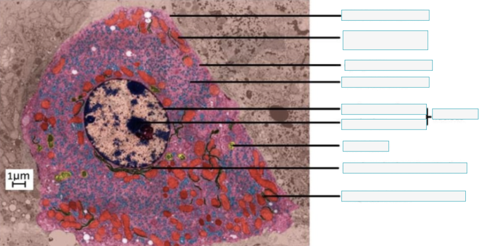
Identify the organelles (1)
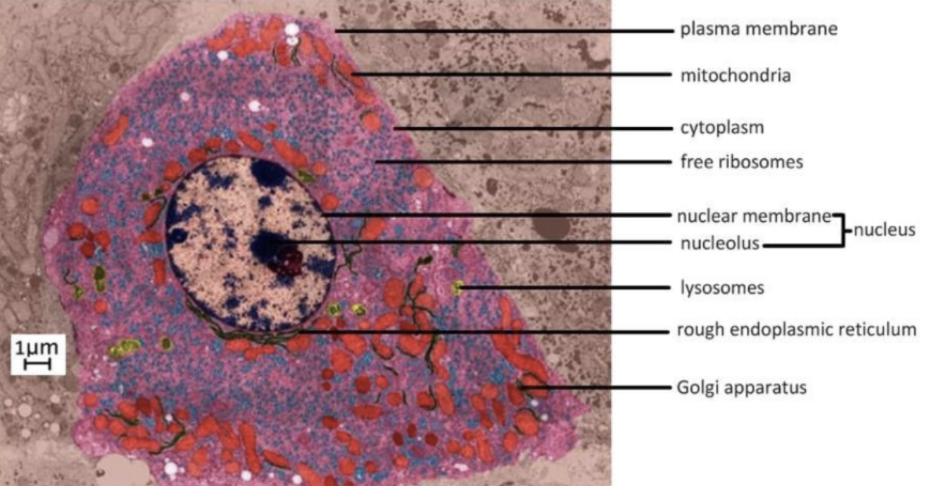
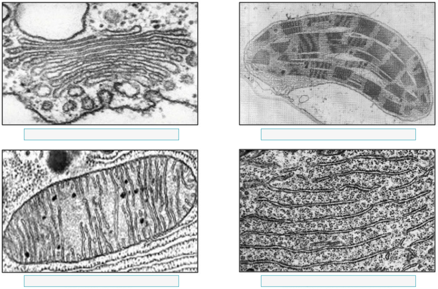
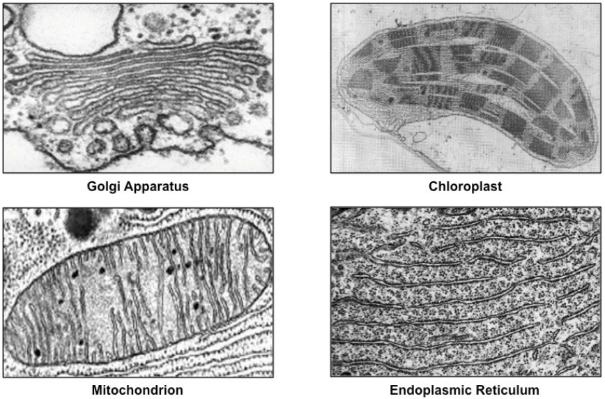
Identify the organelles (2)
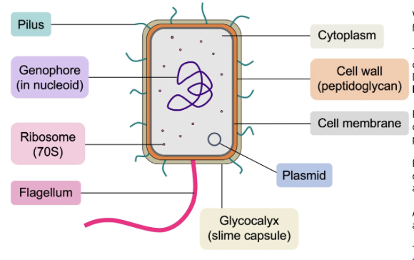
Draw a labelled diagram of prokaryote (9 features!)
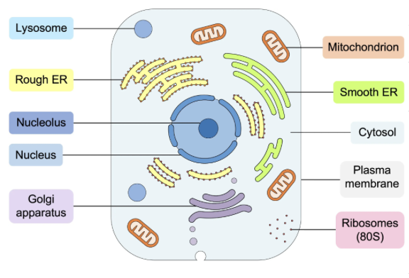
Draw a labelled diagram of animal cell (10 features!)
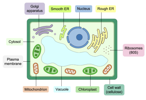
Draw a labelled diagram of plant cell (11 features!)
How to calculate magnification
Magnification (X) = Image Size (Measured by ruler) / Actual Size
Note: All should be in same units
Developments in Microscopy + Advantages
Light Microscopy: View living specimens in natural colors
Advantages –
Inexpensive, simple and easy specimen preparation, live or dead specimen
Fluorescent Stains
Binds specific cellular components to image specific structures; Detailed visibilityImmunofluorescence
Uses antibodies conjugated (bound) to fluorescent probes → Specifically target a cellular component; Often used to detect viral proteins in infected cells
Electron Microscope
Advantages –
Over 500,000X magnification, higher resolution
Scanning Electron Miscrope
Scatter electrons across surface to differentiate depth → 3D MapsTransmission Electron Miscrope
Pass electrons through specimens → Cross-section ImageFreeze Fracture Electron Microscopy
Freeze samples then break sample into small pieces → See internal structure; Visualise structures not normally visibleCryogenic Electron Microscopy
Freeze samples to fix them into firm / stable structure; Improved resolution of image + Reduce damage from electron beam
Origins of Eukaryotic Cells by Endosymbiosis
Larger prokaryotes engulfed smaller ones and formed an endosymbiotic relationship, allowing them to exist inside them. The large one provided protection from environment, and the smaller provided energy.
Evidence for Endosymbiotic Theory
Chloroplasts and mitochondria:
Have double membranes
Can replicate via binary fission
Contain smaller ribosomes
Have small circular strands of DNA
Cell Differentiation
Process for developing specialised tissues in multicellular organisms
Basis for Differentiation
Different patterns of gene expression triggered by environmental changes
Evolution of Multicellularity
Multicellularity has evolved repeatedly (From prokaryotes → single-celled eukaryotes → multi celled eukaryotes, etc.)
Include fingi, eukaryotic algae, and all plants and animals
Advantages of Multicellularity
Allows larger body size and cell specialization
Properties of Stem Cells
Capable to divide endlessly and differentiate along different pathways
Differences Between Totipotent, Pluripotent, and Multipotent Stem Cells
Totipotent: Can become any cell type
Cells in early-stage animal embryosPluripotent: Cab become a range of different checks
Endoderm, Mesoderm, Ectoderm LineMultipotent: Can only become related cell types
Stem cells in adult tissues e.g. bone marrow
Location and Function of Stem Cell Niches in Adult Humans
Stem cell niche can maintain the cells or promote their proliferation and differentiation
Bone Marrow
Can become different types of blood cellsHair Follicles
Produce cells for hair growth, repair, regeneration + Immune cells and nerves
Production of Unspecialised Cells
During sexual reproduction, gametes fuse together
As a result of fertilisation, a zygote is formed
The single-celled zygote divides rapidly and form unspecialised, embryonic stem cells
Impacts of Gradients on Gene Expression (Within early-stage embryos)
Cell’s position in the embryo → Concentration of morphogens → Different gene expression patterns → Forms specialised cell types
At the anterior of the embryo, low retinoic acid → thyroid development
At the posterior, high retinoic acid → colon
Cell Size as an Aspect of Specialization
Sperm (5 μm, 60 μm in length)
Long flagellum provides propulsion to rapidly reach eggRed Blood Cells (7-8 μm in diameter)
Allows it to manoeuvre through capillariesEgg (~100 μm in diameter)
Contain many nutrientsStriated Muscle Fibres (narrow - 20-100 μm, long - 10 cm)
Coordinate contraction + Significant muscle force generationNeurons (narrow - 10 μm, long - 1 m)
Efficient and rapid transmission of nerve impulses from brain to spinal cord to muscles for movement
Surface Area to Volume Ratio
Large SA:Vol ratio → decrease rate of exchanges → Divide to maintain low SA: Vol ratio
Entry and exit of substances in cell; Rate of exchange
Movement of substances within cell from one end to another
Adaptations to Increase SA:Vol Ratios in Cells + Examples
Flattening of cells
Erythrocytes: High SA:Vol ratio → Large SA for oxygen to bind to hemoglobin
Microvili
Apical surface (facing lumen / inside) of proximal convoluted tubule: Large SA to maximize absorption and secretion of solutes (Na+, Cl-, glucose, etc.)
Invagination
Basal side (facing blood at base of cell) of proximal convoluted tubule: Increase rate of solute transfer from epithelial cells into blood
Adaptation for Type I and Type II Pneumocytes in Alveoli
Type I Pneumocytes (~5%)
Extreme thinness + Squamous → Reduce distance for diffusion
Type II Pneumocytes (~95%)
Presence of many lamellar bodies (secretary vesicles) in cytoplasm to discharge surfactant to alveolar lumen → Prevent alveolar walls sticking together; Reduce surface tension
Adaptations of Cardiac Muscle Cells and Striated Muscle Fibres
Both have contractile myofibrils
Cardiac muscle cells
Branched → Create space between muscle cells → Flexible; Allow rapid heart pump
Short cells; One nucleus → Synchronised contractions
Intercalated discs → Facilitate rapid signal transmission → Coordinated contractions + Strong cell-to-cell adhesion → Structural integrity during pumping
Striated muscles (Connected to skeleton)
Unbranched → Provides strength
Long cells → Increase overlap of myosin (converts chemical to mechanical energy) + Actin (for muscle contraction) → Generate movement
Multinucleated → Support constant need for protein production + Tissue repair
Adaptations of Sperm and Egg Cells (HINT: 6 for sperm,
Sperm
Enzymes in acrosome (head) region → Help with entry
Streamline shape → Speed and movement
Lots of mitochondria → Produce ATP for energy to swim
Flexible microtubules in tail → Swimming
Lack of other organelles → Conserve energy to fulfil function
Small size (See before) → Travel efficiently and attach to surface of egg
Egg
Zona pellucida and cortical granules (cell membrane structures) undergoes chemical reactions when sperm enters → Prevent additional sperm from entering
Lots of mitochondria → Produce energy
Cytoplasm contains many nutrients → Support growth of early embryo; Acts as stored food supply
Large SA and size (See before) → Facilitate more opportunities for sperm to enter
Behaviour of Phospholipids and Amphipathic Lipids in Water
Naturally form continuous sheet-like bilayers
How do membranes function as effective barriers between aqueous solutions?
Hydrophobic hydrocarbon chains have LOW permeability to large molecules and hydrophilic particles (IONS, POLAR molecules)
Example of Simple Diffusion Across Membranes
Due to phospholipid bilayers being selectively permeable, only oxygen and carbon dioxide molecules and other small, non-polar molecules are able to pass through between phospholipids
Diverse Structures, Locations, and Functions of Membrane Proteins (HINT: TRACIE)
Integral Proteins (Embedded; Amphipathic; Mono- or polytonic i.e. penetrating one or two layers)
α-helix: For recognition (Receptors)
Helical bundle: Enzymes, transporters, receptors
β-barrel: For transport (Channel proteins)
Peripheral Proteins (Monotopic or surface only; Usually anchored to an integral protein)
Enzymes, transporters, anchorage
Function:
Transport
Receptors
Anchorage
Cell recognition
Intracellular joints (Junctions between cells)
Enzymes
Osmosis and Role of Aquaporins
Passive movement of water across a selective permeable membrane
Occurs due to differences in solute concentrations; Movement occurs until concentrations are equal → Changes cell size
Hypertonic = High solute concentration environment
Plasmolysed cells; Water moves out of cell
Isotonic = Equal solute concentration environment
Normal cells; No net movement
Hypotonic = Low solute concentration environment
Turgid cells; Water moves into of cell
Aquaporins (Integral protein) = Water Channels
As polar molecules cannot pass quickly through membrane, aids faster water transportation in response to solute concentrations
How does the structure of channel proteins make membranes selectively permeable? (Channel & Carrier proteins)
They allow specific ions to diffuse through channels are open and not when they are closed.
Channel proteins
Have pores → Allows specific size + charge of molecules pass
Carry only water-soluble molecules; Specific to ions
Close and open in response to electrochemical / mechanical signals
Carrier proteins
Changes shape to carry ion across membrane
Can transport along (Facilitated Diffusion) OR against concentration gradient (Active Transport)
Types of Cellular Transport
Passive
Simple diffusion
Facilitated diffusion
Carrier proteins
Active
Pump
Carrier proteins
Active transport (Pump proteins)
Protein pumps = Globular proteins; Embedded in membrane
Uses ATP to transfer specific particles across membrane; Movement against concentration gradient
Example of Pump Protein (Sodium-Potassium pump)
Selectivity in Membrane Permeability
Allow selective permeability in membrane:
Facilitated diffusion
Active Transport
Not selective; Depends only on size and polarity of particles:
Simple diffusion
Structure and Function of Glycoproteins and Glycolipids
Glycoproteins: Cell membrane proteins attached to carbohydrate chains
Glycolipids: Phospholipids attached to carbohydrate chains
On extracellular side of membrane
Assist in cell identification + Adhesion
E.g. antigens on blood types
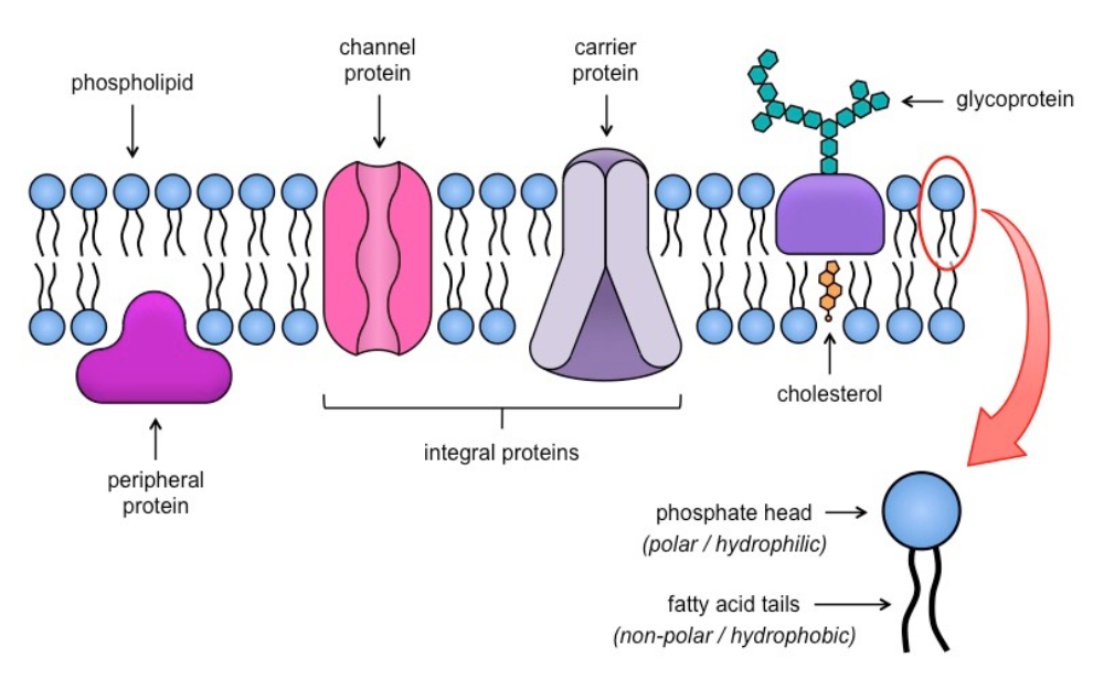
Draw a diagram of the Fluid Mosaic Model of Membrane Structure
Include:
Peripheral + Integral proteins
Glycoproteins
Phospholipids
Cholesterol
+ Indicate hydrophobic + hydrophilic regions
Relationships between fatty acid composition of lipid bilayers and their fluidity
Unsaturated fatty acids (kink) in lipid bilayers → Lower melting points → Fluid → Flexible at temperatures experienced by a cell
Saturated fatty acids (straight) → Higher melting points → Stronger at high temperatures
Example of adaptions in membrane composition in relation to habitat
In unicellular organisms, e.g. bacteria
Have fatty acid desaturase → Speed up reactions; Increase double bonds in fatty acid tails → More unsaturated fatty acids → More flexible structure at different temperatures → Able to withstand changes in temperature
Cholesterol and Membrane Fluidity in animal cells
Acts as a modulator of membrane fluidity → Stabilises membrane at higher temperature / Prevents stiffening at lower temperatures
Many are embedded in membranes
Endocytosis and Exocytosis + Examples
Endocytosis
Invagination of membrane / Membrane is pinched into a vesicle forms flask-like structure → Envelops substances into vesicle; Brings materials into cell
Phagocytosis: Large cells & Particles
Pinocytosis: Water & Liquids
Receptor-mediated: Specific molecules
Exocytosis
Vesicles containing substances from Golgi apparatus travels to membrane → Fuses to bilayer → Releases contents to environment
Insulin released into bloodstream by pancreatic cells
Neurotransmitters released at synapses
Note: Fusion of vesicles in exocytosis ADDS phospholipids to bilayer which offsets those TAKEN during endocytosis
Types of Gated Ion Channels + Examples
Facilitated diffusion; Gated = Open/close in response to chemical or mechanical stimuli
Voltage-gated Ion Channels
Sodium / Potassium Channels
Sodium ions bind to protein pump and move into neurone; Depolarises membrane
Potassium channels open → Potassium ions move outside of cell; Repolarization
Ligand-gated Ion Channels (Neurotransmitters)
Nicotinic Acetylcholine
Acetylcholine binds to a receptor protein on the postsynaptic neurone
Conformational change to channel
Opens channel; Sodium, potassium, and calcium ion to diffuse into cytoplasm of postsynaptic neurone (along electrochemical gradient)
Makes the inside of postsynaptic neurone relatively positive (depolarised) and generates impulse
Exchange Transporters (Sodium-Potassium Pumps)
Helps maintain membrane resting potential
Moves ions against concentration gradient (Movement TOWARDS HIGH solute concentration)
Generally, cells are more negative inside than outside cell
Initially open on intracellular side of cell
THREE sodium ions bind to pump protein → Phosphorylates it; Changes configuration
Pump opens to extracellular side
Sodium ions are released; TWO Potassium ions bind
Pump is dephosphorylated; Reverts configuration
Potassium ions are released into intracellular matrix
More potassium ions INSIDE cell; More sodium ions OUTSIDE cell
Net Movement = Potassium ions leak OUT; Sodium ions leak IN through leakage channels
NOTE: More potassium leakage channels → More permeable to potassium at rest → More potassium leak out → Relatively negative charge inside cell
Indirect Active Transport (Sodium-Dependent Glucose Cotransporters)
Used for glucose (re)absorption by cells in small intestine and nephron
Sodium-potassium pump uses ATP to create electrochemical gradient of sodium ions (High conc. OUTSIDE cell; Low conc. INSIDE cell)
Two sodium ion and one glucose molecule binds to a cotransporter
Ions are transported across membrane ALONG its gradient, carrying glucose molecules as well AGAINST its gradient
Cell-Adhesion Molecules
Different types of CAM → Different types of cell to cell junctions
Desmosomes
Form sturdy + flexible sheets of cells in organs → Hold cells together in tissues during stretchingPlasmodesmata (Plants)
Tubes connecting cytoplasm of adjacent cells → Exchange materials i.e. water, small solutes
Conditions on Early Earth and the Prebiotic Formation of Carbon Compounds
Lack free oxygen → Absence of ozone layer
Higher concentrations of CO2 and CH2 → Higher temperatures + UV light penetration
Caused a variety of carbon compounds to form spontaneously by chemical processes that no longer occur
Smallest units of self-sustaining life
Cells
Differences between living and nonliving (Why are viruses non-living?)
Viruses…
Cannot metabolise
Cannot perform homeostasis
Cannot move
Cannot grow
Cannot reproduce independently
Cannot excrete metabolic waste
Cannot intake nutrients
Spontaneous Origin of Cells
Cells = Highly complex structures; Only produced by division of pre-existing cells
Requirements for Evolution of First Cells
Catalysis
Self-replication
Self-assembly
Emergence of compartmentalisation
Evidence for Origin of Carbon Compounds
Miller-Urey Experiment
Simulated early Earth conditions; Showed organic compounds (hydrocarbons, amino acids) can be formed from inorganic precursor molecules
Water = Ocean
Gas inlet = Addition of methane, ammonia and hydrogen
Heat = Produce water vapor
Electrical sparks = Electrical storms
Criticism of Miller Urey Experiment
Earth’s early atmosphere was different from the composition of gases used in experiment
Glassware, medium of experiment, could have reacted
Formation of Vesicles
By coalescence (merging) fatty acids into spherical bilayers
Needed to allow internal chemistry to become different from outside of compartment
RNA as Presumed First Genetic Material (HINT: 2 Points)
RNA can…
Be replicated
Has some catalytic activity
(Both genetic material + enzyme)
Ribozyme in ribosome catalyses peptide bond formation during protein synthesis
Evidence for Last Universal Common Ancestor (LUCA)
Universal genetic code
Shared genes across all organisms
Other forms of life may have evolved but became extinct due to competition from LUCA and its descendants
Approaches Used for Estimation Dates
Studying fossils via radiometric techniques based on half-life of radioactive isotopes
Genetic dating / Analysis
Evidence for Evolution of LUCA Near Hydrothermal Vents
Fossilised evidence of life from ancient seafloor hydrothermal vent precipitates
Evidence of conserved sequences from genomic analysis; Common genetic sequences in organisms near vents → Suggest single ancestor
Presence of mineral-rich environment + Necessary acidic/basic fluids for chemical reactions
Presence of hydrogen and CO2 → Reducing environment for carbon compound formation
Organelles
Discrete subunits of cells; Adapted to perform specific functions
Which cell components ARE and ARE NOT considered organelles? (HINT: 3 & 4)
NOT considered:
Cell wall
Cytoskeleton
Cytoplasm
CONSIDERED:
Nuclei
Vesicles
Ribosomes
Plasma membrane
Cilia/Flagella
Golgi apparatus
Mitochondria
Chloroplasts
Lyososomes
Advantage of Separation of Nucleus and Cytoplasm into Separate Compartments
Separates activities of gene transcription and translation
Allows for post-transcriptional modification of mRNA before it meets ribosomes in cytoplasm → More possibilities for protein synthesis (this is what I think, not in syllabus)
In prokaryotes, mRNA immediately meet ribosomes (due to lack of separation)
Advantages of Compartmentalisation in Cytoplasm of Cells + Examples
Allows for…
Appropriate concentration of metabolites for optimized function
Pancreatic cells have lots of rough ER → Export many proteins
Liver cells have lots of smooth ER → Break down toxins
Separation of incompatible biochemical processes
Lysosomes + Phagocytic vacuoles = Contain hydrolytic enzymes → Digest cellular debris / Engulf pathogenic materials (Prevents autophagy)
Adaptation of Mitochondria for Production of ATP (HINT: 5 features)
Double membrane
Separates contents of mitochondrion from rest of cell
Outer membrane: Contains transport proteins to move pyruvate into mitochondrion
Inner membrane: Contain ATP synthase required to generate ATP
Small volume of inter membrane space
Allows for a high concentration of proton to fill up quickly → ETC
Large SA in cristae
Tubular regions surrounded by membrane
Increase SA for ETC and ATP syntheses → Increased ATP production
Compartmentalisation of enzymes
Optimise pH conditions for enzymes in Krebs cycle
Substrates of Krebs cycle in matrix
Enzymes for first stage respiration (Link reaction + Krebs cycle)
Adaptations of Chloroplast for Photosynthesis (HINT: 3 features)
Large SA of thylakoid membranes
Greater absorption of light by PS
Small volumes of fluid inside thylakoid
Faster accumulation of protons to create concentration gradient
Compartmentalisation of enzymes and substrates of Calvin cycle in stroma
Store appropriate enzymes + Optimised pH conditions for them to work in Calvin cycle
Functional Benefits of Double Membrane of Nucleus
Nuclear pores = Channel proteins
Controls entry and exit of signalling molecules and transcription factors → Regulation of gene expression
During mitosis and meiosis (cell division)…
Nucleus membrane breaks down to let chromosome separate / movement of DNA structures
When finished, vesicles from ER (with specific lipids and proteins) bind to chromosome → Reform nuclear envelope
Structure and Function of Vesicles
Membrane-bound sacs containing substances for transport or storage
Peroxisomes, lysosomes, transport vesicles, secretory vesicles
Formation of vesicles (Selective and efficient)
Clathrins line coated pits; Specific proteins bind to receptors and become anchored at that site
Forms cage-like structure by polymerisation
Cathrin surrounds membrane invagination → Forming clathrin-coated pit
Eventually seals off to become a vesicle