Psych Exam 2 Flashcards
 Call Kai
 Learn
 Practice Test
 Spaced Repetition
 Match
 Flashcards
 Knowt Play
1/107

There's no tags or description
Looks like no tags are added yet.
108 Terms
Research Methods I + II
What are the ways to image a brain?
Computed Tomography (CT)
Positron Emission Tomography (PET)
Magnetic Resonance Imaging (MRI)
Functional Magnetic Resonance Imaging (fMRI)
Also other methods in animal models!
Computed Tomography (CT)
Series of x-rays are passed through the head
Beams are absorbed in different ways based on tissue density
Helpful to look at basic brain structures
White (axons) vs. Gray (cell bodies) Matter
Often used for diagnostics

Magnetic Resonance Imaging (MRI)
More powerful (and expensive!) than a CT scan (looks at structure mainly)
Better resolution
Images taken from any plane
Uses magnetism instead
Functional MRI (fMRI) also looks at function
Change in brain metabolism during cognitive task
BOLD signal (tracks changes of oxygenated vs. deoxygenated blood)

BOLD (Blood Oxygen Level Dependent) Signal
Used as an indirect measure of neural activity in an fMRI by tracking changes in blood oxygenation
a surplus of oxygen-rich blood goes to active brain areas (b/c they consume oxygen)
oxygen-rich blood has a brighter magnetic appearance, whereas oxygen-depleted blood has a darker magnetic appearance

Positron Emission Tomography (PET)
Patient receives a radioactive tracer molecule
Areas will absorb the substance if it uses it
“Positrons” are released from area and collides with electrons
Resulting gamma waves detected in scanner
Note: a lot of radioactive exposure

Can also look at brains post-mortem (after death) - Histology, Nissl + Golgi Stains
Histology is commonly used to look at anatomical structures within biological tissue
Gross Anatomy = big picture
Microscopic Anatomy = small (histology is microanatomy)
Nissl stains use basic dyes to identify structures rich in RNA
Golgi stains use silver (reveals the entire neuron structure)

Post-Mortem Brain Research (cont.)
Immunohistochemistry: to visualize the location of proteins using antibodies (antibodies bind to proteins)
Can use fluorescent molecules to look at many simultaneously
In-situ hybridization: to look at mRNA location in tissues using a labeled nucleic acid probe
Which gene is ON (epigenetics)
What if we want to assess neural activity in a different way?
Electroencephalogram (EEG)
Measure electric fields as a result of active groups of neurons
Correlate activity to behavior
Magnetoencephalogram (MEG)
Tiny magnetic fields are created by active groups of neurons
Excellent temporal resolution (precise timing)
Also for brain activity

Pre-clinical Models Can be Used to Record From Deep Brain Structures
Stereotaxic surgery:
Targeting a brain region for precise placement of wires (electrode) or cannula for drug delivery
Uses brain atlas for coordinate systems (helps guide stereotaxic surgery)
We have these for humans too!

What about manipulating the brain?
We’ve already seen TMS (transcranial magnetic stimulation) with the somatomotor example!
Can also use direct (tDCS) or alternating (tACS) current instead of magnetism
Deep Brain Stimulation (DBS) as an extreme measure
For “Treatment-resistant” patients (ex: severe depression)
Human stereotaxic surgery

More Sophisticated Research Methods in Animals
Optogenetics: Use of lightsensitive proteins (opsins) to activate or inhibit neural activity (injected into a specific area of the brain)
What do these proteins remind you of??? (ion channels - opsins are essentially light-gated ion channels)
Channelrhodopsin = excites neurons (reacts to blue light)
Halorhodopsins = inhibit neurons (reacts to yellow/orange light)
Superior temporal resolution (superior time precision - real time reactions)

Necessity vs. Sufficiency
Necessity
An event/trait cannot occur/exist without a necessary factor - “loss of function” experiments
Sufficiency
Presence of a sufficient factor triggers or guarantees outcome when it is normally absent - “gain of function” experiments
Additional Sophisticated Techniques in Animals
Chemogenetics: use of “Designer Receptors Exclusively Activated by Designer Drugs” (DREADDs) (scientists place these receptors onto specific brain cells)
These are G-protein coupled receptors (What do those do? - G protein is activated to open ion channels or do other cell stuff)
DREADDs activated by specific compounds (CNO) to either excite or inhibit cells
Longer-lasting
How do you think like a scientist? - Scientific Method
Make observations in the world
Question about the “why” and the “how”
Formulate a hypothesis about a mechanism
Design and execute an experiment
Collect and interpret data
Conclusions & implications
*Much of this is summarized in an abstract*

3 Approaches to Study the Biological Basis of Behavior
Somatic Intervention: Manipulating the body (brain) changes behavior
Behavioral Intervention: Manipulating behavior changes the body (brain)
Correlation: Measure how closely the body and behavior covary

Experiments Need to be Carefully Designed
Control Group: They do not experience any alteration or treatment
AKA: “Vehicle”, “Placebo”, “Sham”
Between-Subject: separate experimental and control groups
Within-Subject: Same set of subjects with alternating treatment

What are Variables?
Independent Variable: The structure or function that is being altered
What you manipulate
Dependent Variable: The response that is being measured
Nuisance Variable: Other factors that could influence results

Understanding the Brain Through Reductionism + Why Use Animal Models (3 R’s)
Reductionism: Breaking a system down into its smaller parts in order to understand it
Many features of the brain are evolutionarily conserved
Shared features w/ animals
Use of animal models due to ethical and technical limitations for behavioral neuroscience in humans (example: genetic mouse model of ASD)
Use of the 3 R’s in animal research: Replace, Reduce, Refine

Example: Genetic Mouse Model of ASD
Identified human genes first
Shank3, Fmr1, Chd8
A gene is inserted into a mouse genome
Mouse will develop with the gene
OR gene is knocked out
Observe the mouse behavior
Excessive grooming, social anxiety
“ASD-like”
Data Can Take Different Forms (Qualitative vs. Quantitative)
Qualitative and/or Quantitative
Qualitative = descriptive, text-based (e.g., survey responses)
Quantitative = measured or expressed in numbers
Prioritize quantitative measures for statistical comparisons
Experimental vs. Control
Can still have qualitative measures depending on the experiment
What Are Some Quantitative Behavioral Metrics?
Duration = amount or % time engaging in a task/behavior
Rate = frequency or # of behaviors
Intensity = strength/magnitude of the behavioral response
Speed = how quickly or slowly does the behavior progress
Latency = Time prior to initiative response
Error = # of mistakes made
Data Visualization: Figures (Experimental Protocol + Representative Data)
Experimental Protocol
Overview of the experiment design
Timelines
Experimental groups
Representative Data
“Punchline” figure
Main differences

Data Visualization: Figures (Combined Data + Summary Figure)
Combined Data
Differences across groups
Highlights statistical differences
Bar plots, line plots, histogram
Summary Figure
Big picture ideas from the study

Hormones
What are hormones?
Chemicals that travel via the bloodstream to act on target organs or tissues
Most secreted from endocrine glands
Endocrine system is distributed throughout the body
Some neurons can secrete/release hormones
CRH neurons in the hypothalamus

Difference Between Hormones and NT Communication
Synaptic transmission (chemical)
Precise locations
Affects nearby cells if they have proper receptors
NTs don’t usually travel far (20- 40 nm)
Rapid, but short-lasting
Hormonal communication (primarily in bloodstream)
Spreads throughout the body
May travel over 1 meter!
Affects any type of cell if they have proper receptors
Messages are slower, but long-lasting

Hormones are Released in Different Ways
Can affect multiple or only one target organ
Neurocrine: released by presynaptic cell, binds to receptors on postsynaptic cell
Endocrine: secreted into the bloodstream, travels throughout the body, affects target tissues with receptors
Neuroendocrine: released into the bloodstream following neurotransmitter communication
Many Chemical Signals - Pheromones + Allomones
Pheromones: Chemicals released from the body to affect others of the same species
Allomones: Chemicals released from the body to affect different species

Hormones are Classified By Structure
Peptide hormone: small proteins made of a string of amino acids
Amine hormone: modified version of a single amino acid; also called monoamine hormone
Steroid hormone: derived from cholesterol and are made of four rings of carbon atoms
Pepride + Amine vs. Steroid Hormones
Peptide and Amine hormones will bind directly to GPCRs
Either excitatory or inhibitory
Acts within seconds to minutes
Steroid hormones are lipid-soluble and can pass through cell membrane
“Selective permeability”
Intracellular receptors
Acts within hours to days
Can be permanent or transient (temporary)

Pituitary Gland = Major Hormone Center (Anterior Pituitary vs. Posterior Pituitary)
Under control of the hypothalamus
Hypothalamus sends efferent projections out towards the pituitary gland
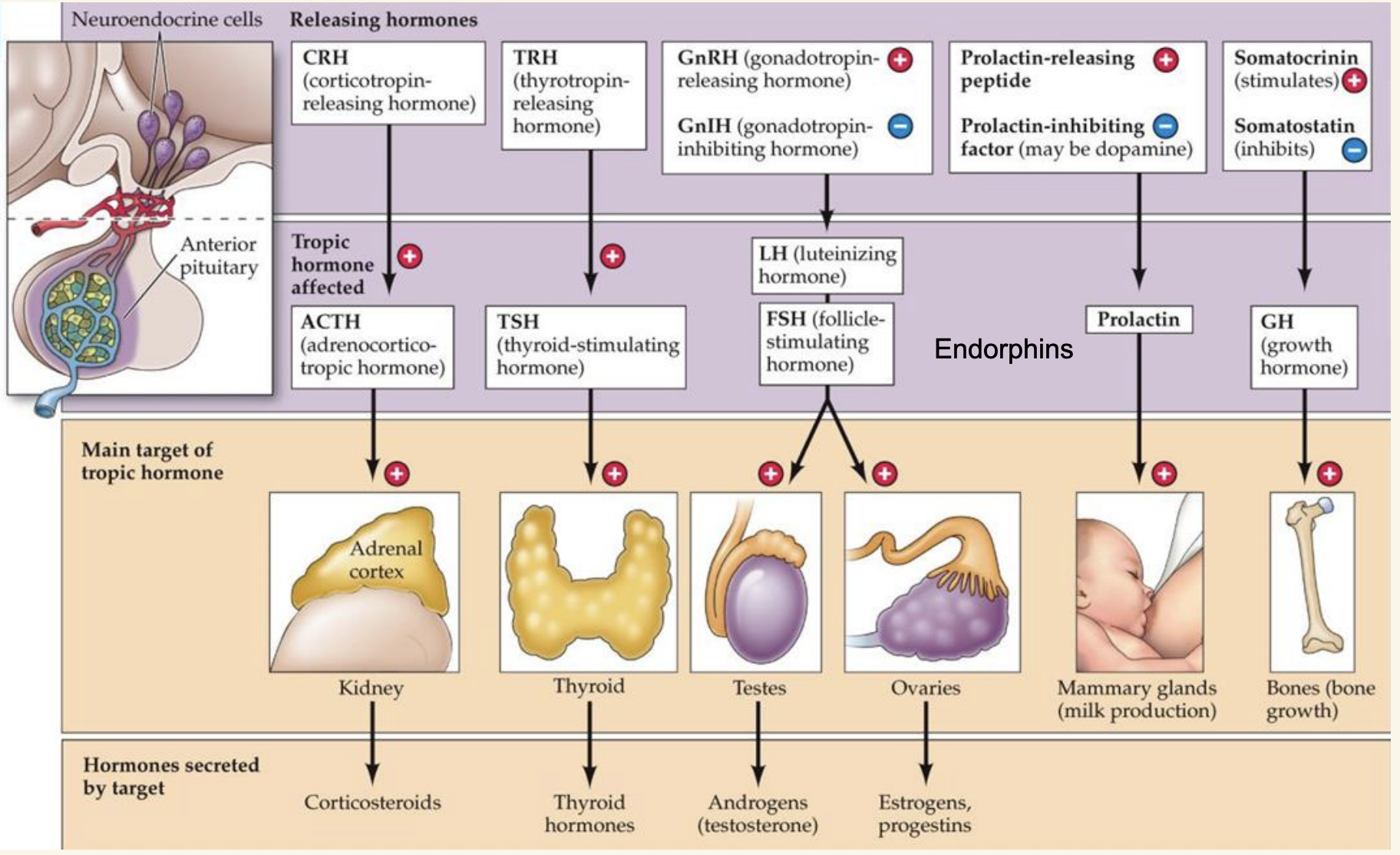
Anterior Pituitary: Regulates other endocrine glands—synthesizes and releases different hormones (tropic hormones)
Releasing hormones from the hypothalamus triggers anterior pituitary to make new (tropic) hormones, which bind to new targets
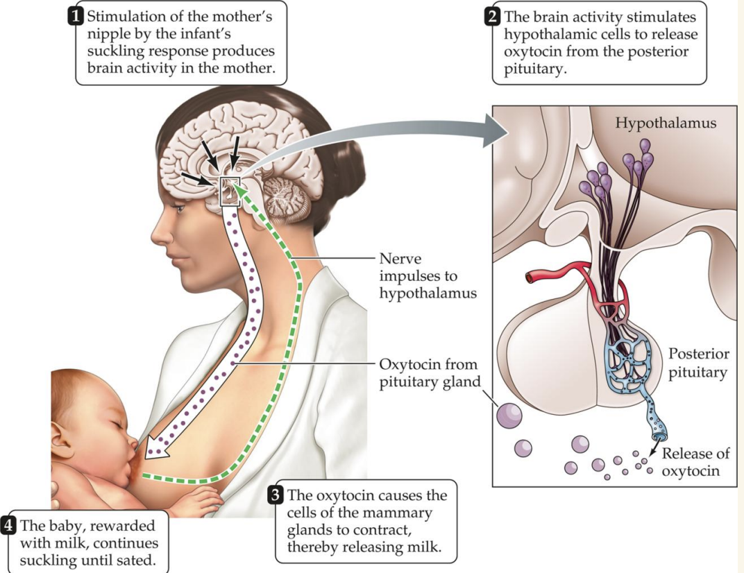
Posterior Pituitary: Does not synthesize hormones, instead is a funnel for neuroendocrine cells to send axons from hypothalamus
Transports Oxytocin + Vasopressin

Anterior Pituitary Hormone Processes/Examples
Hypothalamus creates releasing hormones, which travel to the anterior pituitary
The anterior pituitary then creates tropic hormones that regulate endocrine glands throughout the body
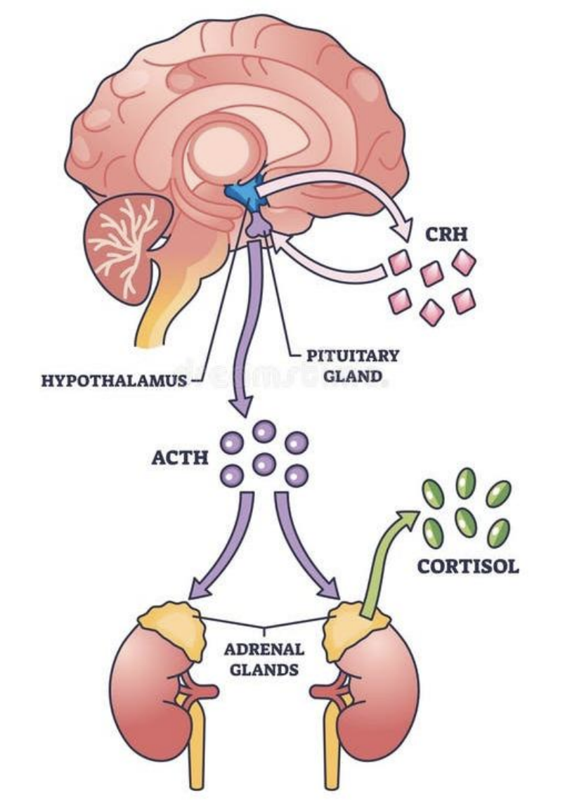
Ex: CRH (in hypothalamus) —> ACTH (in anterior pituitary) —> adrenal cortex —> corticosteroids
FLAT PEG - acronym for anterior pituitary hormones
Follicle Stimulating Hormone
Luteinizing Hormone
ACTH
Thyroid
Prolactin
Endorphins
Growth Hormone
Oxytocin (posterior pituitary)
Oxytocin-producing neurons in the hypothalamus release from posterior pituitary
Can be used by both brain and body
Amygdala, Nucleus Accumbens (NAc), Prefrontal Cortex (PFC), Raphe Nuclei
“Milk Letdown Reflex” (for breast feeding)
Maternal & social behavior
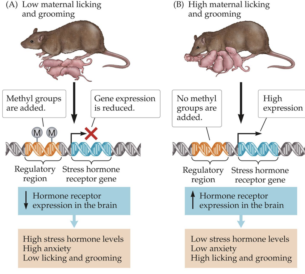
How do Hormones Relate to Maternal Experiments with Rats?
Vasopressin (posterior pituitary)
Hypothalamus will stimulate “thirst” and release vasopressin through the posterior pituitary
Important hormone for regulating blood volume and salt concentration
Direct action on kidneys for water retention and reduces urine production
Also important for social behavior
ACTH (anterior pituitary)
Adrenocorticotropic hormone
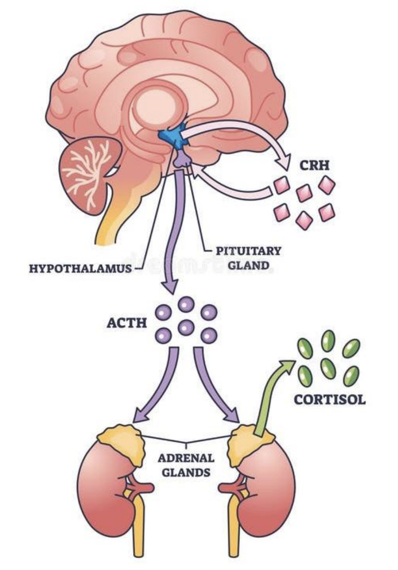
Major player within the HPA axis (hypothalamic-pituitary-adrenal axis: system for body’s stress response/management)
Hypothalamus will secrete corticotropin-releasing hormone
This goes into the anterior pituitary gland
ACTH released by anterior pituitary and is used in the adrenal glands
ACTH then causes release of cortisol (stress hormone)
Gonad Hormones (anterior pituitary)
Gonadotropin-releasing hormone (GnRH) cells in the hypothalamus will travel to the anterior pituitary
Release one or both that act on gonads
Follicle-stimulating hormone (FSH): stimulates growth of follicles or sperm production
Luteinizing hormone (LH): Egg release or testosterone production

Other Anterior Hormones (Thyroid-Stimulating, Prolactin, Growth Hormone)
Thyroid-stimulating hormone (TSH): will stimulate the production of T3 and T4
Low = thyroid needs to work harder!
High = thyroid needs slow down!
Prolactin: breast development and milk production
Growth hormone (GH): physical growth, repair
Negative Feedback
We cannot produce hormones all the time!
Hormone output finds its way back to the hypothalamus
Enough hormones are bound to trigger a biological response
Cue to stop producing more hormones
Process can start again

Drugs and the Brain I + II
Drugs Act on Receptors
Drug = a chemical substance, when taken into the body, alters the structure or function of the body in some way
Exogenous (from outside the body)
Psychoactive drugs have effects on the brain and nervous system
Drugs can bind to receptors (ionotropic or metabotropic)
Mimic NTs - Agonist
Prevents receptor from working - Antagonist
Antagonists can be either competitive (binds to same site as NT) or noncompetitive (binds to a different site as NT)

Four Main Routes of Drug Administration (in order of speed)
Injection: bypasses the digestive tract
Intravenous: in bloodstream (fastest)
Intramuscularly: muscles (2nd fastest)
Subcutaneous: under the skin (slowest)
Ingestion: Oral route
Inhalation: Absorbed in capillaries in the lungs
Absorption: Skin contact
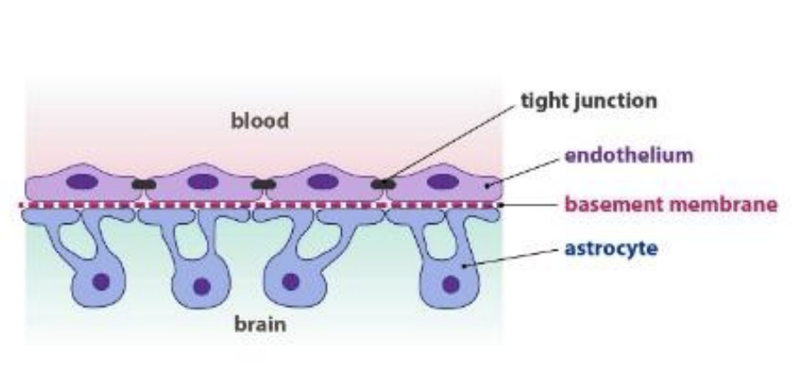
Blood Brain Barrier (BBB)
Prevents most molecules from crossing into the brain from the body (barrier lines blood vessels in the brain)
Astrocytes help maintain structures
Some things can pass through freely
Lipid-soluble molecules
Small molecules
Transporter proteins shuttle things through
Pharmacokinetics vs. Pharmacodynamics
Pharmacokinetics
What the body does to the drugs
All factors from drug entry to body, movement within body, to exit from body
Tells us about a drug’s bioavailability
How much drug is free to act on the body’s targets
Pharmacodynamics
What the drugs do to the body
All processes related to ligand-receptor binding
Tells us about a drug’s mechanism of action
How the drug exerts effects on brain and behavior
Drugs are Broken Down Eventually…
Cleared from the brain by Glymphatic system or the BBB
CSF + glia + blood vessels
The liver is the primary source of metabolism via biotransformation (body altering a compound)
Break down into smaller parts
Kidneys are the primary source of excretion
Small amounts can also be excreted elsewhere
A Drug’s Effects Depend On…
Bioavailability: How much drug is in circulation (to act on targets)
Where it binds in the brain
Affinity: how strongly the drug binds to a receptor
Efficacy: the ability of a ligand (drug) to (in)activate a receptor
Examine a dose-response curve
ED50: Dose needed for half of the maximal effect (lower ED 50 = better binding affinity)
Want to maximize biological effects while minimizing a dose

Efficacy vs. Potency
Maximal Efficacy = maximal extent in which effects appear in clinical populations (maximum effect a drug can achieve)
Graph plateau shows this
Potency = amount of drug required to produce a biological effect
Want to minimize dose while maximizing effects

Therapeutic Index
Separation between useful doses and dangerous doses
Toxic Dose (TD50): 50% animals show signs of toxicity
Lethal Dose (LD50): Fatality of 50% animals
TI = LD50 - ED50
Substance Use Disorder
Patterns of symptoms caused by using a substance that an individual continues taking, despite negative effects
Using more of a substance than intended or using it longer than you’re meant to
Trying to cut down or stop, but being unable to
Experiencing intense cravings or urges to use the substance
Need more of the substance to get the desired effect (tolerance)
Developing withdrawal symptoms when not using substance
Neglecting responsibilities at work, home, or school because of substance use
Using substances in risky settings that put one in danger
Continuing use despite changes in physical or mental health
Withdrawal, Dependence, Relapse
Withdrawal: When drug use is terminated and symptoms are the opposite of the drug’s effects
Dependence: Body has adapted to the drug and requires it to function normally
Severity depends on the type of drug and pattern of use
Shift from impulsive to compulsive behavior
Relapse: Return to active drug use after a period of successful abstinence or recovery

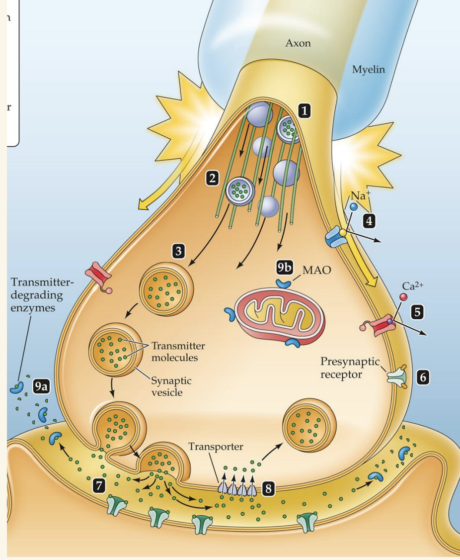
Presynaptic Mechanisms (of Drugs)
Neurotransmitter production
Enzymes that make NTs (1)
Transport materials down microtubules (2)
Neurotransmitter storage (3)
NT Release (7)
Bind to ion channels (4, 5)
Pre-synaptic receptors (6)
NT clearance
Re-uptake proteins (8)
Degradation (9)
Neuroscience IRL: Caffeinated Drinks
Blocks autoreceptors on the presynaptic neuron
Feedback mechanisms for neurotransmitter release
Normally adenosine binds there!
Prevents adenosine from activating sleep-promoting pathways
Can still become dependent and experience withdrawal
Postsynaptic Mechanisms
Receptors
Antagonists (1) prevent receptor from working
Agonists (2) activate the receptors
Other intracellular processes
Receptor regulation (3)
Second messenger systems (4) - ex: GPCRs

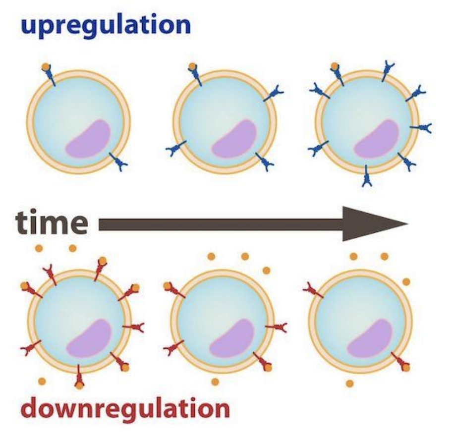
Receptor Regulation
Target tissues can increase or decrease sensitivity to a drug by regulation of the # of receptors
Down-regulation: fewer receptors
Up-regulation: more receptors
The “Reward System”
Mesolimbocortical pathway (VTA → Nucleus Accumbens) of dopamine (for motivation + reward)
Many addictive substances and behaviors cause dopamine release into the nucleus accumbens
Research from the lab shows the importance of this pathway
Necessity and Sufficiency
Neuroscience IRL: social media algorithms

Opioids - examples, theraputic uses, addiction potential
Examples: Morphine (Schedule II), Fentanyl (Schedule II), Carfentanil (Schedule II), Oxycodone (Schedule II) • Heroin (Schedule I)
Therapeutic uses: Pain management, Anesthesia, Cough suppressant (back in the day)
Epidemic in the US
High addiction potential
Opioids (cont.) - effects, administration, mechanism of action, withdrawal, therapeutic uses, withdrawal, treatment for addiction
Acute effects: Euphoria, relaxation, pain relief, respiratory depression, nausea
Can cause coma and death
Administration through either ingestion (pills) or IV injection
Bind to dedicated opioid receptors
Receptor activation will then suppress GABA production (inhibiting an inhibitor)
Indirect release of dopamine
Withdrawal is brutal
Muscle pain, diarrhea, vomiting, nausea, sweating, anxiety, high heart rate, abdominal pain, insomnia
Desire to avoid adds motivation to use
Treatment for addiction
Drugs that directly block opioid receptors (Narcan, Naltrexone)
Medication-assisted treatment (Methadone, buprenorphine)
Harm-reduction strategies (Narcan, test strips, needle disposal/exchange programs)
Cognitive behavioral therapy
Cannabinoids - examples, theraputic uses, addiction potential, acute effects, mechanism of action, withdrawal
Examples: THC (Schedule I), Dronabinol (Schedule II), CBD (not considered a controlled substance
Therapeutic uses: FDA-approved epilepsy treatment (CBD and dronabinol), relieve nausea & vomiting, chronic pain management
Smoking, eating, topicals
Addiction potential: mild to moderate
Acute effects:
Relaxation, mood alterations, increased appetite, impaired judgment, motor deficits, impulsivity
High doses cause hallucinations, synesthesia, paranoia, confusion, respiratory problems, cognitive decline, short-term memory loss, heart attacks
No evidence suggesting brain damage
Mechanism of Action:
Binds to the endogenous cannabinoid receptors - Anandamide (2-AG) (blocks Ca2+ channels), CB1 & CB2, Increase firing of dopamine neurons*
Withdrawal includes irritability, insomnia, decreased appetite and mild anxiety
Stimulants - examples, therapeutic uses, acute effects, addiction potential, withdrawal
Examples: Caffeine (not a controlled substance), Nicotine (not a controlled substance), Cocaine (Schedule II), Amphetamines/ Methamphetamines (such as Adderall, Ritalin, vyvance (Schedule II)) (not a controlled substance)
Smoking, snorted, ingestion
Therapeutic uses: ADHD treatment, Narcolepsy, Local anesthetic (rare; cocaine)
Acute effects: Increased alertness & focus, Elevated heart-rate/blood pressure, Decreased appetite, High doses induce neurotoxicity, psychosis, heart strain, stroke risk, insomnia, anxiety
Strong addictive potential
Withdrawal includes headaches, fatigue, depression, sleep disturbances, irritability, anxiety, difficulty concentrating, cravings
Stimulants Mechanism of Action - Nicotine, Cocaine, Amphetamines
Nicotine
Binds to nicotinic acetylcholine receptors (some of which are found on dopamine neurons) (ionotropic)
Dopamine neurons will become active and release dopamine
Neuroscience IRL: nicotine and memory
Cocaine
Indirect agonist: blocks dopamine reuptake proteins
Dopamine stays in the synaptic cleft longer
Amphetamines
Indirect agonist: Reverses the direction of the transport proteins
Pumps more dopamine OUT of the cell

Alcohol - examples, acute effects, mechanism
Examples: beer, wine, liquor
High potential for misuse
No therapeutic uses, just recreational
Effects are considered biphasic
Initial stimulant, then depressant
Acute effects:
Relaxation, lower inhibition, motor impairment
High doses lead to liver disease, cardiovascular problems, brain atrophy, dependence
Mechanism:
“Depressant” ≠ mood
GABA agonist (mimics GABA - inhibitory)
Glutamate antagonist (NMDA - blocks ionotropic receptor - blocks excitatory)
Alcohol - withdrawal, widely consumed, treatment
Withdrawal is severe
5-6 hrs post-drinking: tremors, nausea, sweating, vomiting, etc.
15-30 hrs: convulsive activity
24-48 hrs: Delirium tremens may last 3-4 days
Most widely consumed psychoactive susbtance
• ~55% heritable alcohol use disorder
Effects almost every tissue in the body
Treatments vary
Benzodiazepines for withdrawal
Naltrexone, acamprosate, disulfiram to curb drinking behavior
Cognitive Behavioral Therapy
Neuroscience IRL: alcohol + memory (brain cannot convert short term memories into long term memories)
Psychedelics - examples, therapeutic research, acute/long term effects, withdrawal, addictive potential
Examples: LSD (Schedule I), Mescaline (Schedule I), Psilocybin (Schedule I), MDMA (Schedule I)
Mode: Ingestion
Current therapeutic research:
LSD = microdosing for anxiety, depression, substance use disorders
Mescaline = substance use disorders
Psilocybin = substance use disorders, PTSD, depression
MDMA = PTSD
Acute effects: Vivid visual hallucinations, slowing the sense of time, disassociation, strong emotional reactions, disruption of logical thought
Long-term effects include “bad trips”, dehydration, serotonin toxicity
Withdrawal includes depression and mood swings
Low addictive potential
Psychedelics Mechanisms of Action - LSD + Psilosybin and MDMA
LSD & psilocybin
Binds to serotonin receptors to activate them
Increased effects
MDMA
Enters neurons that release either serotonin or dopamine
Blocks reuptake
Serotonin and dopamine remain in the synaptic cleft
Emotions
Subjective mental state that is accompanied by distinctive behaviors and physiological changes
Overt behaviors
Motivational states
Physiological arousal
Emotional dysregulation - hallmark of many psychopathologies
Why Are Emotions a Good Thing?
Evolutionary adaptations
Enhance the ability to experience and evaluate environment cues
Can coordinate behavioral responses to solve problems
Central to social communication
Enhance the abilities to interact and communicate with others in appropriate ways
Competing Viewpoints on What Comes First - emotion, physiological arousal, cognitive attribution
Folk psychology: emotion —> physiological reaction
James-Lange Theory:
physiological reaction —> emotion
Cannon-Bard Theory:
physiological reaction and emotion occur at the same time
Schachter + Singer Theory:
physiological reaction —> cognitive appraisal/assessment —> emotion
Are There “Core” Emotions?
There is no unifying list of emotions, but general agreement on six
Fear
Anger
Surprise
Disgust
Happiness
Sadness
Intensity depends on level of arousal

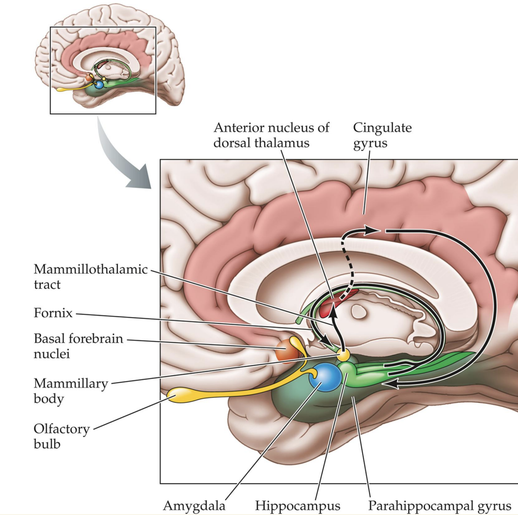
The Limbic System (aka Papez Circuitry)
Set of brain regions that link the cortex with the hypothalamus - important for emotions
Structures:
Hypothalamus, Thalamus, Cingulate Cortex, Hippocampus, Amygdala, Fornix
Amygdala = Seat of Emotion
Important for linking valence to memory
Valence: Is a memory good or bad?
Lesions (damage) produce deficits in emotional processing
Decreased fear and aggression
Decreased facial expressions and vocalization with fear
Poor visual recognition
Lack of social restraint
Hyperorality
Klüver-Bucy syndrome
Stimulation will increase vigilance, anxiety, fear, and aggression
Can do this either electrically (DBS) or with optogenetics (animals only)
fMRI imaging studies reveal greater activation in amygdala BOLD signal in fearful faces than in neutral ones (more oxygenated blood in amygdala w/ fearful faces)
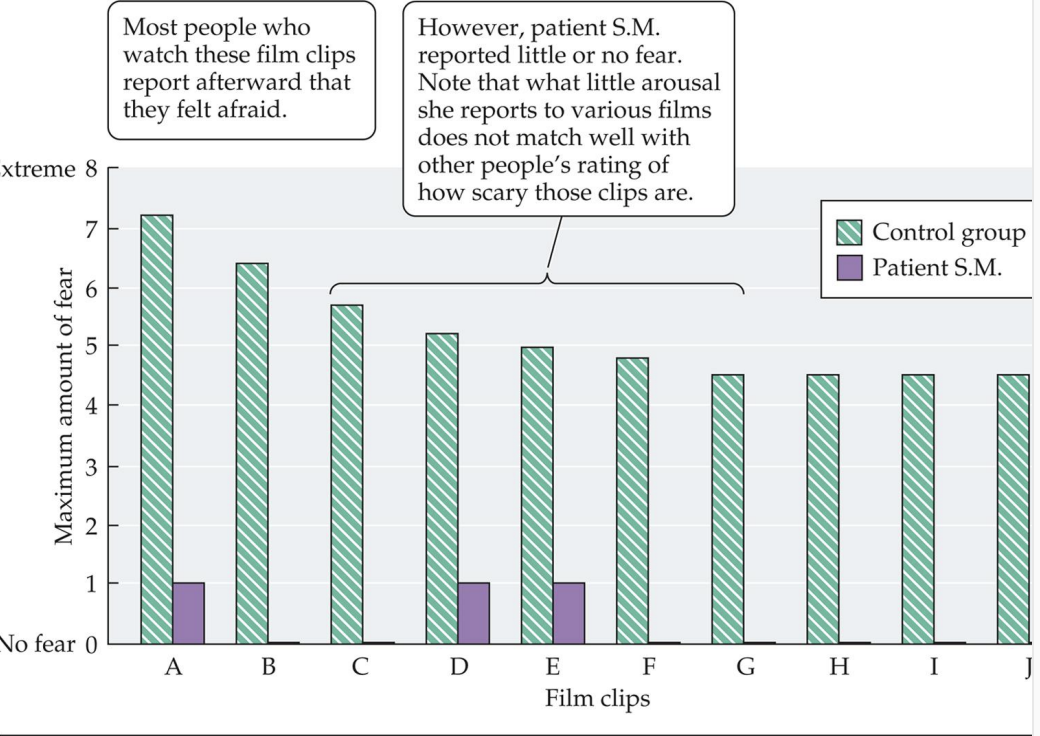
Patient SM - “The woman who was never afraid” (destroyed amygdala)
Most common symptom: Inability to recognize fear in facial expressions
Low self-reporting score on watching film clips
Abnormal eye movement in Patient S.M.
Different Patterns of Brain Activation Across Emotions
Studies use fMRI to study patterns of activation during sadness, happiness, anger, and fear
Both Limbic and non-Limbic areas
Insula (purple)
Cingulate Cortex (both green and blue)
Prefrontal Cortex (yellow)
Amygdala (red)
Network = multiple brain regions working together
Fear Can Also Be Learned
“Fear Conditioning”
CS = Conditioned Stimulus (ex: tone/bell)
CR = Conditioned Response (ex: fear)
US = Unconditioned Stimulus (ex: electrical shock)
UR = Unconditioned Response (ex: fear)

Stress
Stress = A Multidimensional Concept
Stressful stimulus, stress processing, stress response
Acute stress seen as adaptive
Increase alertness, focus and motivation
Stimulate immune system
Stimulate neuronal & astrocyte growth (shown in mice)
Positively impacts memory
Increases resilience
Neuroscience IRL: public speaking (tied to stress hormone)

Reappraising “Stress is Good”
Reappraising (reinterpret its meaning) a stressful experience so that it is seen as helpful and controllable has been shown to improve immediate cognitive, physiological, and behavioral stress responses
Stress on the Body
Activation of two hormonal systems (HPA axis + Sympathetic nervous system) to trigger release of:
Glucocorticoids (cortisol) from the adrenal glands
Epinephrine (adrenaline) and norepinephrine (noradrenaline) from the adrenal glands
Hormones cause the physiological responses of stress
Beta-blockers prevent these during stress!

Stress on the Body - Cortisol + Immune System
Cortisol suppresses the immune system
Short-term defense mechanism allowing body resources to be used elsewhere
Stress does not make you sick, but makes you more likely to get the diseases that make you sick
Once stress levels decrease, immune system back on
Stress on the Body - Long-Term Stress
Our stress response system is not built for chronic stress!!
Long-term stress has devastating consequences
Extreme trauma/stress = PTSD
Physical health decline
Some signs of neurodegeneration
In the wild, animals die under prolonged stress
Connect to Maternal Experiments with Rats Again
Stressed mom —> epigenetic changes in developing mice (methylation, high stress hormone levels and anxiety levels, cannot adapt to cortisol in the same way)

Neuropsychiatric Disorders I and II
Neuropsychiatric Disorders are Highly Prevalent
~15-50% of the global population reports some psychiatric symptoms at some point in life
~19% of US adults experience symptoms each year
Can occur in any age and in any individuals
Rates vary across demographics
Anxiety is Slightly Different From Stress
Both trigger sympathetic nervous system
Some shared symptoms
Difficulty sleeping, focusing
Fast heart rate, breathing
Muscle tension & headaches
Anxiety = Characterized by fear and the occurrence of an inappropriate stress response, even when the stress is not there
Types of Anxiety Disorders
Generalized Anxiety Disorder: persistent excessive anxiety, worry
Social Anxiety Disorder: Fear and avoidance of negative public scrutiny, public embarrassment, humiliation or social interaction
PTSD: Anxiety disorder that results from traumatic experience
Panic Disorder: Recurrent attacks of intense fearfulness
Phobic Disorder: Irrational fears centered on an object, activity, or situation that a person avoids
HPA Axis Revisisted in Anxiety
There is a genetic predisposition to many anxiety disorders
Also, environmental factors play a huge role!
Stress response is triggered by fear and initiated in the hypothalamus
What stress hormone(s)? - CRH, ACTH, Corstisol
“Push-Pull” Regulation of the HPA Axis
Too much amygdala activity stimulates inappropriate hypothalamus activity
Too little hippocampal activity stimulates inappropriate hypothalamus activity
Normally helps with feedback regulation of the hypothalamus to stop cortisol production
Chronic cortisol will cause degeneration/lack of neurogenesis

Hippocampus = Also Affected by PTSD
PTSD Symptoms
Intrusive re-experiencing of the trauma
Avoidance of stimuli associated with the trauma
Negative mood and cognitive capacity associated with the trauma
Excessive arousal and reactivity
PTSD caused a reduced hippocampus size
Severity correlates with size (more severe, smaller size)
Animal Models: “PTSD-Like”
Stress-Enhanced Fear Learning Paradigm
Slightly modified fear conditioning paradigm
One group of animals gets severe shock, the other does not
Can look at behavioral and physiological responses
Freezing, Blood pressure (higher)
Neurogenesis impacted
Developmental consequences
Treatments for Anxiety Disorders
Psychotherapy
Collaborative talk therapy
Benzodiazepines are anxiolytic (reduces anxiety symptoms)
Acts on GABA-A (ionotropic) receptors to increase inhibitory activity (Cl- into cell + inhibits)
Serotonin Agonists
SSRI = Selective Serotonin Reuptake Inhibitor

Extinction Learning
Gradual weakening of the original conditioned response (CR) by re-exposure to the conditioned stimulus (CS)
Like exposure therapy
CS is no longer paired with the unconditioned stimulus (US)
Repeated exposure of cue in “safer” contexts helps decrease the fear
Decreased freezing and blood pressure

Diagnosis With DSM 5
Diagnostic and Statistical Manual 5th version
5 or more symptoms within a 2-week period to get depression diagnosis
Depressed mood (or irritability in children or adolescents) or anhedonia (cannot experience pleasure/joy)
Weight or appetite changes
Sleep disturbances
Psychomotor changes
Fatigue or slow energy
Concentration impairment
Suicidal ideation
What is an Affective Disorder?
Disorder characterized by alterations in emotion or mood
Major Depressive Disorder (MDD): Combination of debilitating symptoms
Dysthymia: Lowered mood, but not as severe as MDD
Bipolar Disorders: Periods of clinical depression alternated by extreme elation or mania
Atypical Depression: Overeating, oversleeping, fatigue - Mood changes in response to events
Psychotic Depression: Major depression along with loss of touch with reality
Postpartum Depression (PPD): Depression after giving birth that outlasts the “baby blues”
Premenstrual Dysphoric Disorder: Severe form of premenstrual syndrome with depression symptoms
Seasonal Affective Disorder (SAD): Depression that occurs each year at the same time, usually winter months
Affective Disorders = Widespread
~7% of the US population has at least 1 depressive episode per year
2019: 15.2% major depressive episodes were between ages 18-25
20% of the population will have a depressive episode in their life
Risk Factors for Depression
Heritable component
“Twin studies”: 60% concordance (identical), 20% concordance (fraternal)
Many genes contribute to making a person susceptible
“Polygenic” condition
Serotonin production/release, HPA axis regulation
BDNF: brain derived neurotrophic factor (protein that helps with neurogenesis + neuroplasticity)
Environmental factors also play a role (diet, substance abuse, etc.)

Alterations in Brain Structure + Function due to Depression
Increased activation in frontal lobes during cognitive tasks
Increased amygdala activation during emotional processing
Decreased blood flow to brain areas implicated in attention
Reduced hippocampal volume and activation during memory tasks

Depression Treatments
Antidepressants
Take a long time to work (can take months)
Examples: SSRIs (prevents serotonin reuptake) and monoamine oxidase inhibitors (prevents NTs from being broken down into metabolites - prevents biotransformation)
Spravato (Esketamine) - (antagonist for Glutamate NMDA receptors)
Nasal spray, works within hours
More potent version of ketamine
For treatment-resistant depression
TMS, DBS (invasive)
Faster relief from symptoms
Cognitive Behavioral Therapy
The Monoamine Hypothesis
Monoamines = serotonin, norepinephrine, dopamine
All drug classes to treat depression increase the level of monoamines that could be used in the synapse
Mood is closely tied to the levels of released monoamines in the brain
Depression as a deficit of neurotransmitters
Mechanism of Action + Examples of Antidepressants (MAO Inhibitors, Tricyclics + Heterocyclics, SSRIs, 2nd Generation Antidepressants + Investigational Drugs)

The Monoamine Hypothesis: Evidence For and Against
Evidence in Support
Drugs are successful in reducing depressive symptoms
Lower levels of serotonin metabolites have been found in CSF
Lower concentrations of serotonin and metabolites in postmortem brain tissue
Depletion of serotonin (tryptophan-free diet) worsens depressive state
Evidence Against
Drugs increase serotonin levels immediately, but people don’t experience reduction in symptoms weeks later
Many people do not respond to drugs
Serotonin depletion does not induce depression in control subjects
Antidepressants do not elevate mood in control subjects
Increased suicide risk in children and teens that use SSRIs
Animal Models Used To Study Depression
Sucrose Preference Test
Tail Suspension Test
Forced Swim Test
