chap. 32 - skin integrity & wound healing
 Call Kai
 Learn
 Practice Test
 Spaced Repetition
 Match
 Flashcards
 Knowt Play
1/88

There's no tags or description
Looks like no tags are added yet.
89 Terms
integumentary system
consists of skin, subcutaneous layer, appendages of skin (glands, skin, hair, nails)
skin
largest organ of the body
unbroken + healthy skin is the first line of defense
adequately nourished + hydrated body cells are resistant to injury and disease
adequate circulation necessary to maintain cell life
if impaired, it cant remove wastes effectively
epidermis
top layer + outermost part made up of epithelial cells — waterproof
dermis
second layer of skin consist of elastic connective tissue made up of collagen
includes = nerves, hair follicles, glands, immune cells, blood vessels
subcutaneous tissue
bottom layer that anchors skin layers to underlying tissue of the body + consists of adipose tissue made up of fat cells and connective tissue
stores fat for energy
serves as heat insulator
provides cushioning for protection
functions of skin
protection — anatomical barrier
sensory perception — pain, temp, touch
regulates body temp — draws heat from body via sweat
vitamin D production — activated by UV rays from sun
psychosocial — cosmetic effect
immunologic — triggers immunologic responses when broken
absorption — meds for local + systemic effects
elimination — sweat excretes small amounts of water, electrolytes, nitrogenous wastes
factors affecting skin integrity
age — children, older adults
lifestyle variables — sun exposure, body piercing
nutrition status — lack of fluid, proteins, vitamin C
changes in health status — dehydration, reduced sensation
illness & therapeutic measures — bed rest, casts, radiation
wound
a break or disruption in the normal integrity of skin + tissues
wound calssification
intentional vs unintentional
open vs closed
acute vs chronic
superficial vs penetrating
partial thickness vs full thickness
unstageable
intentional vs unintentional
intentional → surgical wounds, piercings, IV therapy, lumbar puncture (break of skin through needle)
wound edges clean + smooth (heals faster), bleeding controlled, low infection risk
unintentional → accidents, trauma, stabbing, gunshot, burns
wound edges jagged (harder time healing properly, scar tissue), bleeding uncontrolled, high infection risk
occurs in a non-sterile environment
open vs closed
open → skin surface is broken
anything that causes bleeding — incisions, abrasions, cuts
closed → skin is not broken but soft tissue is damaged
ecchymosis (bruising), hematomas (pooled blood)
acute vs chronic
acute → heals within days to weeks
wound edges approximated (can meet to close wound) — ex: surgical wound
normal healing process
chronic → does not progress through normal sequence of repair
would edges not approximated (can’t meet up to close) — ex: pressure ulcers
stuck in inflammatory phase
partial thickness vs full thickness
partial → all or portion of dermis is intact
full → entire dermis, sweat glands, hair follicles are severed
unstageable
full thickness loss where true depth cannot be determined
types of wounds
incision
contusion — the only closed wound
abrasion
laceration
puncture
penetrating
avulsion
pressure ulcers
venous ulcers
arterial ulcers
diabetic ulcers
incision
clean, intentional cut or wound

contusion
bruise
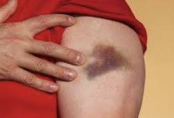
abrasion
friction rubs away epidermis or parts of dermis

laceration
traumatic cut, tear, or opening caused by blunt or sharp trauma
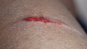
puncture
small hole or perforation made by a sharp, pointed object
needle

penetrating
object pierces the skin + enters underlying tissues or body cavities
knife, bullet, glass shard

avulsion
tissue, body structure, or fragment of bone is forcibly torn away from its normal attachment

types of ulcers
pressure ulcers → immobility
venous ulcers → venous insufficiency
arterial ulcers → PAD causing ischemia + lack of oxygen/blood supply
diabetic ulcers → peripheral neuropathy
first aid for skin trauma
homeostasis = priority — STOP BLEEDING!!
homeostasis achieved spontaneously or through first aid measures
when difficult to acheive:
apply direct pressure → closes capillaries in vessel, preventing blood flow
elevate wound → redirection of blood (gravity)
consider short-term tourniquet for limb injuries
local anesthetic containing adrenaline
4 phases of wound healing
hemostasis
inflammatory
proliferation/repair
maturation/remodeling
hemostasis phase
occurs immediately after injury
stops bleeding + activates WBCs to fight bacteria
blood vessels constrict + platelets activate to form fibrin clot
inflammatory phase
lasts 2-3 days
leukocytes arrive first to ingest bacteria + cellular debris
24 hrs after injury, macrophages enter area + remain for extended amount of time to release growth factors for new epithelial cells and blood vessels
exudate (plasma + blood) result in acute inflammation → heat, redness, swelling
patient may have:
slight temperature
high WBC
general malaise
proliferation/repair phase
lasts several weeks
new tissue built to fill wound — fibroblasts
thin layer of epithelial cells form + blood flow across wound reinstituted
granulation tissue (new tissue) forms scar tissue development
maturation/remodeling
final stage begins ~ 3 weeks after injury — can continue for months to years
collagen makes wound stronger + makes scar eventually become a flat, thin line
local factors affecting wound healing
pressure
dehydration
maceration (skin gets wrinkly — showering too long)
trauma
edema
infection
excessive bleeding
necrosis
biofilm (growth of bacterial microorganisms — protects wound)
systemic factors affecting wound healing
age
circulation
oxygenation of tissues
nutrition status
wound etiology
meds
health status
immunosuppression
adherence to treatment plan
hemorrhage
normal during + immediately after initial trauma
prolonged if coagulation problem or large vessels involved
risk greatest during 1st 24 to 48 hrs post-op
internal or external
assess internal by noting:
distention or swelling of involved body part
change in type of amount
drainage from dressing or drain
signs of hypovolemic shock
hematoma
localized collection of blood underneath tissues
seen as:
swelling
color
sensation
warmth
bluish tinged mass
infection
wound infection is 2nd most common HAI
presence of purulent drainage = infection
higher risk if wound contains:
necrotic tissue
foreign bodies
diminished blood flow to site
may be seen within:
2-7 days post-op in contaminated wounds
4th-5th post-op day in surgical incision
fistula formation
abnormal passage from an internal organ/vessel to outside of body or from 1 internal organ/vessel to another
dehiscence
partial or total separation of wound layers
typically involves abdominal surgical site
c/o “something has suddenly given way”
ALERT → serosanguineous fluid from previously dry wound!!!
occurs after sudden strain:
vigorous coughing
vomiting
sitting up and site is unsupported — teach splinting
patients at risk:
obsese
poor nutritional status
evisceration
total separation of wound layers + protrusion of organs through wound opening — SURGICAL EMERGENCY
evisceration actions
call for help — stay w/ patient + instruct them to remain quiet
place patient in low-fowlers position w/ knees bent
cover wound w/ sterile saline soaked towels — NO ICE PACKS!!
monitor vital signs closely for signs of shock
keep NPO to prep for emergency surgery
document
pressure injuries: stage 1
non-blanchable erythema of intact skin
intact skin w/ a localized area of non-blanchable erythema
pressure injuries: stage 2
partial thickness skin loss w/ exposed dermis
wound bed viable, pink or red, moist, and may present as an intact or ruptured serum-filled blister
fat + deeper tissue are not visible
granulation tissue , slough, eschar not present
pressure injuries: stage 3
full thickness skin loss
fat visible + granulation tissue often present
slough + eschar may be visible
undermining + tunneling may occur
fascia, muscle, tendon, ligament, cartilage, bone are not exposed
pressure injuries: stage 4
full thickness skin + tissue loss
exposed or palpable fascia, muscle, tendon, ligament, cartilage, bone
slough + eschar may be visible
rolled edges, undermining, tunneling occur
depth varies
pressure injuries: unstageable
obscured full thickness skin + tissue loss
damage within ulcer cannot be confirmed b/c it is obscured by slough + eschar
if slough/eschar removed, stage 3 or 4 pressure injury will be seen
pressure injuries risk factors
impaired mobility
alterations in LOC
incontinence
imbalanced nutrition (person w/ low cholesterol at high risk)
age
impaired circulation (oxy. rich blood needed to repair skin + heal wounds)
moisture (can cause maceration)
friction + shear
impaired sensory perception
common pressure injury sites

skin assessment frequency
acute care — on admission + every 24 hrs minimum, every shift especially in critical care
long term care — on admission + weekly for 4 weeks then quarterly or when condition changes
home care — on admission + every visit
assess bony prominences:
blanch test all reddened areas
reassess in 1 hr
inspect + palpate
don’t forget risk areas while sitting
pressure injury assessment: predictive measures
braden scale
severe risk if score <11
pressure injury assessment: mobility
can patient change position independently/voluntarily when in bed? in chair? activity intolerance?
pressure injury assessment: nutritional status
must have initial assessment:
recent weight loss
general appearance
usual eating patterns
serum albumin level
vitamin A + C
trace minerals zinc + copper
monitor daily food + fluid intake
weight patients
pressure injury assessment: moisture & incontinence
assess exposure to body fluids via:
drains or incontinence
sweating/diaphoresis
wounds
measures to reduce exposure
hygiene + skin care
pressure injury assessment: pain
PRN
prior to dressing changes
encourage to increase mobility
pressure injury implementation
topical skin care + incontinence management
inspect skin during bathing or daily personal care
AVOID strong soap + hot water when cleansing
DO NOT scrub — gentle, thorough drying w/ attention to skin folds/creases
apply moisture — DO NOT massage reddened/discolored pressure points → use skin barrier ointment + film sprays
assess skin around drain sites — body fluids on skin = risk for skin breakdown
evaluate + manage urinary/fecal incontinence — bowel/bladder training; use absorbent pads as last resort
positioning
HOB to 30 degrees or less to minimize shearing force
individualized turning schedule, minimally every 2 hrs
heels off pressure
support surfaces
decrease amount of pressure exerted over bony prominences
no foam rings, no donuts
use of appropriate devices to ensure safe patient handling + movement
patient + family education
wound assessment
type — surgical incision, pressure injury, diabetic, venous stasis
location
size — LxWxD (cm)
presence of undermining, tunneling
tissue viability — type + percentage of granulation, slough, eschar in relation to wound base
exudate/drainage — COCA (color, odor, consistency, amount)
pain — meds 30-45 mins prior to dressing change
drains — presence, type, amount, location
wound closures — staples, sutures, tissue adhesive
would cultures — cleanse or irrigate would prior to obtaining specimen (requires HCP order)
serous drainage
composed primarily of clear, serous portion of blood + from serous membranes
clear + watery
sanguineous drainage
consists of large # of RBCs + looks like blood
bright red = indicative of fresh bleeding
darker = older bleeding
serosanguineous drainage
mixture of serum + RBCs
light pink to blood tinged
purulent drainage
made up of WBCs + liquified dead tissue debris + both dead/live bacteria
thick
often has musty or foul odor
color varies — dark yellow or green
drainage amount: scant
wound moist; no measurable drainage (none)
drainage amount: small/minimal
wound very moist; <1/3 of dressing
drainage amount: moderate
wound wet; drainage involves 1/3-2/3 of dressing
drainage amount: large/copious
wound tissue filled w/ fluid; involves >2/3 of dressing
guaze, idoform guaze, NuGuase
dressing packed loosely so wound is allowed to drain
purpose = allow healing from base of wound
ex = infected wounds, after removal of hemorrhoids
penrose drain
open drainage system consisting of a soft rubber tube that provides a sinus tract
purpose = drains blood + fluid
ex = after incision + drainage of abscess, in abdominal surgery
blake drain
closed drain — silicon tube w/ 4 chambers — similar to JP externally
purpose = drains blood + fluid
ex = cardiac surgery in place of a chest tube or JP drain
chest tube drain
closed drain — mediastinal placement (diff from chest tube used in pleural space)
purpose = drains blood
ex = after cardiac surgery
hemovac drain
closed drain — portable negative pressure suction device
purpose = drains blood + fluid
ex = after abdominal, orthopedic surgery
jackson-pratt (JP) drain
closed drain — bulb suction device
purpose = drains blood + fluid
ex = after breast surgery or mastectomy, abdominal surgery
T-tube drain
closed drain — tube placed in common bile duct
purpose = collects bile
ex = after gallbladder surgery
measuring size of wound
measure using greatest measurement:
head-to-toe length
side to side width
depth
measuring depth of wound
moisten sterile cotton-tipped applicator w/ saline + insert gently into wound at 90 degrees
mark point of swab that is even w/ surrounding skin surface or grasp applicator w/ thumb + forefinger at the point of wound’s margin
remove swab + measure depth w/ ruler
measuring would tunneling
determine direction
moisten sterile cotton-tipped applicator w/ saline + gently insert into site where there is tunneling
view direction of applicator as a hand of a clock — direction of patients head = 12:00
moving clockwise, document deepest sites where wound tunnels
determine depth by marking point on swab that is even w/ wound edges
remove swab + measure depth w/ ruler
document both direction + depth of tunneling
cleaning wounds w/ approximated edges
moisten sterile gauze pad or swab w/ prescribed cleaning agent
use new swab or gauze for each downward stroke
clean from top to bottom
work outward from incision in lines parallel to it
wipe from clean to less clean
cleaning wounds w/ edges that are not approximated
moisten sterile gauze pad or swab w/ prescribed cleaning agent + squeeze out excess solution
use new swab or gauze for each circle
clean wound in full or half circles, beginning from center to outer
clean at least 1 in. beyond the end of the new dressing
negative-pressure wound therapy (NPWT)
uses application of sub atmospheric (negative) pressure to a wound through suction
accelerates wound healing
optimizes blood flow
removes wound fluid
maintains moist environment
monitor for bleeding
debridement
removal of devitalized tissue or foreign material from a wound
helps remove cells that are alive but not functioning from the wound bed + edges
5 types = sharp, mechanical, enzymatic, autolytic, biotherapy (maggot) therapy
sharp debridement
use of a sterile, sharp instrument to remove devitalized tissue
mechanical debridement
use of wet-to-dry dressing or hydrotherapy
enzymatic debridement
proteolytic agents break down necrotic tissue w/o affecting viable tissue in the wound
autolytic debridement
use of occlusive, moisture-retaining dressing + body’s own enzymes and defense mechanisms to break down necrotic tissue
biotherapy debridement
use of medical-grade larvae to dissolve dead + infected tissue from wounds
physiological effects of heat therapy
vasodilation
increases tissue metabolism + capillary permeability
reduces muscle tension
relieves pain
reduces blood viscosity
physiological effects of cold therpay
vasoconstriction → “co” for cold
reduces muscle spasms
slows nerve conduction + transmission of pain
reduces edema + inflammation
controls bleeding
rebound phenomenon of heat therapy
produces maximum vasodilation in 20-30 mins
continuation beyond 30-45 mins causes reflex vasoconstriction + tissue congestion
recovery time = 1 hr to restore vasodilation advised before reapplication
STOP BEFORE REBOUND BEGINS (opposite effect)
rebound phenomenon of cold therapy
maximum vasoconstriction occurs when skin temp. reaches 60F (15C) or in about 30 mins to 1 hr
vasodilation begins as a protective mechanism to prevent body tissue from freezing
recovery time = 1 hr is best before reapplication
STOP BEFORE REBOUND BEGINS (opposite effect)
therapeutic uses of heat therapy
infections/inflamed tissue
wounds
muscle tension
chronic pain
therapeutic uses of cold therapy
muscle sprains, strains, spasms
fractures
minor burns
superficial lacerations + puncture wounds
post-injections
joint + soft tissue trauma
chronic pain syndrome
contraindications of heat therapy
for areas of active bleeding
for an acute localized inflammation (appendicitis)
over a large area if a patient has cardiovascular problems
contraindications of cold therapy
if site of injury is edematous (swollen/puffy)
if patient has impaired circulation
in presence of neuropathy
if patient is shivering
heat & cold therapy assessment
assess temp. tolerance
skin + skin integrity
patients response to stimuli
equipment being used
identify any contraindications
bodily responses to heat + cold
local effects of heat + cold
influencing factors of heat + cold tolerance
application of heat + cold therapies — requires HCP order