microbio exam 4
 Call Kai
 Learn
 Practice Test
 Spaced Repetition
 Match
 Flashcards
 Knowt Play
1/104

There's no tags or description
Looks like no tags are added yet.
105 Terms
hygiene hypothesis
decreasing the diversity and levels of microbes in our normal microbiota may negatively affect immune responses
can happen via antibiotic use, diet changes, or lifestyles changes
innate immunity
the immune response we are born with; always the same, regardless of the pathogen
first-line defenses
help prevent pathogen entry
ex: tears rinse/flush microbes away from the eye; mucus in the respiratory tract traps invaders and the cilia sweeps it away
mechanical barriers
part of first-line defenses
rinse/flush/trap pathogens
ex: tears, mucus membranes, urine, earwax
chemical barriers
part of first-line defenses
create hostile environments for pathogens and/or directly attack pathogens
ex: lysozymes, stomach acids, antimicrobial peptides (AMPs)
physical barriers
part of first-line defense
physically block pathogen entry
ex: main one if epithelial tissue (skin, linings of body organs)
second-line defenses
molecules and cells that actively target pathogens
Lymphatic system organs and tissues that collect, circulate and filter fluid throughout the body before that fluid goes back into the blood
primary lymphoid tissue
part of second-line defense
thymus and red blood marrow that help produce immune system cells
thymus: near the heart
bone marrow: is spongy tissue deep inside bones
secondary lymphoid tissue
part of second-line defense
filter lymph and get fluid back into the body
includes lymph nodes
leukocytes
part of second-line defense
white blood cells that serve generalized immune functions
often release molecules that help control or eliminate pathogens
differential white blood cell count
part of second-line defense
rapid test used to see if leukocytes are overrepresented or underrepresented in the blood
neutrophils
part of second-line defense
most common leukocytes and first responders from bloodstream to injured tissues
look to engulf pathogens
eosinophils
part of second-line defense
uncommon but versatile leukocytes that stain red/orange
basophils
part of second-line defense
rarest leukocyte that releases histamine, which makes blood vessels leaky so more immune cells can respond
mast cells
part of second-line defense
relatively stationary so they act as “lookouts” during potential infection
monocytes
part of second-line defense
eventually leave blood and enter tissues, they mature into macrophages (which are experts at phagocytosis)
dendritic cells
part of second-line defense
leukocytes with cytoplasmic extensions that help them “grab” pathogens for phagocytosis; common near portals of entry
lymphocytes
part of second-line defense
small leukocytes that that directly kill infected/damaged host cells
includes natural killer cells, and adaptive cells like B and T cells
cytokines
part of second-line defense leukocytes
signaling molecules that help immune cells coordinate responses
includes chemokines (attract more leukocytes) and interleukins (stimulate the production of more platelets)
interferons
part of second-line defense leukocytes
“alarm” molecules that can either 1) stop nearby cells from making proteins, 2) trigger nearby cells to die, and 3) activate more leukocytes
iron-binding proteins
part of second-line defense
proteins (like hemoglobin) that tend to bind to free-floating iron so invading pathogens cannot use it
complement cascades
part of second-line defense
Over 30 different proteins work together in a cascade fashion to respond to pathogens.
They all ultimately result in opsonization (tagging pathogens for destruction), the creation of a membrane attack complex (MAC) which bursts pathogens open, and inflammation
classical pathway
part of second-line defense
complement proteins are activated by antibodies that are bound to the pathogen
alternative pathway
part of second-line defense
complement proteins are activated by directly interacting with the pathogen
lectin pathway
part of second-line defense
complement proteins are activated by mannose-binding lectin bound to pathogens
inflammation
part of second-line defense
innate response to infection which recruits immune cells, limits spread of pathogens, and delivers vital nutrients to damaged tissues
this process can damage our own cells, but if it is temporary, it is generally a net benefit
vascular changes phase
phase of inflammation
part of second-line defense
blood vessels enlarge and become porous. these changes are brought about by molecules like histamines or kinins
leukocyte recruitment phase
phase of inflammation
part of second-line defense
molecules called chemoattractants draw leukocytes to the injured tissue
resolution phase
phase of inflammation
part of second-line defense
chemicals signal leukocytes to undergo apoptosis (self-destruction), blood vessels are repaired via angiogenesis, and pus (collection of remnant fluid of dead cells) may form
fever
part of second-line defense
abnormally high systemic body temperature in response to pyrogens (fever-inducing agents that can be molecules, viruses, or cells)
adaptive immunity
idk
third line of defense!
antigen presentation
first stage of adaptive immunity, can be cell-mediated or humoral response
antigen-presenting cells grab up antigens and show them to T cells (B cells just grab antigens directly)
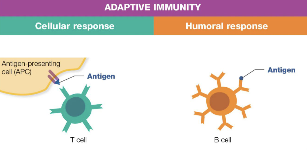
MHC 1
intracellular antigen fragments are carried by MHC 1 to the surface where they bind to T cytotoxic cells

MHC 2
antigen-presenting cells take in extracellular antigens that are broken into fragments. these fragments are carried to the surface where they bind to T helper cells

lymphatic activation
second stage of adaptive immunity, can be cell-mediated or humoral response
cytokines activate leukocytes and activated T cells influence B cell activation

two required steps of activating T cells
aspect of second stage of adaptive immunity, can be cell-mediated or humoral response
primary activation signal (MHC)
secondary activation signal (co-stimulatory proteins)

superantigens
uniquely potent antigens
they bypass the antigen-presentation process and force a direct link from MHC to T cell receptor, which activates the T cell much faster
lymphocyte proliferation and differentiation
third stage of adaptive immunity, can be cell-mediated or humoral response
activated B and T cells rapidly divide to produce either effector cells that respond to antigens or memory cells that will hang out in tissue for later

antigen elimination/memory
fourth stage of adaptive immunity, can be cell-mediated or humoral response
effector cells work to eliminate antigens (and then die off) while memory cells stick around in tissue

apoptosis
programmed cell death
B cells vs T cells
both are the most important adaptive leukocytes
T cells mature in thymus
B cells mature in bone marrow
antigen
any molecule that can trigger an immune response, meaning they are immunogenic
Molecules generally increase in immunogenicity in the following categories: small molecules, lipids, polysaccharides, proteins.
epitope
specific part of an antigen where B and T cell receptors bind to in order to recognize antigens

T helper cells
these do not directly seek and destroy pathogens, but rather release cytokines that recruit other leukocytes to the area

T 1 helper cell
recruit other T cells, macrophages, and natural killer cells

T 2 helper cell
stimulate B cells to make antibodies

T 3 helper cells
coordinate function of other leukocytes

T cytotoxic cells
the “foot soldiers of adaptive immunity”
they directly destroy infected, damaged, foreign, or cancerous cells
gene shuffling
idk, slide 20
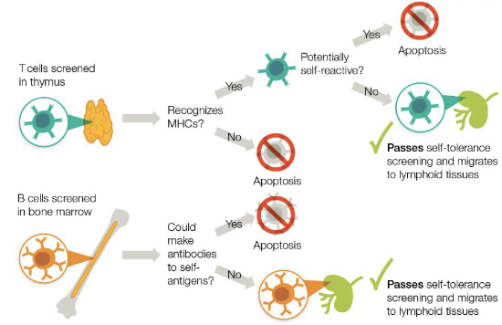
cell mediated response
organized by T helper cells and carried out by T cytotoxic cells
they go through the stages of adaptive immunity
cell-mediated adaptive immunity stages
antigen presentation
T-cell activation
T cell proliferation and differentiation
effector and memory cell activity
STEP 1: cell-mediated adaptive immunity stages
antigens are “presented” to T cells via major histocompatibility complexes (MHCs)
STEP 2: cell-mediated adaptive immunity stages
when T cells bind to antigen-presenting cells, this activates them (two required steps)
STEP 3: cell-mediated adaptive immunity stages
activated T cells divide rapidly (mitosis) and will differentiate into types that act as memory cells, recruitment cells, or seek/destroy cells
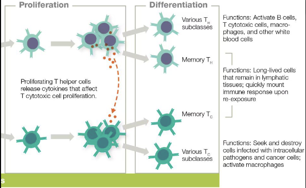
STEP 4: cell-mediated adaptive immunity
T cytotoxic cells will “seek and destroy” cells displaying specific antigens. T helper cells recruit, stimulate, and coordinate other leukocytes activity

humoral adaptive immunity stages
B cells are antigen-presenting cells so they can grab antigens themselves
T-independent antigens can fully activate B cells by themselves, whereas T dependent antigens require a helper T cell to fully activate the B cells
activated B cells divide rapidly (mitosis) and will differentiate into plasma cells that release antibodies, or memory cells that can assist in future immune responses
plasma cells secrete antibodies (immunoglobulins). antibodies bind to pathogen receptors to “block” them, initiate the complement cascade, and increase phagocytotic activity
Immunoglobulin G (IgG)
one of five major isotopes of antibodies
most abundant antibody in blood and can cross placenta to protect the fetus
Immunoglobulin A (IgA)
one of five major isotopes of antibodies
found primarily in mucosal secretions and breast milk
Immunoglobulin M (IgM)
one of five major isotopes of antibodies
created early in infection, but large and cannot migrate quickly
Immunoglobulin E (IgE)
one of five major isotopes of antibodies
involved in allergic reactions and defense against parasitic worms
Imunoglobulin D (IgD)
one of five major isotopes of antibodies
found on B cells, function still poorly understood
Humoral Memory
during primary antigen exposure, IgM is produced first, followed by IgG. due to memory capacity of memory B cells, if secondary antigen exposure occurs, these memory B cells produce tons of IgG very quickly

natural active immunity
immunity from previous infections
natural passive immunity
antibodies pass across the placenta
this is only temporary
artificial active immunity
vaccines used to trigger immune response to make antibodies
artificial passive immunity
patient receives antibodies via antiserums
this is only temporary
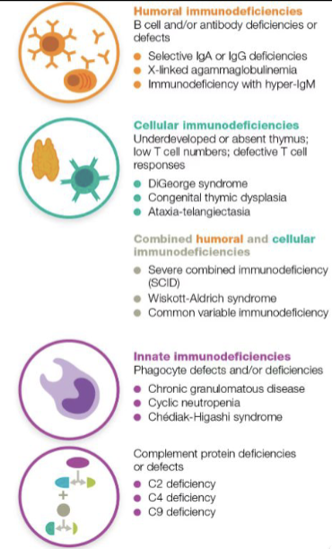
immunodeficiency
lack of properly functioning immune system

primary immunodeficiency
genetic errors lead to deficient immunity

secondary immunodeficiency
natural decline in immune function or external causes like infections or medical interventions
ex: leukocytes declines with age
ex: some drugs suppress immune function
ex: some pathogens have virulence factors that limit immune function. some even target immune cells (HIV infects helper T cells)
autoimmunity
immune system attacks healthy self-tissues
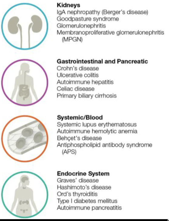
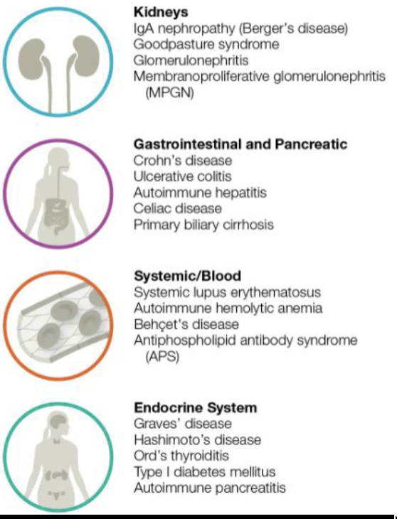
systemic vs localized autoimmunity
s: attack tissues throughout body
l: attacks specific type of tissue
hypersensitivity
inappropriate secondary immune response (inappropriate B and T cells activation)
more common in developed countries
hygiene hypothesis
clean food/water and higher antibiotic usage alters the normal healthy microbiota
type 1 hypersensitivity
part of secondary immune response
allergies; immune response to a harmless threat

allergens
antigen that can trigger IgE production and lead to an allergy
sensitization
part of secondary immune response
IgE antibodies attach to mast cells

post-sensitization
part of secondary immune response
antibody-coated mast cells bind to allergen and release pro-inflammatory chemicals like histamine

anaphylaxis
part of secondary immune response
severe and rapid allergic response to an allergen
can be localized (isolated reactions) or systemic (whole-system reactions)

allergen desensitization
part of secondary immune response (type I hypersensitivity)
body shifts its immune response to an allergen. likely due to more IgG antibodies being formed over time than IgE, which limits how much inflammation can occur

type 2 hypersensitivity
part of secondary immune response
cytotoxic reaction

how blood types work
There are four antigen types in blood. Three are carbs (A, B, O) and one is proteins (Rh factor). Rh is denoted by a “+” symbol if it is present and a “-” symbol if it is absent.
a patient can only accept blood types that have the same or fewer antigens present as their own blood

type 3 hypersensitivity
part of secondary immune response
immune complex
IgG or IgM bind to soluble antigens to make an excessive number of antibody-antigen complexes

type 4 hypersensitivity
part of secondary immune response
delayed hypersensitivity
T cells target host cell antigens instead of antibodies. these responses manifest slower than the previous types
ex: 12-72 hours like touching a poisonous plant

vaccine
biological preparation that provides active acquired immunity to a pathogen
herd immunity
enough people in a population are vaccinated so that the pathogen cannot spread effectively among non-vaccinated individuals
live attenuated (active vaccines)
active virus or living cells that have been altered to be non-pathogenic
these vaccines tend to produce the most effective long-lived memory in the immune system
however, these pathogens are the most likely to potentially cause disease or mutate into a form that is pathogenic
vector (vaccine)
genetically modified harmless virus that delivers genetic instructions and information from the pathogen to your body’s cells
whole-agent
inactive vaccine
vaccine contains the entire dead cellular pathogen/whole inactivated virus that has been rendered inactive by heat, chemicals, or radiation
these vaccines are safer but do not necessarily confer as long-term immunological memory
subunit
inactive subunit vaccine
vaccine contains either purified component of the pathogen or genetically engineered components that resemble the pathogen’s components
purified subunit
inactive subunit accine
antigens are a key component
toxoid
inactive subunit vaccine
inactivated protein toxin is the key component
conjugate
inactive vsubunit accine
linked polysaccharides are the key component
mRNA inactive vaccine
vaccine contains mRNA that when translated in host cells produces a protein or antigen that causes an immune response (without actual pathogen present)
mRNA directs the production of a specific protein found in or on the pathogen
Because cells typically only have the mRNA temporarily, additional vaccine administrations are needed to confer effective immunological memory
serology
form of diagnostic testing
study of what is in a patient’s serum (liquid, non-cellular/non-platelet portion of the blood)
serum is obtained vis centrifuge, which spins liquid at high speeds
agglutination
form of diagnostic testing
since antibodies can bind to more than one antigen at one time, they can clump
these tests are used to determine if antibodies are present in a sample
plaque reduction neutralization test (PRNT)
form of diagnostic testing
add viruses to a series of serum samples and then add those samples to a dish with cells
if antibodies are present, plaques (lysed cell residue) will not form. if antibodies re not present, plaques will form
enzyme-linked immunosorbent assays (ELISAs)
form of diagnostic testing
tests that use enzymes to bind to antibodies within antibody/antigen interactions
direct ELISAs
form of diagnostic testing
enzyme-linked antibodies are added to solution in wells that have antigens at the bottom
a substrate is added and if the substrate molecules bind to the antibodies, a signal occurs (light/color)

indirect ELISAs
form of diagnostic testing
similar to direction ELISA while also adding patient antibodies that connect the antigens and enzyme-linked detection antibodies

sandwich ELISAs
form of diagnostic testing
the wells contain a “capture” antibody, patient serum with potential antigens added, and enzyme-linked detection antibodies “sandwich” the antigen between both antibody types
