A&P Quiz 3
 Call Kai
 Learn
Learn Practice Test
 Spaced Repetition
 Match
Match Flashcards
 Knowt Play
1/32

There's no tags or description
Looks like no tags are added yet.
Name | Mastery | Learn | Test | Matching | Spaced | Call with Kai | Chat |
|---|
No analytics yet
Send a link to your students to track their progress
33 Terms
3 types of cartilage
Hyaline
Provides support, flexibility, and resilience
Most abundant type
Collagen fibers
Articular (joints), costal (ribs), respiratory (larynx), nasal cartilage (nose tip)
Elastic
Similar to hyaline, but contains elastic fibers
External ear and epiglottis
Fibrocartilage
Thick collagen fibers
Has great tensile strength
Menisci of knee, intervertebral discs
Growth of cartilage
Cartilage grows in 2 ways:
Appositional growth - cartilage-forming cells in perichondrium secrete matrix against external face of existing cartilage.
New matrix laid down on surface of cartilage.
Interstitial growth - chondrocytes within lacunae divide and secrete new matrix, expanding cartilage from within
New matrix made within cartilage
Calcification of cartilage occurs during normal bone growth in youth, but can also occur in old age
Hardened cartilage is not the same as bone
Functions of bones
Mechanical functions
Support - for body and soft organs
Protection - of brain, spinal cord, and vital organs
Movement - levers for muscle action
Warehouse
Mineral and growth factor storage - calcium and phosphorus, and growth factors reservoir
Blood cell formation - hematopoiesis occurs in red marrow cavities of certain bones
Fat storage - fat, used for an energy source, is stored in bone cavities
Hormone production - osteocalcin secreted by bones helps to regulate insulin secretion, glucose levels, and metabolism. Does not affect the bone directly.
Classification of bones
Long bones
Longer than they are wide
Limb bones
Short bones
Cube-shaped bones (in wrist and ankle)
Sesamoid bones form within tendons (ex patella). Vary in size and location between individuals
Flat bones
Thin, flat, slightly curved
Sternum, scapulae, ribs, most skull bones
Irregular bones
Complicated shapes
Vertebrae and hip bones
bone structure slide 12
Gross anatomy - compact and spongy bone
Compact bone - dense outer layer on every bone that appears smooth and solid
Spongy bone - made of of a honeycomb of small, needle like or flat pieces of bone called trabeculae
Open spaces between trabeculae are filled with red or yellow bone marrow
Gross anatomy - structure of short, irregular, and flat bones
Thin plates of spongy bone (dipole) covered by compact bone
Compact bone sandwiched between connective tissue membranes:
Periosteum covers outside of compact bone
Endosteum covers inside of compact bone
Bone marrow is scattered throughout spongy bone; no defined marrow cavity
Hyaline cartilage covers area of bone that is part of a movable joint

Gross anatomy - structure of a long bone
Structure of typical long bone
All long bones have a shaft (diaphysis), bone ends (epiphyses), and membranes
Diaphysis: tubular shaft that forms long axis of bone
Consists of compact bone surrounding central medullary cavity that is filled with yellow marrow in adults
Epiphyses: ends of long bones that consist of compact bone externally and spongy bone internally
Articular cartilage covers articular (joint) surfaces
Between diaphysis and epiphysis is epiphyseal line
Remnant of childhood epiphyseal plate where bone growth occurs

gross anatomy - membranes
Periosteum - white, double-layered membrane that covers external surfaces except joints
Fibrous layer - outer layer consisting of dense irregular connective tissue and perforating fibers (Sharpey’s fibers) that secure the periosteum to the underlying bone matrix
Osteogenic layer - inner layer abutting bone with osteogenic stem cells that give rise to most bone cells
Endosteum - connective tissue membrane covering internal bone surface and trabeculae of spongy bone
Lines canals that pass through compact bone
Like periosteum, contains osteogenic cells that can differentiate into other bone cells
Gross anatomy - hematopoietic tissue and bone markings
Hematopoietic tissue in bones
Red marrow is found within trabecular cavities of spongy bone and dipole of flat bones, such as sternum
In newborns, medullary cavities and all spongy bone contain red marrow
In adults, red marrow is located in heads of femur and humerus, but most active areas of hematopoiesis are flat bone dipole and some irregular bones (such as the hip bone, vertebral bodies)
Bone markings
Sites of muscle, ligament, and tendon attachment on external surfaces
Areas involved in joint formation or conduits for blood vessels and nerves
Three types of markings:
Projection: outward bulge of bone
May be due to increased stress from muscle pull or is a modification for joints
Depression: bowl- or groove-like cut-out that can serve as passageways for vessels and nerves, or plays a role in joints
Opening: hole or canal in bone that serves as passageways for blood vessels and nerves
Microscopic anatomy of bone - cells of bone tissue
1) Osteogenic cells - mitotically active (divide) stem cells in periosteum and endosteum
Also called osteoprogenitor cells
When stimulated, they differentiate into osteoblasts or bone-lining cells
Some remain as osteogenic stem cells
2) Osteoblasts - bone-forming cells that secrete unmineralized bone matrix called osteoid
Osteoid is made up of collagen and calcium-binding proteins
Collagen makes up 90% of bone protein
Osteoblasts are actively mitotic
3) Osteocytes - mature bone cells in lacunae that no longer divide
Maintain bone matrix and act as stress or strain sensors
Respond to mechanical stimuli such as increased force on bone or weightlessness
Communicate information to osteoblasts and osteoclasts to remodel the bone
4) Bone-lining cells - flat cells on bone surfaces, help maintain matrix (along with osteocytes)
On external bone surface, lining cells are called periosteal cells
On internal surfaces, they are called endosteal cells
5) Osteoclasts - derived from hematopoietic stem cells that also become macrophages
Giant, multinucleate cells function in bone resorption (breakdown of bone)
Cells have ruffled membrane to increase surface area for enzyme degradation of bone

Microscopic anatomy of compact bone - osteon, lamellae, central canal, perforating canals, lacunae, canaliculi, osteoblasts, interstitial lamellae, circumferential lamellae
Compact bone (also called lamellar bone) consists of:
Osteon (Haversion system)
Structural unit of compact bone
Elongated cylinder parallel to the long axis of bone
Acts as tiny weight-bearing pillars
Made of several rings of bone matrix called lamellae
Lamellae contain collagen fibers that run in different directions in adjacent rings
Withstands stress and resist twisting (torsion)
Bone salts are found between collagen fibers
Canals and canaliculi
Central (haversion) canal runs through core of osteon
Perforating (Volkann’s) canals - canals lined with endosteum that occur at right angles to central canal
Connect blood vessels and nerves of periosteum, medullary cavity, and central canal
Lacunae - small cavities that contain osteocytes
Canaliculi - hairlike cannals that connect lacunae to each other and to central canal
Allow communication between all osteocytes of osteon (nutrients and waste transport)
Osteoblasts that secrete bone matrix maintain contact with each other and osteocytes via cell projections with gap junctions
Interstitial and circumferential lamellae
Interstitial lamellae
Lamellae that are not part of osteon
Some fill gaps between forming osteons; others are remnamts of osteons cut by bone remodeling
Circumferential lamellae
Just deep to periosteum, but superficial to endosteum, these layers of lamellae extend around entire surface of diaphysis
Help long bone to resist twisting

Spongy bone
Appears poorly organized but is actually organized along lines of stress to help bone resist stress
Trabeculae, like cables on a suspension bridge, confer strength to bone
No osteons are present, but do contain irregularly arranged lamellae and osteocytes interconnected by canaliculi
Capillaries in endosteum supply nutrients
Chemical composition of bone - organic and inorganic
Organic components
Includes osteogenic cells, osteoblasts, osteocytes, bone-lining cells, osteoclasts, and osteoid
Osteoid is 1/3 of organic bone matrix, secreted by osteoblasts
Consists of ground substance and collagen fibers, which contribute to high tensile strength and flexibility of bone
Resilience of bone is due to sacrificial bonds in or between collagen molecules that stretch and break to absorb shocks and prevent fractures
Bonds re-form
Inorganic components
Hydroxyapatites (mineral salts)
Makeup 2/3 of bone by mass
Consist mainly of calcium phosphate crystals in and around collagen fibers
Responsible for hardness and resistance to compression
Bone is half as strong as steel in resisting compression and as strong as steel in resisting tension
Lasts long after death bc of mineral composition
Bone development - ossification + types
Ossification (osteogenesis) - the process of bone tissue formation
Formation of bony skeleton begins in month 2 of fetal development
Postnatal bone growth occurs until early adulthood
Bone remodeling and repair are lifelong
From week 8, fibrous membranes and hyaline cartilage of fetal skeleton are gradually replaced with bone tissue
Types:
Endochondral ossification
Bone forms by replacing hyaline cartilage
Bones are called cartilage (endochondral bones)
Form most of skeleton
Intramembranous ossification
Bone develops from fibrous membrane
Bones are called membrane bones
Formation of the bony skeleton - endochondral ossification and main steps
Endochondral ossification
All bones inferior to base of skull, except clavicles
Uses previously formed hyaline cartilage models
Requires breakdown of cartilage prior to ossification
Begins at primary ossification center in center of shaft
Blood vessels infiltrate perichondrium, converting it to periosteum
Mesenchymal cells specialize into osteoblasts
5 main steps:
Bone collar forms around diaphysis of cartilage model
Central cartilage in diaphysis calcifies, then develops cavities
Periosteal bud invades cavities, leading to formation of spongy bone
Diaphysis elongates, and medullary cavity forms; secondary ossificaiton centers appear in epiphyses
Epiphyses ossify; hyaline cartilage remains only in epiphyseal plates and articular cartilages
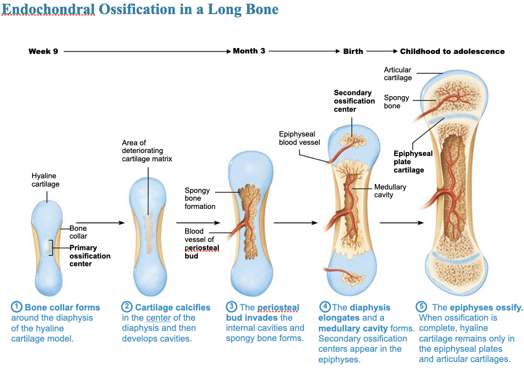
Formation of the bony skeleton - intramembranous ossification and main steps
Intramembranous ossification
Begins with fibrous connective tissue membranes formed by mesenchymal cells
Forms frontal, parietal, occipital, temporal, and clavicle bones
4 major steps:
Ossification centers are formed when mesenchymal cells cluster and become osteoblasts
They secrete osteoid, which is then calcified
Trabeculae are laid down between blood vessels
Lamellar bone replaces woven bone under the periosteum (compact bone) and red marrow appears
Postnatal bone growth
Long bones grow lengthwise by interstitial (longitudinal) growth of epiphyseal plate
Bones increase thickness through appositional growth
Bones stop growing during adolescence
Some facial bones continue to grow slowly through life
Growth is in length and thickness
Growth in length of bones
Interstitial growth requires presence of epiphyseal cartilage in the epiphyseal plate
Epiphyseal plate maintains constant thickness
Rate of cartilage growth on one side balanced by bone replacement on other
Epiphyseal plate consists of 5 zones:
Resting (quiescent) zone
Proliferation (growth) zone
Hypertrophic zone
Calcification zone
Ossification (osteogenic) zone
Growth in Length of Long Bones
1) Resting quiescent zone
Area of cartilage on epiphyseal side of epiphyseal plate that is relatively inactive
2) Proliferation (growth) zone
Area of cartilage on diaphysis side of epiphyseal plate that is rapidly dividing
New cells formed move upward, epiphysis is pushed away, causing lengthening
3) Hypertrophic zone
Area with older chondrocytes closer to diaphysis
Cartilage lacunae enlarge and erode, forming interconnecting spaces
4) Calcification zone
Surrounding cartilage matrix calcifies; chondrocytes die and deteriorate
5) Ossification zone
Chondrocyte deterioration leaves long spicules of calcified cartilage
Spicules are then eroded by osteoclasts and covered with new bone
Ultimately replaced with spongy bone
Medullary cavity enlarges as spicules are eroded
Growth in length of long bones - the end
Near end of adolescence, chondroblasts divide less often
Epiphyseal plate thins, then is replaced by bone
Epiphyseal plate closure occurs when epiphysis and diaphysis fuse - connected at the epiphyseal line
Bone lengthening ceases
Females: occurs around 18 years of age
Males: occurs around 21 years of age
Growth in width (thickness)
Appositional growth (growth in width) can occur throughout life
Bones thicken in response to increased stress from muscle activity or added weight
Osteoblasts beneath periosteum secrete bone matrix on external bone
Osteoclasts remove bone on endosteal surface
Usually more building up than breaking down which leads to thicker, stronger bone that is not too heavy
Hormonal regulation of bone growth
Growth hormone: most important hormone in stimulating epiphyseal plate activity in infancy and childhood
Thyroid hormone: modulates activity of growth hormone, ensuring proper proportions
Testosterone (males) and estrogen (females) at puberty: promote adolescent growth spurts
End of growth by inducing epiphyseal plate closure
Excesses or deficits of any hormones cause abnormal skeletal growth
Bone remodeling, bone resorption, bone deposit
Bone remodeling
About 5–7% of bone mass is recycled each week
Bone remodeling consists of both bone deposit and bone resorption
Occurs at surfaces of both periosteum and endosteum
Bone resorption
Resorption is function of osteoclasts
Dig depressions or grooves as they break down matrix
Secrete lysosomal enzymes and acids that digest matrix
Acidity converts calcium salts to soluble forms
Osteoclasts also phagocytize demineralized matrix and dead osteocytes
Once resorption is complete, osteoclasts undergo apoptosis
PTH (parathyroid hormone) activates osteoclasts
Bone deposit
New bone matrix is deposited by osteoblasts, stimulated mechanically, and by availability of materials (proteins, Ca2+ and phosphate ions)
Control of remodeling
Remodeling occurs continuously but is regulated by genetic factors and two control loops: hormonal and mechanical
Hormonal controls
Negative feedback loop that controls blood Ca 2+ levels
Free (ionic) calcium (Ca 2+) is required for vital processes
99% of 1200-1400 grams of calcium are found in bone
Intestinal absorption of Ca 2+ requires vitamin D
Parathyroid hormone (PTH) - produced by parathyroid glands in response to low blood calcium levels
Stimulates osteoclasts to resorb bone
Calcium is released into blood, raising levels
PTH secretion stops when homeostatic calcium levels are reached
Calcitonin - produced by parafollicular cells of thyroid gland in response to high levels of blood calcium levels
Effects are negligible, but at high pharmacological doses it can lower blood calcium levels temporarily
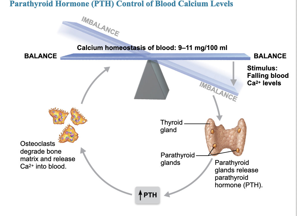
Clinical - homeostatic imbalance
Even minute changes in blood calcium levels can cause severe neuromuscular problems
Hypocalcemia: low levels of calcium cause hyperexcitablility
Hypercalcemia: high levels of calcium cause nonresponsiveness
Sustained high blood calcium levels can lead to deposits of calcium salts in blood vessels or kidneys and formation of kidney stones
Hormonal controls (cont.)
Other hormones play a role in bone density and turnover
Glucocorticoids (cortisone) - increase osteoclast activity
Sex hormones (also steroids) - decrease (favor deposition)
Control of modeling - mechanical + wolf’s law
Response to mechanical stress
Bones react to stresses, like when weight bears on them or muscles pull on them
Wolf’s law states that bones grow or remodel in response to demands placed on them
Stress usually tends to bend bones
Bending compresses one side, stretches other side
Diaphysis is thickest where bending stresses are greatest
Wolf’s law also explains:
Handedness (right- or left-handed) results in thicker and stronger bone of the DOMINANT upper limb
Curved bones are thickest where most likely to buckle
Trabeculae form trusses along lines of stress
Large, bony projections occur where heavy, active muscles attach
Weight lifters have enormous thickenings at muscle attachment sites of most used muscles
Bones of fetus and bedridden people are featureless because of lack of stress on bones
Different roles of hormonal and mechanical controls
Hormonal controls determine if and when remodeling occurs in response to changing blood calcium levels, but mechanical stress determines where it occurs
Fracture classification
Fractures are breaks
During youth, most fractures result from trauma
In old age, most result from weakness of bone due to bone thinning
Fracture classification
Position of bone fragments after fracture
Nondisplaced - retain normal position
Displaced - out of alignment
Completeness of break
Complete - broken all the way through
Incomplete - not broken all the way through
Whether skin is penetrated
Open (compound) - skin is penetrated
Closed (simple) - skin is not penetrated)
Can also be described by location of fracture, external appearance, and nature of break
Common types of fractures
The old
Comminuted - bone fragments into 3+ pieces; common in the aged, whose bones are more brittle
Compression - bone is crushed; common in porous bones (ie osteoporotic bones) subjected to extreme trauma, as in a fall
The young
Spiral - ragged break occurs when excessive twisting forces are applied to a bone; common sports fracture
Epiphyseal - epiphysis separates from the diaphysis along the epiphyseal plate; tends to occur where cartilage cells are dying and calcification of the matrix is occurring
Children
Depressed - broken bone portion is pressed inward; typical of skull fracture
Greenstick - bone breaks incompletely, much in the way a green twig breaks. Only one side of the shaft breaks; the other side bends. Children
Fracture treatment and repair
Treatment involves reduction, the realignment of broken bone ends
Closed reduction - physician manipulates to correct position
Open reduction - surgical pins or wires secure ends
Immobilization of bone by cast or traction is needed for healing
Time needed for repair depends on break severity, bone broken, and age of patient
4 major stages of repair:
1) Hematoma formation
Torn blood vessels hemorrhage, forming mass of clotted blood called a hematoma
Site is swollen, painful, and inflamed
2) Fibrocartilaginous callus formation
Capillaries grow into hematoma and bring fibroblasts
Fibroblasts secrete collagen fibers to span break and connect broken ends
Cells (fibroblasts and chondroblasts) begin reconstruction of bone
Create cartilage matrix of repair tissue
This mass of repair tissue is called fibrocartilaginous callus
3) Bony callus formation
Osteoblasts form spongy bone (trabeculae) within matrix of fibrocartilaginous callus
Callus is converted to bony (hard) callus of spongy bone (about 2 months)
4) Bone remodeling
Begins during bony callus formation and continues for months
Excess material on outside and inside the bone is removed
Compact bone is laid down to reconstruct shaft walls
Final structure resembles original structure, responds to same mechanical stressors

Bone disorders
Imbalances between bone deposit and bone resorption underlie nearly every disease that affects the human skeleton.
Three major bone diseases:
Osteomalacia and rickets
Osteoporosis
Osteomalacia - bones are poorly mineralized
Osteoid is produced, but calcium salts not adequately deposited
Results in soft, weak bones, pain upon bearing weight
Rickets - osteomalacia of children
Results in bowed legs and other bone deformities because bone ends are enlarged and abnormally long
Causes for both: vitamin D deficiency or insufficient dietary calcium
Osteoporosis - group of diseases in which bone resorption exceeds deposit
Matrix remains normal, but bone mass declines
Spongy bone of spine and neck of femur most susceptible
Vertebral and hip fractures common
Risk factors for osteoporosis
Most often aged, postmenopausal women
Affects 30% of women aged 60–70 years and 70% of women by age 80
Estrogen plays a role in bone density, so when levels drop at menopause, women run higher risk
Men are less prone due to protection by the effects of testosterone (which last throughout life)
Additional risk factors for osteoporosis:
Insufficient exercise
Diet poor in calcium and protein
Smoking, Genetics, Hormone-related conditions
Hyperthyroidism
Diabetes mellitus
Consumption of alcohol or certain medications
Osteoporosis - treatment
Traditional treatments
Calcium
Vitamin D supplements
Weight-bearing exercise
Hormone replacement therapy (estrogens) – discouraged, now coming back??
Slows bone loss but does not reverse it
Controversial because of increased risk of heart attack, stroke, and breast cancer. NOW idea is until 65 it's OK!
Recent drugs for osteoporosis are osteoclast inhibitors:
Bisphosphonates: decrease osteoclast activity and number
Partially reverse osteoporosis in spine
Denosumab
Monoclonal antibody shown to reduce fractures in men with prostate cancer
Improves bone density in elderly by preventing activation of osteoclasts by osteoblasts