Frontiers in Physiology
 Call Kai
 Learn
 Practice Test
 Spaced Repetition
 Match
 Flashcards
 Knowt Play
1/421

There's no tags or description
Looks like no tags are added yet.
422 Terms
Inheritance of Cancer
Only up to 10% of cancers are inherited
Are there any curable cancers?
Cancer can go into remission.
Prognosis (if a patient will ge better or worse) is typically measured using a 5-year survival rate
Classification of Cancers
Carcinoma, sarcoma, myeloma, leukaemia, lymphoma, and mixed types
Carcinoma
Malignancy of epithelial origin or cancer of the internal or external lining of the body ( two major types: adenocarcinoma and squamous cell carcinoma)
Sarcoma
Malignancy of the supportive and connective tissues (e.g. bones, tendons, cartilage, muscles): osteosarcoma- bone cancer, glioma- neurogenic connective tissue in brain, chondrosarcoma- cancer of the cartilage
Myeloma
Cancer develops in the plasma cell in bone marrow
Leukaemia
Cancer of the blood cells: myeloid leukaemia- cancer of the myeloid and granulocytic cells, lymphocytic leukaemia- cancer of the lymphoid cells
Lymphoma
Cancer of the glands or nodes of the lymphatic system
Cancer Non-Preventable Risk Factors
Biomedical Factors - genetic susceptibility and hormonal factors in females
Ageing
Ethnicity
Gender
Cancer Preventable Risk Factors
Lifestyles Factors: smoking, alcohol consumption, physicial inactivity and obesity, chronic infections, diet
Environmental Factors: sunlight, radiation, occupational exposure, pollution
Normal Physiology vs Cancer
Large, variably shaped nuclei
Many dividing cells
Disorganized arrangement
Variation in size and shape
Loss of normal features
Hallmarks of cancer cells
Sustaining proliferative signalling, evading growth suppressors, avoiding immune destruction, enabling replicative immortality, tumor-promoting inflammation, activating invasion and metastasis, inducing or accessing vasculature, genome instability and mutation, resisting cell death, and deregulating cellular metabolism.
What drives cancer initiation and progression?
Alterations to the DNA (genetic and epigenetics)
Alterations to the RNA molecules
Leading to the loss or gain of functions of proteins that promote cancer initiation and progression
DNA Mutations in Cancer
Different types of cancers are affected by different combinations of DNA mutations.
One mutation is not sufficient to cause cancer.
Epigenetic Alterations in Cancer
DNA methylation
Histone acetylation
Non-coding RNA
These cause changes in cell proliferation, cell cycle control, apoptosis, metastasis, invasion, DNA damage, and senescence
How is cancer is a heterogenous disease?
This means that there are often subtypes and it differs from one patient to the next
The cancer affects the same cells in the same location but the cause and behaviour is different
The subtypes can indicate prognosis
Tumor Heterogeneity
Tumor heterogeneity also exists within a single tumor - one cells may respond more to drugs and others may be more resistant
Why is cancer so difficult to manage?
Hundreds of potential genetic and non-genetic alterations
Heterogeneity of cancer
Tumour microenvironment – determine how they grow, invade, metastasise, interaction with immune cells
Combinatorial therapy is often needed to treat/manage cancer
There is still much to be learnt about precision therapy for cancer
Mutations in DNA/RNA that Cause Cancer
DNA: mutations such as point, frameshift, etc. and epigenetic alterations
RNA: coding RNA alterations such as alternative splicing and RNA modifications and non-coding RNA alterations such as miRNAs, etc.
Accumulation of Mutations to Cause Cancer
Mistakes that cause cancer (e.g. mutations) accumulate over time.
Each mutation provides a selective advantage → clonal expansion.
Involves multiple changes in cellular properties, uncontrolled growth and proliferation, evade apoptosis, deregulation of cell cycle, cell-cell interaction.
Normal Cell Proliferation
Growth factor binds to receptor on cell membrane
Transiently activates signal-transducing proteins
Signal is transmitted from cytoplasm to nucleus
Transcription of genes to promote cell division
Cell enter and progress through the cell cycle
Sustaining Proliferative Signalling
Gain of function mutations: proto-oncogenes to oncogenes that promote cell growth
Growth factors: secreted by tumour or surrounding stroma cells (e.g. EGF)
Amplification of growth factor receptor (e.g. HER2)
Constitutive activation of signal transducing molecules (e.g. RAS)
Overexpression of nuclear transcription factors (e.g. MYC, FOS)
Evading Growth Suppressors
Loss of function mutations of tumour suppressor genes
Retinoblastoma (RB) : stop normal cell’s passage through the restriction point at G1 phase
p53: Guardian of the genome – arrest cell cycle upon the detection of DNA damage
APC: negative regulator of WNT signalling that activates G1 effectors.
Resisting Cell Death
Normal cells: DNA damage, hypoxia, disruption to metabolic pathways apoptosis (programmed cell death)
Cancer cells: mutations/ altered gene expression
Disrupt key players of apoptosis
Increased ability to withstand intrinsic stresses
Key Players of Apoptosis
Proapoptotic proteins = BAX, BAK, promote mitochondrial permeabilisation
Antiapoptotic proteins = BCL2, BCLXL
BH3 domain only proteins = BAD, BID, PUMA; enhance BAX, BAK
Inhibitors of apoptosis proteins (IAPs), inhibit Caspase-9, stop apoptosis
Revise apoptosis pathway (lecture 2)
Enabling Replicative Immortality
Normal somatic cells:
Have a finite number of replication cycle: ~70 replications
This limit is called the Hayflick’s limit
Shortening of the ends of chromosomes (telomeres) after each mitotic cell division
Telomeres too short cellular senescence.
Cancer cells:
Lengthening of telomeres
2 Mechanisms of Telomere Lengthening
Increase telomerase
Alternative lengthening of telomeres
Activating Invasion and Metastasis
Cause of 80-90% of cancer-related death
Initiated by the breakdown of cell-cell adhesion (E-cadherin)
Degradation of extracellular matrix (MMPs)
Increase motility or chemotaxis
Secretion of factors that stimulate cancer cells to migrate
Inducing or accessing vasculature- angiogenesis
Continuously growing and requires oxygen, nutrients, more efficient ways to get rid of waste.
Needs to develop blood vessels - Vascular Endothelial Growth Factor (VEGF)
Deregulating Cellular Metabolism
Increase in growth factor signalling
Increase in glucose uptake
Build up glycolytic intermediates for DNA, RNA, protein synthesis
RAS signalling
Increase the activity of glucose transporters and glycolysis enzymes enhance glycolysis
Increase factors that stimulate protein synthesis
MYC signalling
Increase expression of genes involved in anabolic metabolism and cell growth (glutaminase) - for mitochondrial utilization of glutamine glycolysis
Avoiding Immune Destruction
Secrete immune suppressive factors (e.g. TGF-β) to paralyse cytotoxic T cells
Recruitment of immunosuppressive cells including myeloid-derived suppressor cells (MSDCs)
Evasion via checkpoint molecules
Evasion Via Checkpoint Molecules
Normal cells
PD-L1/PD-1 shut down T cells to prevent autoimmunity
Cancer cells
Evolve this ability to express these checkpoint molecules to escape recognition and killing by T cells.
Express a high level of checkpoint molecules
Tumour-Promoting Inflammation
Tumour promoting inflammatory cells: macrophages, neutrophils, B-cells
MDSCs: secreted factors promote angiogenesis, promoting tumour growth.
Subsets of myeloid progenitor cells, typically involved in wound-healing: major source of the angiogenic, epithelial, stromal growth factor and matrix-remodelling enzymes
Genome Instability and Mutation
Genomic instability: high tendency/ frequency of mutations or other genetic changes during cell division.
Caused by:
DNA damage/ DNA replication stress induced by oncogenes
Involves the mutations of DNA repair genes.
e.g MYH, base excision repair
Telomere damage – loss of protection to the ends of chromosomes chromosome fusion
Epigenetic dysregulation – global DNA hypomethylation activation of oncogenes
Centrosome amplification

Centrosome Amplification
Centrosome: cellular structure that organises microtubules during cell division
Important for the segregation of chromosomes
Lead to altered karyotypes
Hallmarks of Cancer: New Additions
Nonmutational epigenetic reprogramming
Unlocking phenotypic plasticity
Polymorphic microbiomes
Senescent Cells
Unlocking Phenotypic Plasticity
Escaping terminal differentiation, becoming stem/progenitor cell-like (e.g. in acute myeloid leukaemia, accumulation of myeloid progenitor cells/ poorly differentiated cells.
Polymorphic Microbiomes
Gut microbiome can modulate susceptibility and pathogenesis of human cancer (e.g. colorectal-cancer)
Senescent Cells
Hide away, stay dormant and therapeutic resistant
Mutation vs Polymorphism
Mutation : rare change in the DNA and are associated with a higher risk of developing a disease. Occur in < 1% of the general population.
Polymorphism : DNA sequence variation, which is common (>1%) in the general population and typically not associated with any disease (e.g. Single nucleotide polymorphisms).
Both mutation and polymorphism are genetic alterations.
Disease causing genetic alterations = mutations
Causes of Mutations
Inherited from parent(s)
Carcinogens: UV radiation, chemicals, viruses, smoking, etc.
Random mistakes that occur during the cell cycle
Types of Mutations
Point mutation: change in a single DNA base and includes missense, nonsense, and silent
Frameshift mutation: insertion or deletion that shifts the DNA sequence
Point Mutation Types
Missense: change in a single bp causing an amino acid change
Nonsense: change in a bp causing a stop codon
Silent: change in a bp that doesn’t change the amino acid
Gene Mutation Overview
A change in the nucleotide sequence
Small scale
Can sometimes be corrected
Only a slight structural change
Comparatively low influence
Chromosomal Mutation Overview
A change in the nucleotide sequence of a segment of a chromosome
Large scale
Difficult to correct
Major structural change
Can be lethal
Types of Chromosomal Mutations
Translocation: breakage and removal of a large segment of DNA from one chromosome, followed by the segment's attachment to a different chromosome.
Inversion: DNA segment of the chromosome was turned 180° and reinserted.
Deletion: loss of a piece of DNA segment from a chromosome.
Duplication: production and inclusion of one or more copies of any segment of the chromosome.
Somatic vs Germline Mutations
Somatic mutations
Occur in non germline tissue
Cannot be transferred to an offspring
Germline mutations
Present in egg or sperm
Are heritable
Cause cancer family syndrome (e.g. Familial Adenomatous Polyposis Syndrome)
All somatic cells in the offspring are affected
Inheritance of Cancer Causing Mutations
Inheriting a cancer causing mutation doesn’t guarantee that the person will get cancer
With hereditary cancers, the first mutation is inherited and already present at birth. Additional mutations can build up over time, leading to cancer.
Cancer Promoting Missense Mutations
Gain of function mutations: proto-oncogenes to oncogenes that promote uncontrolled cell growth
Example BRAF V600E (50% of all Melanoma)
Loss of function mutations: tumour suppressor genes
Example: p53 (guardian of the genome)
Cancer Causing Nonsense Mutations
Loss of function: tumour suppressor genes
Inducing premature stop codons – TGA, TAA, TAG
Cancer Causing Silent Mutations
Loss or gain of function due to alternative splicing, changes in RNA stability, and translation
Ex. KRAS - RNA stability
Cancer Causing Frameshift Mutations
Deletion at codon 1309 of APC in colorectal cancer - very frequent in individuals with Familial Adenomatous Polyposis Syndrome
Cancer Causing Translocation
An abnormality of chromosome 22, whereby a part of chromosome 9 is transferred and inserted into it : t(9;22) (q34; q11)
Hallmark of chronic myeloid leukaemia (CML)
Resulted in the BCR-ABL fusion gene
Constitutively active protein tyrosine kinase that promotes cell proliferation and suppresses apoptosis
Inhibits DNA repair, causing genomic instability
Multistep Progression of Cancer

Example of Multistep Progression of Cancer
Adenoma-carcinoma for colorectal cancer is well understood that these mutations occur to cause cancer

Driver vs Passenger Mutations
Driver mutations: Mutations that are necessary for the development, growth and survival of cancer cells
Passenger mutations: Neutral genetic changes that do not provide any selective advantage to the development, growth and survival of cancer cells.
Approach to confirm that a genetic alteration is causing cancer
Identify the genetic alteration/mutation
Validate the role of the genetic alteration/ mutation in vitro and in vivo
“Rescue” or correct the genetic alteration/ mutation to stop the cancer
How to identify the genetic mutation causing cancer?
Gene sequencing
How to validate the effect of a mutation in vitro?
Derive a new cancer cell line from patient with the mutation
Insert the mutation in an existing cancer cell line (e.g with CRISPR)
Determine changes in cancer growth and behavior.
How to validate the effect of a mutation in vivo?
Use mouse xenograft models
Inject cancer cells and engraft them in mice
Measure tumour growth, size, and volume, followed by histology
How to “rescue” cancer in vitro and in vivo?
Using small molecule inhibitors, DNA editing, antisense oligonucleotides, viral-loaded siRNA, and short hairpin RNAs.
Epigenetics
Changes to the DNA and gene expression that do not involve any changes to the DNA sequence itself
Include modifications to the DNA, histone proteins, and RNA

DNA Methylation
The addition of methyl groups to DNA (mainly on cytosine residues). Predominantly at CpGs (CG in the code) in mammals.
CpGs
Most CpGs in the DNA are methylated (because DNA methylation silences retroviral repeat sequences that need to maintain genomic stability)
The exception being CpG rich regions called CpG islands.
CpG islands are present in 50-60% of mammalian gene promoters
Unmethylated cytosines in CpG island allows binding of transcription factors to the promoter of genes to enable the gene to be actively transcribed/expressed.
Methylated cytosines in the promoter region can lead to gene silencing important for tissue specific gene expression
How can DNA methylation go wrong in cancer?
DNA hypermethylation
DNA hypomethylation
DNA Hypermethylation
Can induce silencing of tumour suppressor genes (TSGs). This occurs by hypermethylation at the promoter sites of TSGs that prevent transcription factor binding.
DNA Hypomethylation
Local effects:
Activation of oncogenes (e.g. Cyclin D2 in colorectal cancer).
Loss of imprinting that drives the proliferation of cancer cells (e.g. IGF2/H19 locus in colorectal cancer).
Global effects:
Activation of repetitive sequences increases susceptibility to chromosome breakage and therefore genomic instability.
Reactivation of retrotransposon disrupt the normal gene structure and function/ chromosomal rearrangement
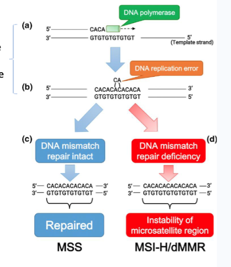
MLH1 methylation in sporadic microsatellite unstable colorectal cancer
MLH1: a mismatch repair (MMR) gene
MLH1 promoter methylation occurs in about 10-20% of all sporadic colorectal cancers (that exhibit microsatellite instability)
MLH1 is a tumour suppressor gene
MLH1 methylation leads to a loss of tumour suppressor function
MLH1 methylation can be transmitted from a parent to an offspring
Chromatin exists in which two states
Heterochromatin (closed): highly condense, gene poor, transcriptionally silent
Euchromatin (open): less condense, gene-rich, more easily transcribed.
How can histone modification imbalance cause cancer?
Over modification of histone proteins causes chromatin to condense and can silence tumour suppressor genes. The opposite can cause activation of oncogenes.
Chromatin remodelling occurs when histone modification go awry and can cause:
DNA replication stress
Metastasis
Tumour immunity
Angiogenesis
Senescence
DNA damage response and repair
Two Classes of Histone Modifications
Repressive
Closed chromatin
Transcription repressed
Active
Open chromatin
Transcription activated
Interplay Between Cancer and Epigenetics
Cancer development and progression involve a combination of genetic and epigenetic changes.
Genetic mutation can occur in genes that regulate DNA methylation
DNMT3A – de novo methyltransferase 3A is responsible for methylating DNA.
Mutations of DNMT3A occur in approximately 20% of acute myeloid leukaemia cases
Alteration of genes that regulates histone modification/chromatin remodelling is frequent in cancer
Approaches to Study Epigenetics Dysregulation in Cancer
Bisulfite Sequencing and Detecting Histone Modifications
Bisulfite Sequencing
DNA methylation detection
Bisulfite Conversion
Sodium bisulfite converts unmethylated cytosine to uracil
Cannot convert methylated cytosine
Process
Extract DNA from cancer sample
Bisulfite conversion
PCR amplification
Sequencing, restriction digestion, quantitative PCR, etc
Detecting Histone Modifications
Antibodies specific to a histone modification are used to immunoprecipitate the histone-DNA complex
DNA is purified
DNA is analysed by microarray, next generation sequencing, or quantitative PCR
How to manipulate DNA methylation and histone modifications to study effect in cancer?
Using inhibitors (e.g. 5-Aza-deoxycytidine for DNA methylation – global effect).
Deleting proteins that regulate DNA methylation/histone modifications – global effect.
CRISPR-based epigenome editing approaches - target specific
Coding RNA
mRNA → protein
Types of coding RNA alterations in cancer
General increase or decrease of mRNA encoding specific genes (e.g. oncogenes, tumour suppressor genes, metastasis-promoting genes)
Alternative splicing
Polyadenylation and RNA export
Modifications to mRNA (epitranscriptomics)
Increase in mRNAs encoding oncoproteins
Increase in transcription factor activity
Increase in transcription co-activator activity (e.g CBP and p300)
Increase in gene enhancer activity such as high c-Myc
Aberrant genetic and epigenetic changes
Decrease in mRNAs encoding tumor suppressors
Loss of transcription factor activity
Loss of transcription co-activator activity
Decrease in gene enhancer activity
Aberrant genetic and epigenetic changes
Aberrant regulation by non-coding RNAs
Alternative Splicing
A cellular process in which exons from a single gene are joined in different combinations, leading to different, but related, mRNA transcripts
Alternatively-spliced mRNAs are called isoforms
A key process in promoting protein diversity and allowing tissue specific gene function (e.g. brain may require isoform 1 of gene X whereas skin requires isoform 2 of gene X
Intron Retention (IR)
Inclusion of an intron in the mature mRNA
The type of alternative splicing that is least studied and understood.
Intron retention is coupled with nonsense-mediated decay (NMD) to repress genes that will otherwise inhibit granulocyte differentiation.
It is a previously overlooked mechanism of gene expression control
IR inactivates tumour suppressor genes
Modifications to mRNA (epitranscriptomics)
Regulate mRNA splicing, maturation, localization, translation, and degradation
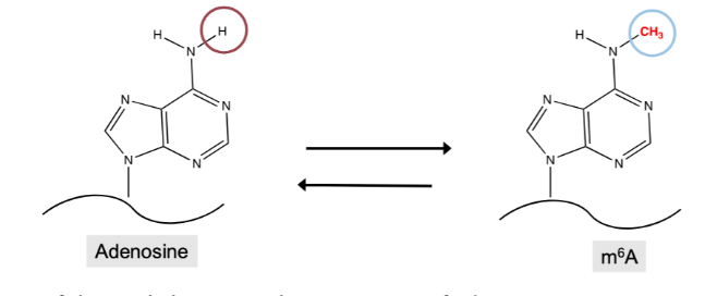
N6-Methyladenosine (m6A) RNA modification
Addition of the methyl group to the N6 position of adenosine.
Most pervasive and prevalent “reversible” internal modification to mRNAs.
Occurs co-transcriptionally (i.e. added to mRNA when transcription is happening in the nucleus).
Regulates all aspects of mRNA metabolism: transcription, splicing, stability, export, translation.
Proteins that regulate in m6A RNA modification
writers : deposit m6A on mRNAs- METTL3 is the catalytic enzyme
erasers: remove m6A on mRNAs
readers : recognise m6A to direct splicing, degradation, export, translation
METTL3 and m6A RNA Modification
AML is the type of cancer showing highest levels of METTL3 overexpression
METTL3 overexpression leads to increased m6A on mRNAs encoding:
MYC- promotes cell cycle progression, maintenance of stem cells
BCL2- prevent apoptosis (programmed cell death)
PTEN- regulates cell division
A strategy proposed to kill AML cells is to deplete METTL3
VIRMA
The largest protein in the m6A writer complex
Acts as a scaffold to hold the other proteins in place and allow METTL3 to function efficiently
Exists as two different isoforms:
Full-length (FL)
N-terminal isoform (N-term)
VIRMA Cancer Hypothesis
VIRMA is aberrantly regulated/expressed in human cancers.
Introduce more full-length and N-terminal VIRMA (N-term) into cancer cells
Measure m6A changes
Measure phenotypic changes to cancer cells in terms of their proliferation and growth
VIRMA Results
VIRMA FL but not N-term promotes proliferation and clonogenicity (more colonies) of cancer cells in vitro
Increased expression of VIRMA FL but not N-term promotes tumour growth in MDA-MB-231 xenografts
Overexpression of FL but not N-term VIRMA increases m6A methylation on mRNAs
Types of Non-Coding RNA
Divided into small (<200 nt) or long non-coding RNAs (lncRNAs) (>200 nt).
Possess structural/housekeeping or regulatory function.
Best studied is microRNAs (miRNAs).
lncRNA
miRNA
18-25 nucleotide long single- stranded RNA
Regulates gene expression post-transcriptionally
Exists in animals and plants
According to miRBase version 22.1, there are 38589 miRNAs discovered to date.
Can be derived from introns, exons, other non-coding RNAs (e.g. snoRNAs).
Once processed, miRNAs will be incorporated into RNA-induced silencing complex (RISC).
miRNA Canonical Function
Mature miRNA in RISC will bind (with partial complementarity) to sequences in the 3’ untranslated region (3’UTR) of target genes.
Triggering
mRNA cleavage
Translation repression
mRNA deadenylation
miRNA Non-Canonical Function
Mature miRNA bind regions of the genome/transcriptome other than 3’ UTR:
Promoter region- trigger gene activation or silencing
lncRNA- disrupt the function of lncRNA
pri-miRNAs- prevent normal miRNA biogenesis
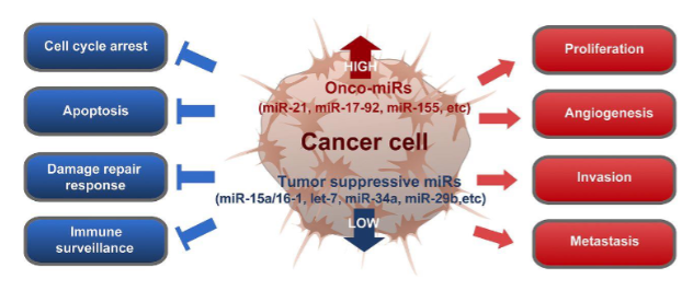
miRNA deregulation in cancer
Onco-miRs – target tumour suppressor genes
Tumour suppressive miRs- target oncogenes
Role in metastasis: miRNAs can be involved in regulating epithelial to mesenchymal transition (EMT), invasion and angiogenesis at the secondary site.

Example: miR-200 family and cancer metastasis
High expression of E-Cadherin is critical to maintain the epithelial state.
ZEB1/2 are transcription regulators that repress the expression of E-Cadherin and promote EMT.
ZEB1/2 are cognate target of miR-200 family members (miR-200a,miR-200b, miR-200C, miR- 141, miR-429).
miR-200b targets Quaking 1 (QKI)
QKI is a critical regulator of blood vessel formation- mice lacking QKI gene die in utero because blood vessels cannot form.
In cancer (e.g. lung cancer), miR200b level decreases, allowing QKI to be expressed at high levels to promote angiogenesis
circRNA
Single-stranded RNA, which unlike linear RNA, forms a covalently closed continuous loop.
Generated due to back-splicing of mRNAs - exons join together to form a circle
To date, over 180,000 circRNAs have been identified in humans
Functions of circRNA
Transcriptional regulation: interact with transcription factor/RNA pol II transcription complex to promote transcription.
Splicing regulation: compete with the splicing of linear mRNAs leading to altered gene expression.
Post-transcriptional regulation: act as miRNA sponges to interfere with miRNA action.
Translational regulation: binds to translational initiation-related proteins (e.g. eIF4G and PABP) to suppress translation. Some circRNA contains start codon and competes with linear mRNA to access the ribosome for translation : mRNA traps.
ciRNA in Cancer
Good cancer biomarkers
Can be associated with different cancer hallmarks
Example: circRNA-10720 and cancer metastasis
High expression of E-Cadherin is critical to maintain the epithelial state.
Twist1 is another transcription regulator that represses E-Cadherin.
In hepatocellular carcinoma (liver cancer), Twist1 upregulates the expression of circRNA-10720, which is derived from Cul2 gene.
circRNA-10720 sponges miRNAs that targets vimentin.
Vimentin is a filament protein that is crucial for the establishment of the mesenchymal phenotype.
Given that miRNAs targeting vimentin are sponged by circRNA-10720, vimentin is released from miRNA-mediated repression and becomes highly expressed in metastatic cancer cells → promotes EMT and metastasis