MICB 3301 Lab Quiz 2
 Call Kai
 Learn
Learn Practice Test
 Spaced Repetition
 Match
Match Flashcards
 Knowt Play
1/164

There's no tags or description
Looks like no tags are added yet.
Name | Mastery | Learn | Test | Matching | Spaced | Call with Kai | Chat |
|---|
No analytics yet
Send a link to your students to track their progress
165 Terms
Bacteria aren't well visualized under small magnification because...
The refractive index of bacteria is almost the same as the water it is suspended in
What is needed in order to visualize bacteria more clearly?
-Greater magnification and resolution
-Contrast between cells and background
What is a dye?
An organic compound containing CHROMOPHORE and AUXOCHROME chemical groupings attached to benzene rings
What structures make up a dye?
Chromophore, auxochrome, benzene ring
What is a chromophore
The specific part of a molecule responsible for absorbing light and producing/reflecting color for humans to see.
What role do chromophores play in dye?
Color of stain
What are the four common chromophores?
NN (azo), NO (nitrose), NO2 (nitro), CS (thio)
What role do auxochromes play in dye?
Increase solubility of molecule and increase effects of chromophore groups (color of stain)
What are the two common auxochromes?
-NH2 (amino), OH (hydroxyl)
What determines if a dye is acidic or basic?
Whether a salt is formed with a base or acid
Acidic dye (salt dissociation)
Salt dissociates into negatively charged chromogenic ion (anion) and metal cation
Acidic dye (binding properties)
Negatively charged, bind to positively charged microbial cell structures like cytoplasm
Basic dye (salt dissociation)
Salt dissociates into positively charged chromogenic ion (cation) and anion
Basic dye (binding properties)
Positively charged, bind to negatively charged nucleic acids and proteins and acidic waxes of acid fast bacteria
What kind of dye (basic/acidic) is often used with bacteria? Why?
Basic dye, surface of bacteria is often negatively charged
What is benzene?
A colorless, cyclic, 6-carbon compound with NO staining properties (can be altered to form dye colors)

What is the structure of benzene?

What color dye does trinitrobenzene form?
Yellow
What is the structure of trinitrobenzene?

What group makes trinitrobenzene get its color?
3 -NO2 groups
What is a chromogen?
Substance that can be converted into a dye, but insoluble in water until altered
a colorless or faintly colored chemical compound that turns into a colored compound, pigment, or dye through a chemical, enzymatic, or oxidation reaction
What is the structure of picric acid?
Substitute one -H from trinitrobenzene to an
-OH group

Is picric acid an acidic or basic dye?
Acidic
Chromogenic portion of the molecule
What is simple staining?
One dye stains a particular component of a microbe
What dye is commonly used for simple staining?
Methylene blue
Is methylene blue acidic or basic?
Basic
Which components of a cell stain more deeply with methylene blue?
Acidic components (granules or volutin)
What kinds of things are methylene blue useful for?
-Diagnosing diptheria
-Staining raw milk
-MB + eosin for blood smears
-MB + eosin + lactose agar for medium inhibitory to G+ bacteria
-differentiate between E. coli and Enterobacter aerogenes
What is methylene blue mixed with for dying blood smears?
Eosin
What is methylene blue mixed with for creating a medium inhibitory to Gram + bacteria?
Eosin + Lactose agar
What two microbes are differentiated using methylene blue + eosin + lactose agar?
Escherichia coli, Enterobacter aerogenes
What microbe was used in Exercise 3A: Simple Staining?
Saccharomyces cerevisiae (yeast)
What stain was used in Exercise 3A: Simple Staining?
Loeffler's methylene blue
What is the procedure for simple staining (3A)?
1. Put water on slide with inoculating loop
2. Flame and add yeast culture, mix
3. Fix slide with flame
4. Apply Loeffler's methylene blue on staining rack and observe with immersion oil
What method of staining is also known as "Gram's Method"?
Differential staining
What is differential staining?
Uses 2 or more dyes to categorize cells into two separate catergories: Gram-Positive, Gram Negative
Gram Positive
Purple due to thick multi-layered peptidoglycan wall that can hold onto purple stain.

Gram Negative
Pink due to thin peptidoglycan wall that can’t hold onto purple stain so counterstain is needed.

Gram-variable
Microorganisms that do not consistently stain as either Gram-positive or Gram-negative, resulting in a mixed appearance of both purple (Gram-positive) and pink or red (Gram-negative)
Why is age a important determinant of the response to Gram’s Method
Many gram-positive bacteria react gram-negatively in the late growth stage as older bacterial cultures (typically older than 16 to 24 hours) undergo cell wall degradation and thinning of the peptidoglycan layer, causing Gram-positive cells to lose the crystal violet dye more easily and appear Gram-negative
What are the four main reagents in the differential staining process?
1. crystal violet (primary stain, basic dye)
2. Iodine (mordant)
3. Decolorizer (95% ethyl alcohol)
4. safranin O (counterstain, basic dye)
What is a mordant?
A chemical that fixes a dye to a substance and forms an insoluble compound
What is counterstain?
A stain that has a color that contrasts the primary stain
What color is Safranin O dye and what color does it contrast?
Red, contrasts purple of crystal violet
What microbes were utilized in Exercise 3B: Differential Staining?
-Staphylcoccus epidermidis, Bacillus subtilis, Escherichia coli
What is the procedure for Differential Staining/Gram Staining (3B)?
1. Prep smear of each microbe and one mixed with all the microbes on one slide; fix with flame
2. Apply crystal violet dye for 1 min, rinse
3. Apply iodine for 1 min, rinse
4. Apply decolorizer with slide tilted 2-5 seconds; rinse
5. Counterstain with Safranin O for 30 sec, rinse
Blot and examine under oil immersion
What is Negative Staining (3C) used for?
Visualizing capsules - Typical staining methods led to the destruction of the material which is why negative staining is used.
Capsule
The layer of well-organized polysaccharide material outside the cell. Contributes to the moist, glistening appearance of a bacterial colony.
How does Negative Staining work?
Stains the background NOT the organism, negatively charged dye repels negatively charged microbes
India Ink or 1% Nigrosin is used

What two dyes are often used in Negative Staining?
India ink, Nigrosin
What microbe (genus) was used in the Negative Staining (3C) demonstration?
Klebsiella
What are endospores?
Dormant cell produced by bacteria that are resistant to heat, radiation, and harsh chemicals

Sporulation
A survival mechanism that allows the bacterium to produce a dormant cell type that can survive until nutrients are available again and vegetative growth can resume
What are two examples of microbes which form endospores?
Bacillus, Clostridium
When does endospore formation begin?
when cell growth stops due to lack of nutrients
What kinds of dyes can (sometimes) penetrate endospores?
Basic dyes (the dye is then removed from the cells with decolorizer but not the spores)
What microbe was utilized in Exercise 3D: Spore Staining?
Bacillus subtilis
What two dyes were utilized in Exercise 3D: Spore Staining?
5% aqueous malachite green, 0.5% aqueous safranin O
What are the four locations of spores within cells?
Terminal
Subterminal
Central
Swollen sporangium
What is the procedure for Spore Staining (3D)?
1. Swab Bacillus subtilis and place into tube with 10-15 drops of malachite green stain
2. Put tube in boiling bath for 10 min
3. Remove swab and rub onto slides, heat fix
4. Decolorize slide with water
5. Counterstain with safranin O for 10 to 30s, rinse
6. Examine under oil immersion
7. Prepare Gram and examine under oil immersion and compare
Can more than one spore be created from a bacteria
No only one bacteria can create one spore
What was used to stain flagella in 3E?
Mordant (precipitates onto flagella to increase diameter and size within range of the microscope)
Why is the flagella require special staining technique
The extremely small diameter of the flagella + extremely fragile. Rather the best method is through electron microscopy
What technique was used for Exercise 3E: Staining Flagella?
Leifson's Technique (mordant and dye are mixed and added at same time)
Why do some microorganisms need to be stained with a more rigorous method than Gram’s
Because their unique chemical or physical structures block standard water- or alcohol-based dyes used in the Gram stain
What technique was used for Exercise 3F: Acid-Fast Staining?
Ziehl-Neelsen staining
The pink dye (Ziehl’s Carbol Fuchsin)
What stains are used in the Ziehl-Neelsen Acid-Fast Staining (3F)?
Ziehl's Carbol Fuchsin, counter-stain with methylene blue, decolorize with acidified alcohol (3%HCl)
How do you know if a microbe is Acid-Fast?
resists decolorization with acidified alcohol
What is an example of a microbe (genus) that is Acid-Fast?
Mycobacterium
(has a thick Gram + cell wall, contains waxes/mycolic acids which are impervious to Gram staining, high lipid content)
What disease is Acid-Fast Staining good for identifying?
Tuberculosis
What are the two microbes used in Exercise 3F: Acid-Fast Staining?
Mycobacterium phlei, Staphylcoccus epidermidis
Why is acid-fastness a characteristic of the members of the genus Mycobacterium?
Mycobacterium have a gram-postive cell wall (thick stacked peptidoglycan layer) but it also has a very high lipid content and contains waxes known as mycolic acids that gram staining doesn’t work on (hydrophobic).
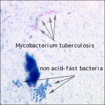
What is the procedure for Acid-Fast Staining (3F)?
1. Add swab with S. epidermidis to carbol fuchsin tube, rotate and remove swab
2. Add swab with M. phlei to carbol fuchsin tube, place in boiling bath
3. Roll swab onto slides, fix with heat
4. Decolorize with acid alcohol, rinse
5. Counterstain with methylene blue for 10 to 30s, rinse, air dry
6. Prepare Gram stain
What clinical application does Acid-fast staining have?
Useful in the diagnosis of the disease tuberculosis caused by mycobacterium tuberculosis
What color does Acid-fastness affected bacteria display compared to their counterparts?
Appear bright red or brilliant pink (Ziehl’s carbol fuchsin) while non acid-fast bacteria appear blue → absorb the counterstain (typically methylene blue)
What is the bacteria that stains blue during acid-fast staining (3F)
Staphylococcus Epidermidis

What is the bacteria that stains pink or red during acid-fast staining (3F)
Mycobacterium Phlei

What microbe was used during Exercise 3G: Granule Staining?
Corynebacterium xerosis
Granules are stained with methylene blue and counterstained with neutral red

What are metachromatic granules (volutin)?
Reserves of polymerized meta phosphates
What can Granule Staining (3G) be used for?
Diagnosing diphtheria caused by Corynebacterium diphtheriae
What are metachromatic granules
Considered to be a reserve of polymerized metaphosphates and are sometimes called volutin
Found in Corynebacterium Dipheriae
Can change colors
Corynebacterium Dipheriae
Has the diagnostic feature of Metachromatic granules
Causes the disease diphtheria
Albert Stain
A type of differential stainused for staining metachromatic granules and can be used for the presumptive diagnosis of diphtheria
Made up of two basic dyes toluidine blue and malachite green
Cells of the bacteria Corynebacterium Dipheriae stain green while the granules stain dark blue
What color do metachromatic granules appear?
Violet-red
Protoplast
Cells devoid of cell walls
Lysozyme
Attacks peptidglycan by hydrolyzing the glycosidic bond that accents N-Acetylmuramic acid with N-acetylglucoasmine
What enzyme is used to remove the cell wall of a Protoplast (3H)?
Lysozyme
What substance is used to help maintain protoplasts in water?
Sucrose
What bacteria was used the demonstration for 3H: the Protoplast
Bacillus Subtilis
two groups of fungi
yeasts and molds
Fungi that are decomposers of organic material
Chemoorganoheterotrophs

How do Fungi feed?
Secrete exoenzymes into environment and absorb decomposed nutrients
What are the main characteristics of yeasts?
Unicellular, reproduce sexually (spore formation) or asexually (budding)
What are two examples of microbes that are yeasts?
Candida albicans, Saccharomyces cerevisiae
What are the main characteristics of molds?
Multicellular, Filamentous, form branching network of cells (mycelium)
Hyphae with cross walls
Septate

Hyphae without walls
Aseptate

sporangiospores
Spores that develop WITHIN sacs at end of hyphae
conidiospores
spores that are not enclosed in a sac, located at the tips or sides of the hyphae
What are the four groups of fungi?
Chytridiomycota, Zygomycota, Ascomycota, Basidiomycota