topic 1
 Call Kai
 Learn
 Practice Test
 Spaced Repetition
 Match
 Flashcards
 Knowt Play
1/90

There's no tags or description
Looks like no tags are added yet.
91 Terms
although the variety of life, both past and present is extensive but what is the same
the biochemical basis of life is similar for all living things
what are monomers
smaller units from which larger molecules are made
polymers
polymers are molecules made from a large number of monomers joined together
what are examples of monomers
-monosaccharides
-amino acids
-nucleotides
condensation reaction
a condensation reaction joins two molecules together with the formation of a new chemical bond and involve the elimination of a molecule of water
hydrolysis reaction
breaks a chemical bond between two molecules and involves the use of a water molecule
monosaccharides
are monomers from which larger carbohydrates are made
examples of common monosaccharides
glucose
galactose
fructose
carbohydrates
most are polymers
-they are made of the elements C,H,O
-the monomers they are made from are monosaccharides
condensation reaction
a condensation reaction between two monosaccharides forms a glycosidic bond
disaccharides
are formed by the condensation of two monosaccharides
maltose
maltose is a disaccharide formed by condensation of two glucose molecules
sucrose
is a disaccharide formed by the condensation reaction of a glucose molecule and fructose molecule
lactose
is a disaccharide formed by the condensation of a glucose molecule and galactose moelcule
two isomers of glucose and with structures
(glucose fructose and galactose are isomers meaning same molecular formulae but a different arrangement of atoms in space)

polysaccharides
are formed by the condensation of many glucose monomers
how are glycogen and starch formed
by the condensation of alpha glucose
how is cellulose formed
condensation.of beta glucose
how to test non-reducing sugars
-add acid and boil (acid hydrolysis)
-cool solution and then add alkali to neutralise
-add benedict’s reagant and heat
-a positive test means solution from blue to red (precipitation)
a-glucose
-main substrate for respiration
-the form in which carbonhdyrate is transported in mammalian blood
-forms the polysaccharides starch and glycogen
b-glucose
forms the polysaccharide cellulose
fructose
-found in nectar and many fruits, it is sweeter than glucose and helps to attract animals for polination and fruit dispersal
galactose
with glucose, forms disaccharides lactose
maltose
formed from the breakdown of starch in germinating seeds where it provides energy for the growing embryo
sucrose
forms in which sugars are transported in phloem in plants
lactose
sugar found in milk;energy source for suckling mammals
starch
-monomer
-bond between monomer
-function
-location
-structure
-explanation of how structure leads to function
-formed by the condensation of alpha glucose
-1-4 glycosidic bonds in amylose and 1-4 and 1-6 in amylopectin ( in starch formation molecules of alpha glucose can be synthesised into amylose and amylopectin)
-storage for excess glucose in plants- main carbohydrate in plants
-plant cells
-made of 2 polymers amylose-an unbranched helix of alpha glucose. it gets its helical structure from the bond angles of the glycosidic bonds. amylopectin-a long branched molecule of alpha glucose
-compact due to the helical structure of amylose lots of glucose can be stored in a small space. this can make it an efficient energy store
many branches( in amylopectin) increases number of points where enzymes can attach meaning many glucose monomers can be released rapidly
insoluble= doesnt affect water potential so water isnt drawn into or out of cells by osmosis
cellulose
-monomer
-bond between monomers
-function
-location
-structure
-explanation of how structure leads to function
-beta glucose-made of long unbranched chains of beta glucose. the bonding of beta glucose forms a straight chan
-1-4 glycosidic bonds
-supports and strenghten the cells
-cell wall
-these chains run in parallel to each other and are linked by hydrogen bonds to form microfibrils. many microfibrils are arranged in parallel groups called fibres
-many hydrogen bonds provide collection strength, insoluble-wont affect water potential
glycogen
-monomer
-bond between monomers
-function
-location
-structure
-explanation of how structure leads to function
-alpha glucose
-1-4 and 1-6 glycosidic bonds ( mostly 1-6 glycosidic bonds)
-energy storage in animals (storage in animals) - same structure as amylopectin just more brnaches
-liver and muscle cells
-highly branched molecule
-many branches- increases number of points where enzymes can attach so many glucose monomers can be released rapidly
compact=lots of it can be stored in a small space efficient energy store
why can benedict’s reagent can be used for the presence of reducing sugars
because reducing sugars can donate an electron to the benedict’s reagent - add benedicts solution and heat in a water bath thats been brought to boil for 2 mins and positive result (coloured brick-red precipitate forms)
which are reducing sugars and how to test for them
all monosaccharides and some disaccharides are reducing sugars ( sucrose is non reducing)
-do benedicts test (solution stays blue) then add dilute HCl acid and heat in a water bath thats been brought to boil
-add sodium hydrogen carbonate to neutralise
-repeat benedicts test
positive result -a brick red precipitate
( we need to add the acid at the start to hydrolyse the non reducing sugar into its monosaccharides. only then can these reduce cu2 ions to cu+ ions giving us the brick red precipitate
how to separate a mixture of monosaccharides and identify their compounds
-could use chromotography with known standard solutions
how to test for starch
add iodine dissolved in potassium iodide solution
-solution changes colour from brown-orange to blue/black
how could students identify the unknown concentration of glucose in an unknown substancce
-produce a dilution series of glucose solution and use colorimetric techniques to produce a calibration curve with which to identify the concentration of glucose in an unknown solution
lipids
-non polar and hydrophobic
-contains C,H,O atoms only(and sometimes P and N)
what are the two groups of lipids
triglyceride (main component of fats and oils)
phospolipids (type of lipid molecule that is a major component of cell membranes)
how are triglycerides formed
formed by the condensation of one molecule of glycerol and three molecules of fatty acid
-act mainly as energy stores in plants and animals
-fatty acids have general formula of RCOOH so are carboxylic acids and glycerol is an alcohol (-OH functional group)
condensation reaction between glycerol and a fatty acid (RCOOH) what type of bond does it form
an ester bond
why are lipids insoluble in water
the hydrocarbon tails of fatty acids are hydrophobic making it insoluble
the R-group of a fatty acid can be what
saturated or unsaturated
what is the difference between triglyceride and phospholipid
In phospholipids, one of the fatty acids of a triglyceride is substituted by a phosphate-containing group.
how does triglyecerides structure relates to function
-mainly used as energy storage molecules
insoluble-dont affect water potential which could cause water to move into or out of cells by osmosis which could cause cells to swell burst shrink
-high ratio of energy-storing C-H bonds to carbon atoms and are therefore excellent sources of energy
-low mass to energy ration good storage molecules as much energy can be stored in a small volume
high ration of H to O atoms-release water when oxidised and therefore providing an important source of water especially for organisms living in dry deserts
phospholipid
a hydrophilic head
hydrophobic tails
make up the bilyaer of cell membranes this what can enter and leave the cell
polar molecules( hydrophobic and hydrophillic ends) forms a bilayer in an aqueous environmentn
-this forms a hydrophobic barrier at the centre of the bilayer so water-soluble substances cannot pass through
phsopholipid structurea allows to form glycolipids by combining with carbohydrates within the cell surface membrane. glycolipids are important in cell recognition
structure
A typical phospholipid consists of:
one glycerol molecule
two fatty acid chains
These are non-polar and hydrophobic
one phosphate group
This is polar and hydrophilic
Sometimes attached to an additional polar group (e.g. choline)
The fatty acids are bonded to glycerol by ester bonds, while the phosphate group is attached to glycerol via a phosphoester bond

The structure of a phospholipid
monolayers and bilayers
As a result of having hydrophobic and hydrophilic parts, phospholipid molecules form monolayers or bilayers in water

In the presence of water, due to the hydrophobic and hydrophilic parts, phospholipids will form monolayers or bilayers.
Function of phospholipids
Hydrophobic fatty acid tails create a hydrophobic core when a phospholipid bilayer forms
This acts as a barrier to water-soluble molecules
The hydrophilic phosphate heads form H-bonds with water, allowing the cell membrane to be used to compartmentalise
This enables the cells to organise specific roles into organelles, helping with efficiency
The composition of phospholipids contributes to the fluidity of the cell membrane
If there are mainly saturated fatty acid tails, then the membrane will be less fluid
If there are mainly unsaturated fatty acid tails, then the membrane will be more fluid
Phospholipids control membrane protein orientation
Weak hydrophobic interactions between the phospholipids and membrane proteins hold the proteins within the membrane but still allow movement within the layer
benefit of carrying out an emulsion test
The emulsion test can be carried out quickly and easily in a lab to determine if a sample contains lipids
drawback of carrying out an emulsion test
This test is qualitative so it does not give a quantitative value as to how much lipid may be present in a sample
emulsion test steps
-shake sample with ethanol
filter the liquid into a test tube of water leaving any solids behind
positive result- lipid will precipitate forming a white emulsion
proteins
polymers made from the monomers of amino acids. amino acids contains C H O and N atoms and sometimes S
proteins are essential in cells because they form the following
Enzymes
Cell membrane proteins (e.g. carrier)
Hormones
Immunoproteins (e.g. immunoglobulins)
Transport proteins (e.g. haemoglobin)
Structural proteins (e.g. keratin, collagen)
Contractile proteins (e.g. myosin)
amino acids
Amino acids are the monomers of proteins
There are 20 amino acids found in proteins common to all living organisms
the general structure of an amino acid
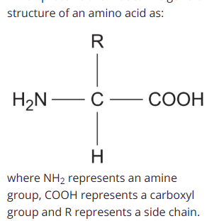
how do the twenty amino acids that are common in all organisms differ
The twenty amino acids that are common in all organisms differ only in their side group.
what does a condensation reaction between two amino acids form
a peptide bond
dipeptide
Dipeptides are formed by the condensation of two amino acids
Polypeptides
Polypeptides are formed by the condensation of many amino acids. The bonds formed between amino acids are called peptide bond s
what may a functional protein may contain
one or more polypeptide
chromotography of amino acids
A spot of the unknown amino acid sample mixture is placed on a line at the bottom of the chromatography paper
Spots of known standard solutions of different amino acids are then placed on the line beside the unknown sample spot
The chromatography paper is then suspended in a solvent
Each amino acid will be more or less soluble in the mobile phase than others and will therefore separate, travelling with the solvent at different times/distances from the line, depending on their:
charge
size
identification of amino acids
The unknown amino acid(s) can then be identified by comparing and matching them with the chromatograms of the known standard solutions of different amino acids
If a spot from the amino acid mixture travels the same distance as a spot from a known standard, it indicates that the mixture contains that amino acid
To view the spots from the different amino acids, it may be necessary to first dry the chromatography paper and then spray it with ninhydrin solution; this chemical reacts with amino acids, producing an easily visible blue-violet colour
protein structure
There are four levels of structure in proteins
Three are related to a single polypeptide chain
The fourth level relates to a protein that has two or more polypeptide chains
primary structure of proteins
The sequence of amino acids bonded by peptide bonds is the primary structure of a protein
DNA of a cell determines the primary structure of a protein by instructing the cell to add certain amino acids in specific quantities in a certain sequence, during translation. This affects the shape and, therefore, the function of the protein
The primary structure is specific for each protein (one alteration in the sequence of amino acids can affect the function of the protein)
secondary structure
The secondary structure of a protein is held together by hydrogen bonds that form between the -NH region of one amino acid and the -C=O region of another
The hydrogen of -NH has an overall positive charge, while the oxygen of -C=O has an overall negative charge
Hydrogen bonds are relatively weak, so they can be broken easily by high temperatures and pH changes
Two shapes can form within proteins due to the hydrogen bonds:
α-helix
β-pleated sheet
The α-helix shape occurs when the hydrogen bonds form between every fourth peptide bond
The β-pleated sheet shape forms when the protein folds so that two parts of the polypeptide chain are parallel to each other, enabling hydrogen bonds to form between the folded layers
tertiary structure
Further conformational change of the secondary structure leads to additional bonds forming between the R groups (side chains)
The additional bonds are:
hydrogen bonds between R groups
disulfide bonds between cysteine amino acids
ionic bonds between charged R groups
weak hydrophobic interactions between non-polar R groups
This structure is common in globular proteins such as enzymes and antibodies
quaternary structure
Occurs in proteins that have more than one polypeptide chain working together as a functional macromolecule, for example, haemoglobin
Each polypeptide chain in the quaternary structure is referred to as a subunit of the protein
functions of proteins
Proteins perform a wide range of essential roles in all living organisms due to their diverse structures
They are therefore vital for structure, transport, communication, defence, movement, and catalysis in all living cells:
Enzymes – biological catalysts that speed up metabolic reactions (e.g. amylase, DNA polymerase)
Transport proteins – carry substances (e.g. haemoglobin transports oxygen; channel proteins in membranes)
Structural proteins – provide support (e.g. collagen in connective tissues; keratin in hair and nails)
Hormones – regulate processes (e.g. insulin controls blood glucose levels)
Antibodies – part of the immune response, recognising and neutralising pathogens
Contractile proteins – enable movement (e.g. actin and myosin in muscles)
disulfide
Disulfide bonds (also known as disulfide bridges) are strong covalent bonds that form between two cysteine R groups (this is the only amino acid with an available sulfur atom in its R group)
These bonds are the strongest within a protein, but occur less frequently, and help stabilise the proteins
They can be broken by reduction
Disulfide bonds are common in proteins that are secreted from cells e.g. insulin
ionic bonds
Ionic bonds form between positively charged (amine group -NH3+) and negatively charged (carboxylic acid -COO-) R groups
Ionic bonds are stronger than hydrogen bonds, but they are not common
These bonds are broken by pH changes
hydrogen
Hydrogen bonds form between strongly polar R groups
These are the weakest bonds that form, but the most common, as they form between a wide variety of R groups
biochemical test; proteins
a few drops of biuret solution
a blue ring forms at the surface of the solution which disappeares upon shaking and turn lilac purple
this test can be used for enzymes as they are proteins
enzymes
are globular proteins
each enzyme lowers the activation energy of the reaction it catalyses they do that by providing an alternative reaction pathway of lower activation energy
lock and key model for enzymes
the tertiary structure of the enzyme is such that the shape of the enzymes active site is complementary to that of the substrate so the substrate can fit into the enzyme’s active site to form an enzyme-substrate complex
what is the specifity of an enzyme determined by
The specificity of an enzyme is a result of the complementary nature between the shape of the active site on the enzyme and its substrate(s)
The active site of an enzyme has a specific shape to fit a specific substrate
Extremes of heat or pH can change the shape of the active site, preventing substrate binding – this is called denaturation
the properties of an enzyme related to the tertiaty structure of its active site and its ability to combine with complimentary substrate to form enzyme-substrate complex
The shape of the active site is determined by the complex tertiary structure of the protein that makes up the enzyme:
Proteins are formed from chains of amino acids held together by peptide bonds
The order of amino acids determines the shape of an enzyme
If the order is altered, the resulting three-dimensional shape changes
induced fit model
in order to completely form an enzyme substrate complext the enzyme and substrate have to alter their shape slightly. this weakens specific bonds in the substrate needing to be broken, which lowers the activation energy required for the reaction to happen increasing the rate of reaction
development of induced fit model
Scientists often use models to explain their observations from experiments
As technology and research advances within a field, new models can be developed and old ones disproven
The lock and key model covered at GCSE was originally thought to be an accurate model of enzyme action
It was suggested that the rigid shape of the active site of the enzyme is a precise fit for the specific shape of the substrate
New techniques have allowed scientists to discover that proteins are not rigid structures
Experiments showed that multiple regions of an enzyme molecule moved in response to the environment
Many of these movements were minimal, but some of them were more significant
The larger movements occurred when the substrate bound to the enzyme
These findings led to the now widely accepted induced fit model
There is evidence to support the induced fit model:
X-ray diffraction techniques allow for 3D pictures of molecules to be formed
This technique was used to produce pictures of the enzyme hexokinase before and after it bound to its substrate glucose
The images confirmed that the active site of the enzyme changed shape after the substrate bound
the effect of temperature
as the temperature increases the rate of reaction increases
this is because enzyme and substrate molecules have more kinetic energy so frequency of collisions between these molecules increases so more enzyme-substrate complexes are formed per unit time
however after optimum temperature the rate begins to decrease. this is because the rise in temperature causes hydrogen and ionic bonds (which holds the enzymes tertiary structure in place to break altering the tertiary structure of the enzyme changing the shape of the active site too
after a certain temperature the shape of the active site is no longer complementary to that of the substrate. the enzyme is said to be denatured
the effect of ph
as you move away from the optimum pH rate of reaction begins to decrease
this is because the change in concentration of H plus and OH minus ions interferes with hydrogen and ionic bonds that hold the enzyme’s tertiary structure in place, altering the shape of the active site too
if Ph deviates too far from the optimum ph the shape of the active site is changed too much that it is no longer complementary to that of the susbtrate. the enzyme is said to be denatured
the effect of enzyme concentration
the more enzymes that are in a solution the more likely a substrate molecule is to collide with one to form an ES complex
af first enzyme conc is directly proportional to rate of reaction
however, after a certain point the amount of substrate becomes limiting so increasing the enzyme concentration has no further efect on increasing the rate of reaction so rate levels off and remains constant
effect of substrate concentration
the more substrate there is in a solution the more likely an enzyme is to collide with one and form an enzyme substrate complex
at first substrate concentration is directly proportional to rate of reaction
however after a certain point the amount of enzymes becomes limiting because all active sites are occupied and therefore increasing the substrate concentration will have no further effect on increasing the rate pf reaction the rate levels off and remains constant
competitive inhibitors
are molecules that have a similar shape to that of the enzymes normal substrate=they can ft into the enzyme’s active site to form an enzyme-inhibitor complex. this prevents substrate molecules from b inding to the active site
competitive inhibitors compete with substrate molecules for available active sites. the difference in concentrations of the inhibitor and substrate molecules determines how many enzymes are inhibited
-the higher the concentration of competitive inhibitor the lower the rate of reaction because there is more competition for available active sites so fewer es complexses formed
non competitive inhibitors
work by binding to enzymes in a place other than the active site. this changes the tertiary structure of the enzyme altering the shape of the active site too meaning that it is no longer complementary to that of the substrate. substrate molecules can therefore no longer bind to the active site
-they are often foreign to the body and toxic. this is an irreversible reaction and the active site shape is changed forever
-rate of reaction is decreased as non competitive inhibitors reduce the number of functional enzymes available. substrate and inhibitor do not compete for the active site so increasing the substrate concentration has no influence on the effect of the inhibior
DNA
Deoxyribonucleic acid
holds genetic information in all living cells
RNA
ribonucleic acid
transfers genetic information from DNA to ribosomes
similarities of DNA and RNA
polynucleotides
-phosphate group
pentose sugar
nitrogenous containing organic base (2:02)
how to do nucleotides be joined together
nucleotides themselves can be joined togethere by a condensation reaction between a phosphate group of one nucleotide and the pentose sugar of another
this forms phosphodiester bond between twoo nucleotides which consissts of one phosphate group and two ester bonds
structure of dna
two DNA polynucleotide strands which are joined together by H bonds between complementary base pairs
complementary means specific base pair with each other
Adenine (A) complementary Thymine (T) two hydrogen bonds form between these bases
Cytosine is complementary with Guanine 3 hydrogen bonds
due to the complementary base pairing the quantity of A is same as T and the wuantity of C is the same as G
the two DNA strands are antiparallel=they run in opposite directions (one strand runs in the 5’ to 3’ direction and whilst the other runs in the 3’ to 5’ direction
why did scientists at first doubt the DNA could carry the genetic code
because it had a simple chemical composition
-some argued that genetic information must be carried by proteins which are chemical varied. however in 1953 experiments showed that dna was the true carrier of the genetic code
how does DNA structure relate to function
The two DNA polynucleotide strands are only joined together by relatively weak H bonds meaning that the strands can sepearate during DNA replication
base pairs are protected from being corrupted by being within the helical cylinder of the sugar-phosphate backbone
very large molecule so can carry lots of genetic information
RNA
much shorter than DNA
made from. single polynucleotide chain
3 main types of RNA that we need to know about:
-mRNA =transfers genetc information from DNA to ribosomes
tRNA=involved in protein synthesis
rRNA =make up part of the ribsome structure