Cardiovascular system
 Call Kai
 Learn
 Practice Test
 Spaced Repetition
 Match
 Flashcards
 Knowt Play
1/43

There's no tags or description
Looks like no tags are added yet.
44 Terms
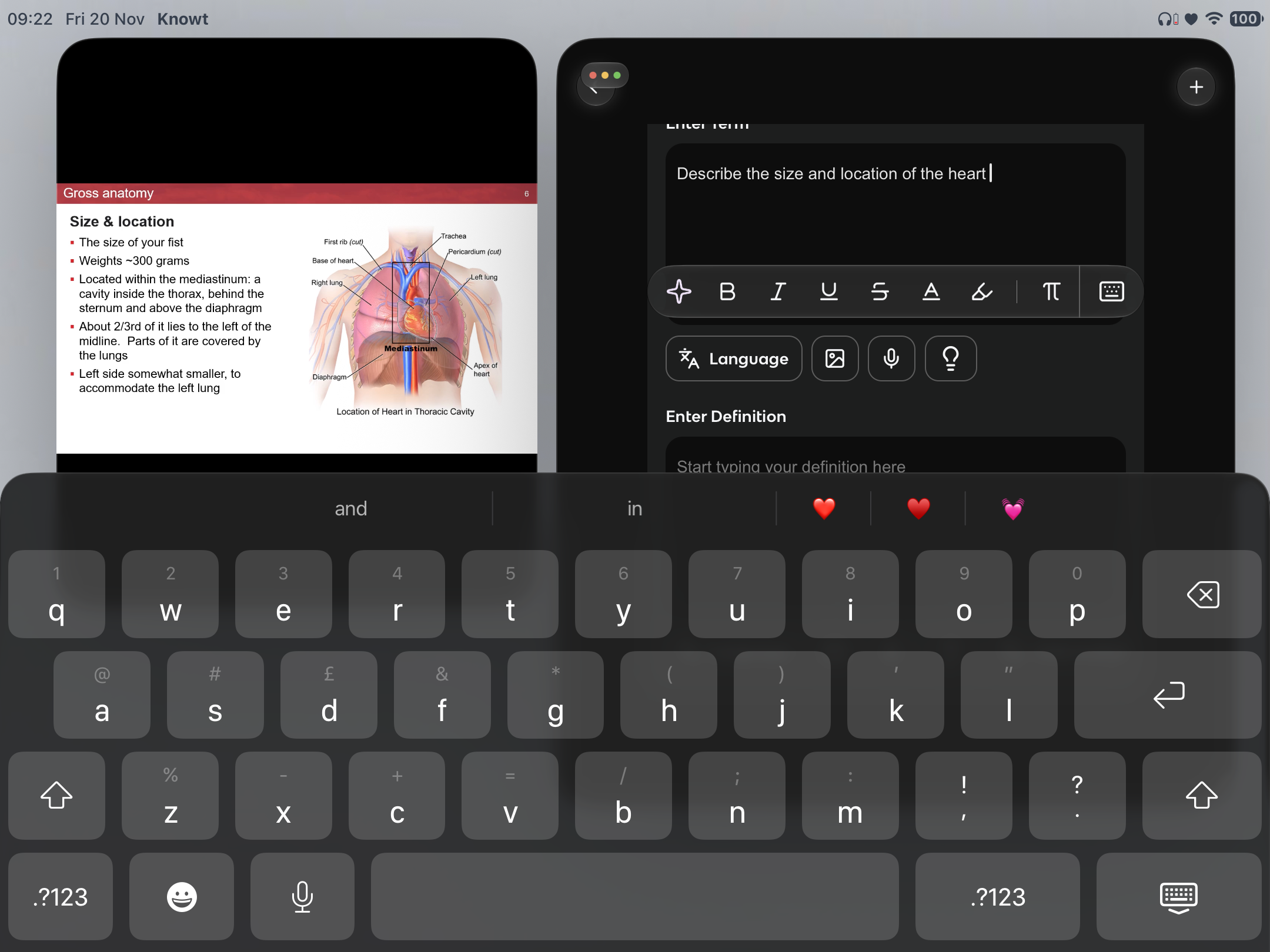
Describe the size and location of the heart
Describe the pericardium

Identify and describe the 3 layers if the heart wall

How is the LV more developed and why?
more myocytes
Must push at higher pressure
Describe the right atrium

Describe the right ventricle

Describe the valves in the lungs

Describe atrioventricular valves
Prevent back flow from ventricle to atrium, when ventricle contracts
Right AV valve → tricuspid (has 3 flaps)
Left AV valve → bicuspid (has 2 flaps)
Describe the AV valve flaps both at rest and during contraction
Rest → flaps hang down limply into ventricle below, blood flows freely from atrium
Ventricle contracts → intra ventricular pressure rises , forcing the blood on the ventricle superior LH against valve flaps → flap edges meet , closing the valve and preventing blood flow
How are the flaps reinforced?

Describe the semilunar valves
Aortic valve & pulmonary valve
Prevent back flow from the 2 large arteries (aorta and pulmonary tract ) back into ventricles
Consists of half moon cusps
How do semi lunar valves work?

Are there valves in the veins?

What are some defects if the valves and how can they be solved ?

What is ‘diastole’ and ‘systole’?
diastole, relaxation
Systole , contraction
Describe the cardiac diastole
blood flows into atria until full , AV valves shut
AV valve open and 70% of blood leaves passively
describe atrial systole and ventricular diastole
remainder squeezed out by atria contracting
Describe atrial diastole and ventricular systole
ventricles contract, shutting AV valves
Semilunar valves open and blood leaves ventricles
Describe cardiac diastole
ventricles relax , and bavkflow of blood shuts semilunar valves
How are cardiac muscles different to skeletal muscles?

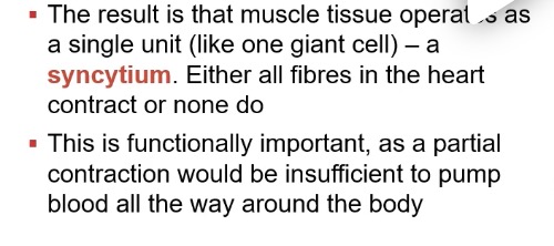
What are intercalated discs?

What is the result if intercalated discs and why is it important?
Where are the 2 large,y independent synctia located, formed by cardiac muscles?

What does the prevalence of mitochondria and myoglobin reflect about cardiac muscle?

What is controlling the heart?
It’s myogenic
Independent if other systems
How is the heart myogenic? (with the atria)
Sinoatrial node sends a wave if depolarisation across the atria
(SAN has many Na+ leakage channels , always open, depolarising lowly and reaches threshold)
Rapidly spreads through 2 atria (due to branching and intercalated discs
The atria depolarise simultaneously
Atria and ventricles covered by soereate muscke sheets, so potential in the atria cannot immediately reach the ventricles
How is the heart myogenic? (With the ventricle)

What hapoens once the wave if depolarisation travels down the bundle of his?

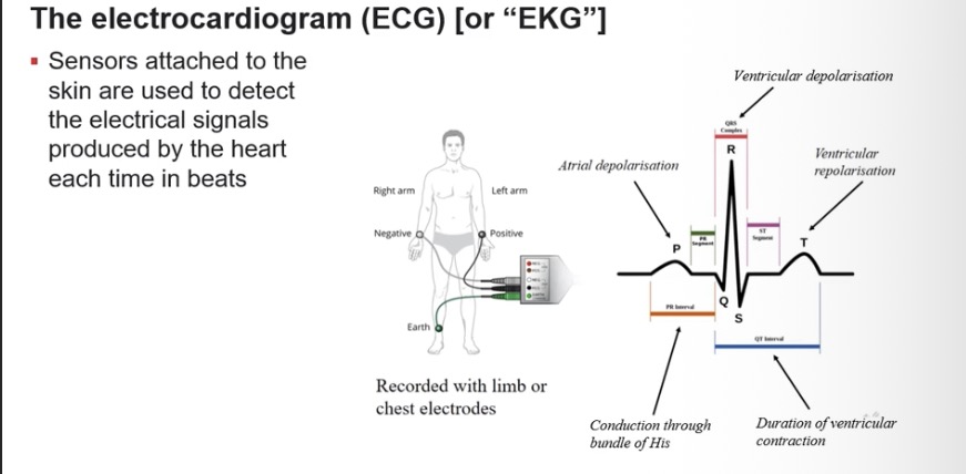
How can we measure the hearts electrical activity?
What can defects in the myogenic system cause?

What haooens if another part if the system takes over SAN as pacemaker?

What is a break in the bundle of his or AVN a called?

How can pathologies of heart be fixed?
With artificial pacemaker
How can the ANS fine tune the heart’s pace?

What is blood pressure and how is it measured?

What is one way blood pressure changes.
Cardiac cycle:
Systolic pressure : pressure in system when heart contracts
Diastolic pressure : pressure when heart is relaxed
Always given in pairs for BP → systolic/diastolic
How else can look pressure vary?

What are some additional ways bp vary?
between people
Age
Time of day
Feeling
What are the three types of blood vessel?
arteries
Veins
Cappillaries
For veins and arteries, what is the structure if the lumen?
Tunica externa
Tunica media
Tunica intima

how do the structure if veins and arteries vary?

What do veins contain that arteries don’t?
Valves
Prevent back flow, die to low pressure
Skeletal muscle helps to propel blood towards heart
Describe cappillaries

Identify and describe the 3 main types of cappillaries
