Practice Exam 3 AP I
 Call Kai
 Learn
 Practice Test
 Spaced Repetition
 Match
 Flashcards
 Knowt Play
1/185

There's no tags or description
Looks like no tags are added yet.
186 Terms
List two of the seven criteria listed in your lab manual that are used to name muscles?
direction of muscle fibers
relative size
location
number of origins
location of origin and insertion
shape
action

Identify the type of muscle tissue shown?
cardiac

Identify the indicated layer of connective tissue.
perimysium
A bundle of skeletal muscle cells within a skeletal muscle is called:
Fasicle
A flat, sheet-like bundle of connective tissue that connects a muscle to another muscle or to a bone is called:
aponeurosis
A strong, cord-like bundle of connective tissue that connects a muscle to another muscle or to a bone is called:
tendon
What do we call muscles that immobilize the origin of another muscle so that all of the tension is exerted at the insertion?
FIXators
→ “FIX” the origin in place
Origin = start: stays still.
Insertion = end: moves when the muscle contracts.
Which of the seven criteria used for naming a muscle applies to the brachioradialis muscle?
location of origin and insertion
Brachioradialis → location of origin and insertion.
Brachio = arm → originates on the humerus.
Radialis = radius → inserts on the radius.
Memory trick: “Arm to radius”—the name tells you its attachment points.
Which of the seven criteria used for naming a muscle applies to the biceps femoris muscle?
number of origins and location
Biceps femoris → number of origins and location.
Bi = two, ceps = heads → two heads (origins).
Femoris = thigh → location.
Memory trick: “Two heads in the thigh.”
Name one action of the sternocleidomastoid muscle.
rotation of the head
Name one action of the tibialis anterior muscle.
dorisflexion of the foot at the tarsal joints
Name one action of the trapezius muscle.
elevation of the scapula
Name one action of the flexor carpi radialis muscle.
flexion of the hand at the carpal joints
Name one insertion of the rectus femoris muscle.
tibial tuberosity
Name one insertion of the gracilis muscle.
medial surface of the tibia
Name one insertion of the platysma muscle.
lower margin of the mandible
Name one insertion of the pectoralis major muscle.
intertubercular sulcus of the humerus
Name one origin of the rectus femoris muscle.
anterior inferior iliac spine
Name one origin of the external oblique muscle.
lower eight ribs
Name one origin of the tibialis anterior muscle.
lateral condyle of the tibia
Name one origin of the supraspinatus muscle.
supraspinous fossa of scapula

Identify the indicated structure.
linea alba

Identify the indicated muscle.
rectus abdominis

Identify the indicated muscle.
orbicularis oculi
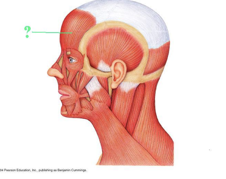
Identify the indicated muscle.
frontalis

Identify the indicated muscle.
platysma

Identify the indicated muscle.
masseter
You locate a muscle that extends from the inner margin of the anterior part of the mandible to the hyoid bone, and from the hyoid bone to the mastoid process of the temporal bone. The muscle you have located is the:
Digastric
Think of one muscle under your jaw that has two fleshy sections. That’s the digastric.
The question describes its path:
Starts inside your lower jaw.
Goes down to the hyoid, a small bone under your chin.
Goes back up toward the bone behind your ear.
It makes a V shape, with the hyoid at the bottom.
For your test, remember: “Jaw → hyoid → ear = digastric.”
The name helps too: di = two, and it has two muscle bellies (sections).
You locate a muscle that is visible in the V-shaped opening between the infraspinatus and the teres major. This muscle extends from the posterior surface of the scapula close to the point formed by the inferior and medial margins, and from there to the greater tubercle of the humerus. The muscle you have located is the:
teres minor
You locate the muscle segments in between the ribs on the lateral portion of the rib cage. These segments are more superficial and posterior than the adjacent muscle groups that lie between the ribs. The fibers of these muscles run diagonally from superior to inferior and from posterior to anterior. The muscle group you have have located is the:
External Intercostal
You locate a large sheet of muscle on the lateral surface of the abdomen. The fibers of this muscle run diagonally from superior to inferior and posterior to anterior. This muscle extends from the anterior surface of the lower eight ribs to the linea alba, the pubic crest and tubercles, and the iliac crest. The muscle you have located is the:
External oblique abdominis
You locate a muscle on the posterior surface of the thigh that lies on the medial side. This muscle is the more superficial of the two muscles in this location. This muscle extends from the tuberosity of the ischium to the medial surface of the body of the tibia. The muscle you have located is the:
Semitendinosus
You locate a muscle filling the medial surface of the ilium. This muscle extends from the medial margin of the ala of the ilium to the lesser trochanter of the femur. This muscle you have located is the:
Iliacus
You locate a thin, strap-like muscle wrapping around the anterior surface of the thigh from lateral to medial and proximal to distal. This muscle extends from the anterior superior iliac spine to the medial surface of the tibia just below the tibial tuberosity. The muscle you have located is the:
sartorius
You locate a muscle that covers the lateral surface of the lower leg. This muscle has fibers that run parallel to the bone over which it lies. This muscle extends from the head of the fibula, around the lateral malleolus, to the medial cuneiform and first metatarsal. The muscle you have located is the:
Fibularis longus

Identify the indicated muscle.
sternohyoid

Identify the indicated muscle.
sternocleidomastoid

Identify the indicated muscle.
diaphragm

Identify the indicated muscle.
external intercostals
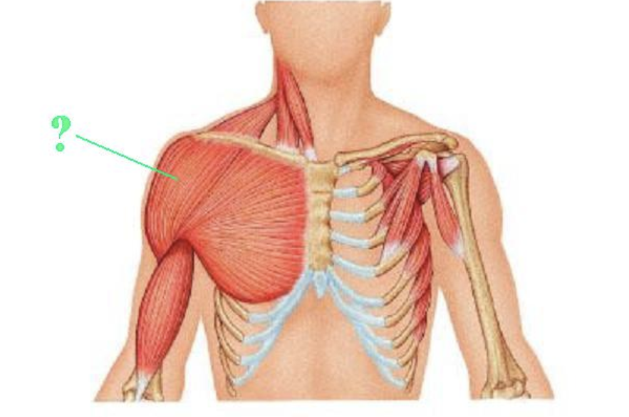
Identify the indicated muscle.
deltoid
List two of the seven criteria listed in your lab manual that are used to name muscles?
THINK:
Really Determined Lions Never Lose Strength Again
Really → Relative size
Determined → Direction of muscle fibers
Lions → Location
Never → Number of origins
Lose → Location of origin and insertion
Strength → Shape
Again → Action
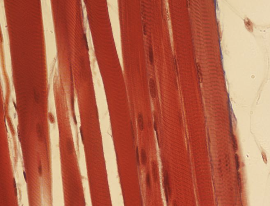
Identify the type of muscle tissue shown?
skeletal

Identify the indicated structure.
muscle fiber (in lab manual says myofibril)
The connective tissue sheath that forms around an entire skeletal muscle is called:
epimysium
A strong, cord-like bundle of connective tissue that connects a muscle to another muscle or to a bone is called:
tendon
The connective tissue sheath that forms around an individual skeletal muscle is called:
endomysium
What do we called muscles that oppose or reverse a particular movement?
antagonists
Which of the seven criteria used for naming a muscle applies to the biceps femoris muscle?
THINK: Really Determined Lions Never Lose Strength Again
Number of origin and location
Which of the seven criteria used for naming a muscle applies to the rectus femoris muscle?
THINK: rectus = straight
REALLY DETERMINED LIONS NEVER LOSE STRENGTH AGAIN
direction of fibers and location
Name one action of the brachialis muscle.
flexion of the forearm at the elbow
Name action of the platysma muscle.
tenses the skin of the neck
Name one action of the rectus femoris muscle.
Flexion of the thigh at the hip.
Name one action of the deltoid muscle.
abduction of the arm at the shoulder.
→ moving AWAY from the body
Name one insertion of the masseter muscle
angle and ramus of the mandible
Name one insertion of the vastus medialis muscle
tibial tuberosity
Name one insertion of the zygomaticus minor muscle
skin and muscle at the corner of the mouth
Name one insertion of the buccinator muscle
orbicularis oris muscle (around the mouth)
Name one origin of the zygomaticus minor muscle
zygomatic bone
Name one origin of the masseter muscle
zygomatic arch
Name one origin of the rectus abdominis muscle
pubic crest and symphysis

Identify the indicated muscle.
external oblique abdominal
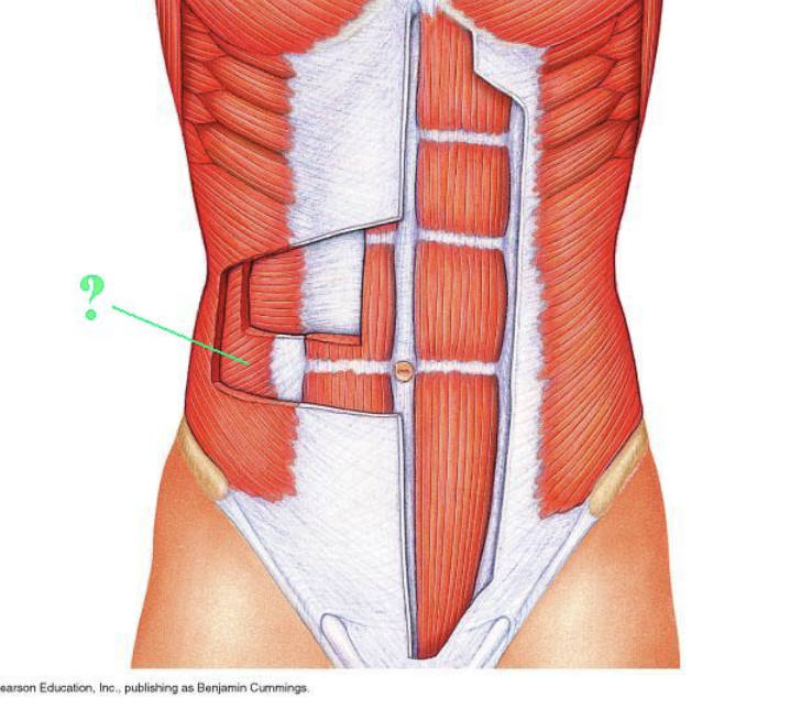
Identify the indicated muscle.
internal oblique abdominal
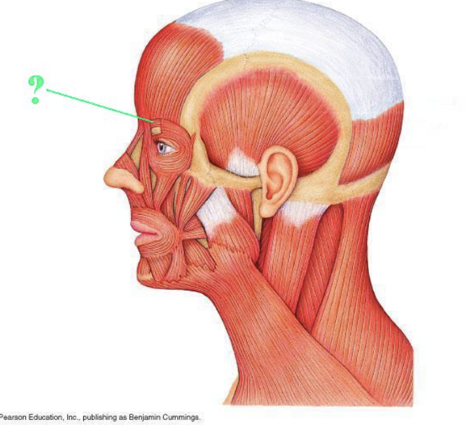
Identify the indicated muscle.
corrugator supercilli

Identify the indicated muscle.
mentalis

Identify the indicated muscle.
depressor labii inferioris
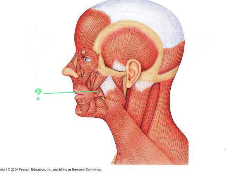
Identify the indicated muscle.
buccinator
You locate a muscle on the posterior surface of the neck, extending from the occipital bone and the spinous processes of cervical vertebra 7 and the thoracic vertebra to the acromion and spine of the scapula and the lateral third of the clavicle. The muscle you have located is:
trapezius
You locate a muscle that extends from the tip of the chin to the skin just below the lower lip. The muscle you have located is the:
mentalis
You locate a muscle that fills the space above the spine of the scapula on the posterior side. This muscle extends from this surface to the greater tubercle of the humerus. The muscle you have located is:
supraspinatus
You locate a series of muscle segments following the pattern of the ribs along the lateral surface of the chest. These muscles extend from the back to the lateral surface of the chest. The muscle group you have located is the:
serratus anterior
You locate a large muscle on the anterior side of the arm. This muscle extends from the coracoid process and the supraglenoid tubercle on the margin of the glenoid cavity to the radial tuberosity. The muscle you have located is the:
biceps brachii
You locate a muscle on the proximal and lateral (thumb) side of the forearm. This muscle is adjacent to the V-shaped gap at the bend of the elbow, wrapping around the forearm from the posterior side to the lateral side of the wrist. The muscle you have located is the:
Brachialis
You locate a muscle on the posterior side of the forearm whose distal tendon extends to the wrist on the pinkie side. This muscle extends from this tendon to the lateral epicondyle of the humerus. The muscle you have located is the:
Extensor carpi ulnaris
You locate a muscle at the proximal end of the long, strap-like tendon that runs along the lateral side of the thigh to the knee. The muscle you have located is the:
tensor fasciae latae
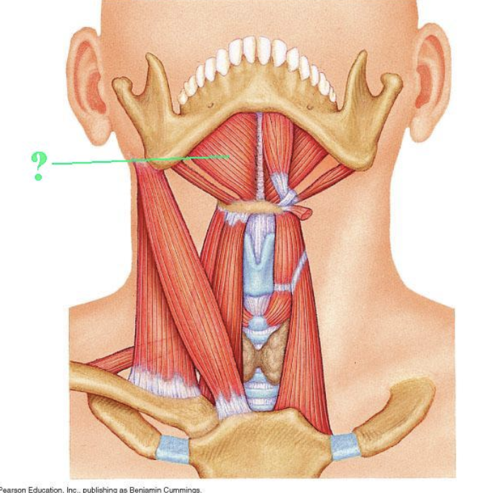
Identify the indicated muscle.
mylohyoid

Identify the indicated muscle.
sternothyroid

Identify the indicated muscle.
teres major

Identify the indicated muscle.
supraspinatus

Identify the indicated muscle.
flexor carpi radialis
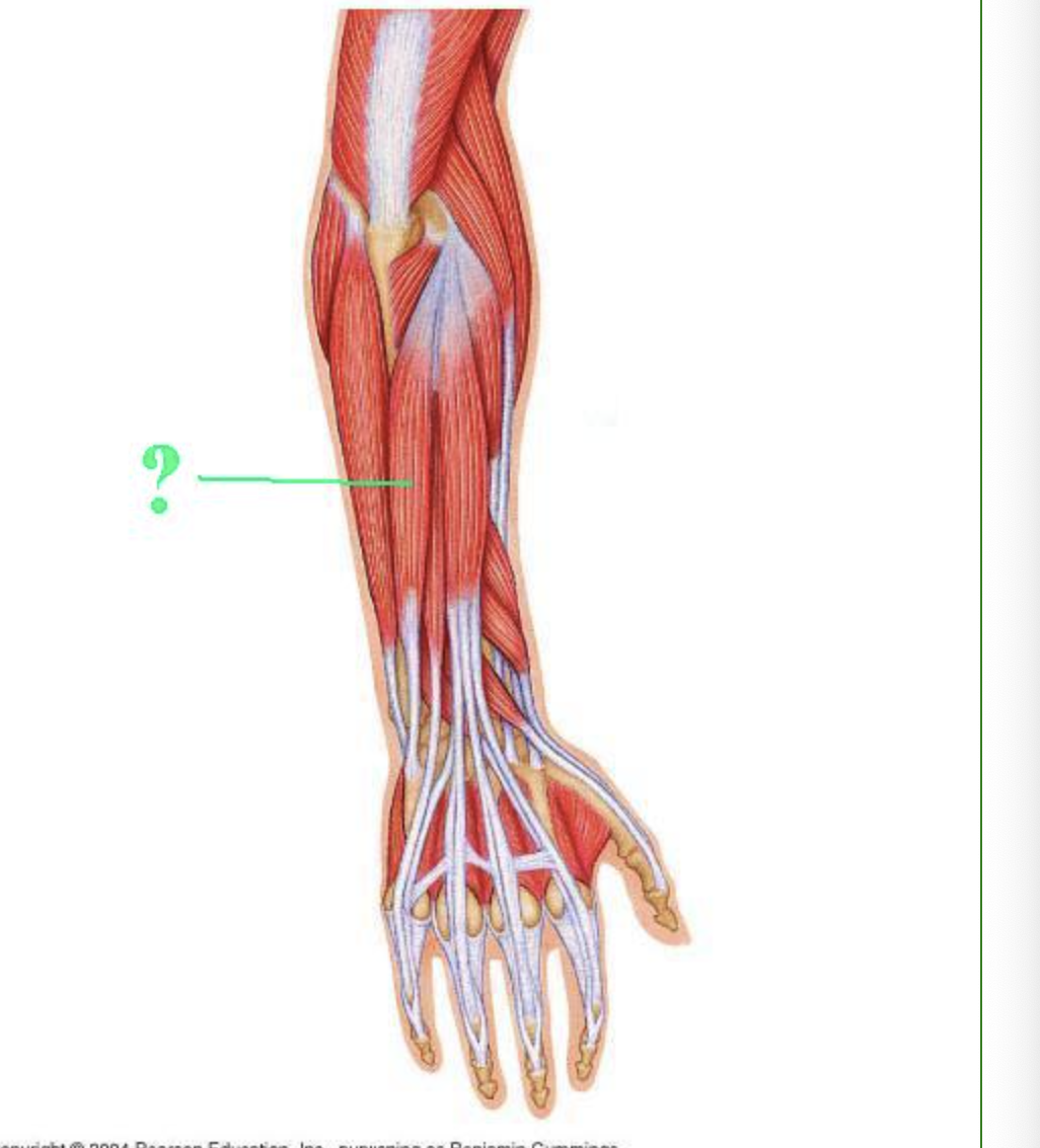
Identify the indicated muscle.
extensor carpi ulnaris

Identify the indicated muscle.
tensor fasicae latae

Identify the indicated muscle.
psoas major
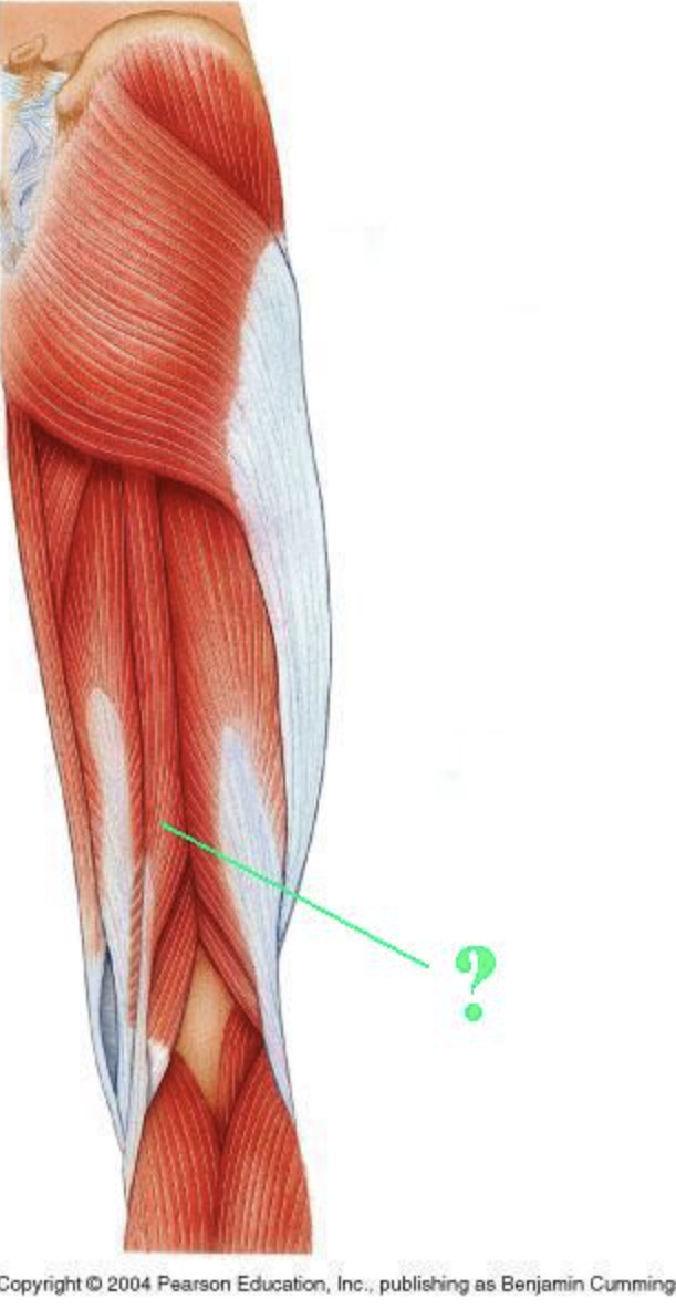
Identify the indicated muscle.
semitendinosous
What is the O and I of the zygomaticus major/minor?
O: zygomatic bone → your cheekbone
I: skin and muscle at the corner of your mouth
Memory trick: “A smile pulls your mouth toward your cheekbone.”
Cheekbone = anchor (origin); mouth corner = moving attachment (insertion).
What is the O and I of the masseter?
O: zygomatic arch and zygomatic bone
I: angle and ramus of the mandible
THINK: cheekbone → jawbone:
it pulls your jaw upward to close your mouth
Which of the seven criteria used for naming a muscle applies to the brachioradialis muscle?
location of origin and insertion
Which of the seven criteria used for naming a muscle applies to the tibialis anterior muscle?
location
Name one action of the rectus femoris
flexion of the thigh at the hip
Name one action of the zygomaticus major muscle
elevation of the lateral corners of the mouth
Name one action of the semimembranosus muscle
extension of the thigh at the hip
Name one insertion of the gastrocnemius muscle
calcaneus
Name one insertion of the rectus abdominis muscle
xiphoid process of the sternum
Name one insertion of the buccinator muscle
orbicularis oris muscle
Name one insertion of the brachialis muscle
coronoid process of the ulna
Name one origin of the gastrocnemius muscle
medial and lateral condyles of the femur
Name one origin of the buccinator muscle
maxillary bone
Name one origin of the semitendinosus muscle
ischial tuberosity
Name one origin of the triceps brachii
posterior humerus

Identify the indicated muscle.
levator labii superioris
You locate the muscle segments in between the ribs on the lateral portion of the rib cage. These segments are more superficial and posterior than the adjacent muscle groups that lie between the ribs. The fibers of these muscle run diagonally from superior to inferior and from posterior to anterior. The muscle group you have located is:
external intercostals