CFPC 2027-Hemato
 Call Kai
 Learn
 Practice Test
 Spaced Repetition
 Match
 Flashcards
 Knowt Play
1/51

There's no tags or description
Looks like no tags are added yet.
52 Terms
% blasts
< 20% = SMD or chronic
> 20% = acute
Acute leukemia vs. chronic leukemia
Acute: immature cells, blasts > 20% BMO
Chronic: mature/differentiated, blasts < 10%
Lymphoid vs. myeloid
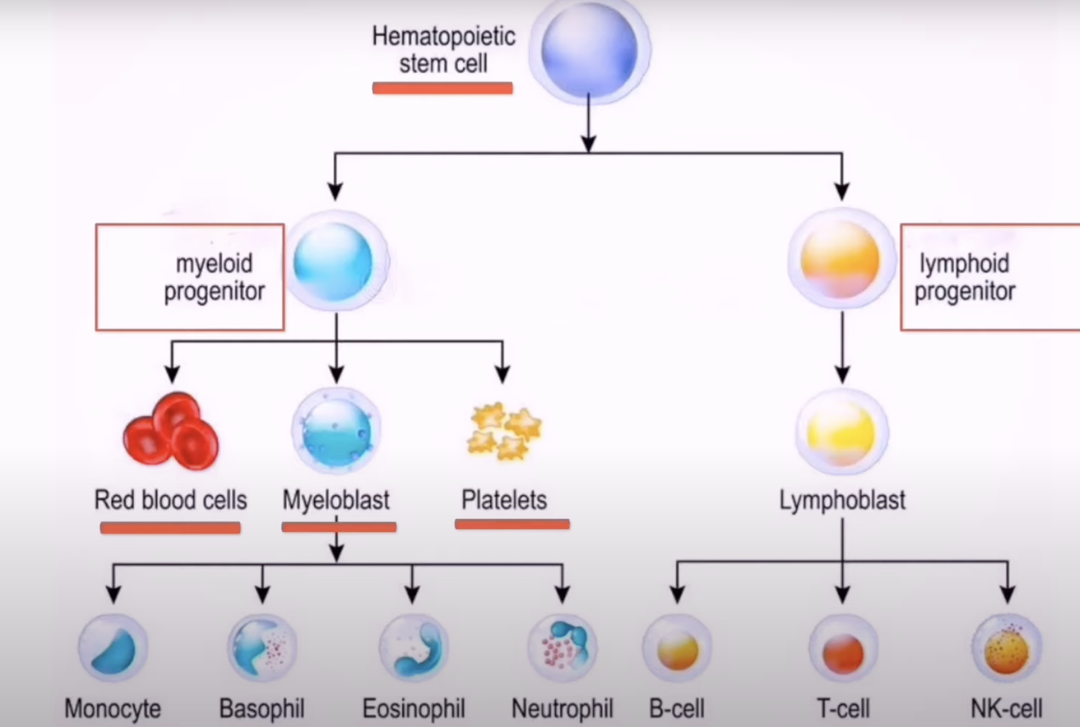
ALL, AML, CLL, CML
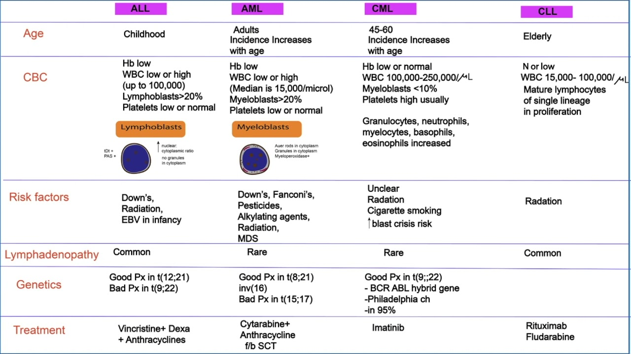
Leukemia tests
FSC (leucocytose) + différentielle, frottis, bx moelle
**they all present with leucocytosis (except CLL that can sometimes present with leucopenia)
What can SMD turn into
AML
How to differentiate between AML and CML?
Thrombocytopnenia is more common in AML.
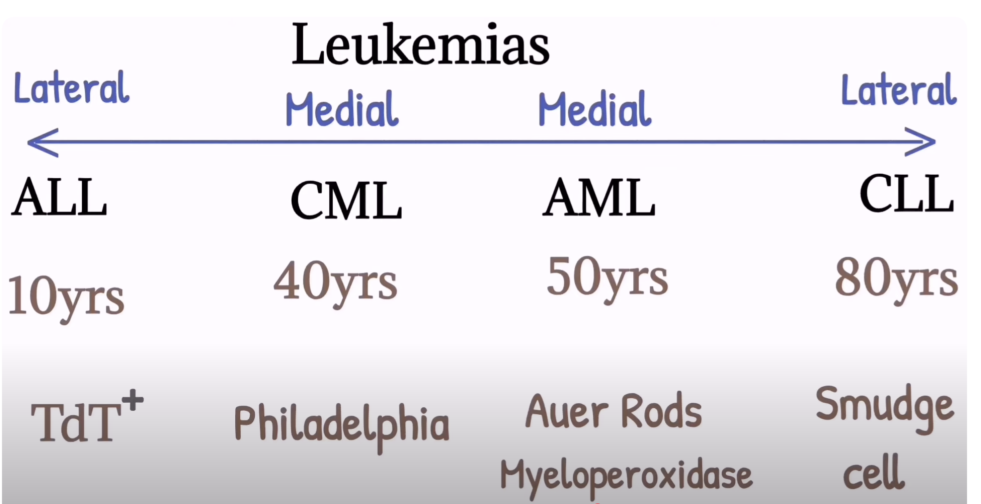
Tips for differentiating leukemias
ALL: children
CML: Chromosome (philadelphia), Massive splenomegaly
AML: Auer rods
CLL: Crushed Little Lymphocytes (smudge cells)
Réactions aiguës post-transfusion
-Allergie (urticaire, anaphylaxie)
-Réaction hémolytique aigue: fièvre, dlr abdo, hypoTA.
-Fébrile non hémolytique
-TRALI: OAP + SDRA
-TACO: fluid overload
Réactions tardives post transfusion
-Delayed hemolytic transfusion reaction: jour 5, ictère, fièvre.
-Iron overload
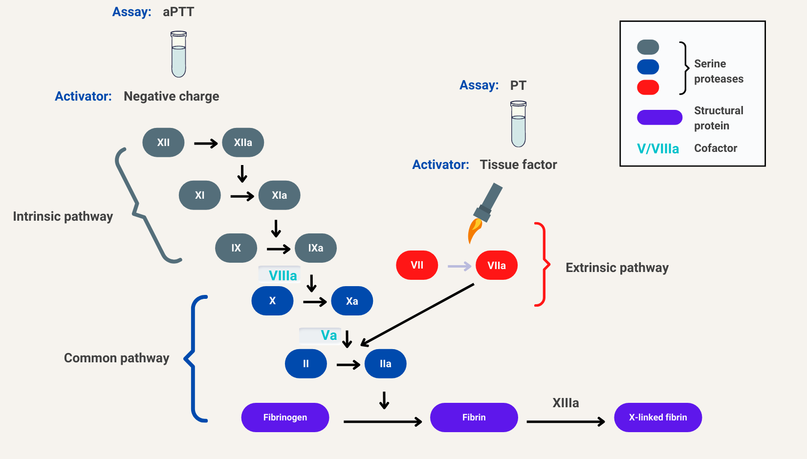
Cascade de coagulation
DVT treatment
HBPM x ≥ 5 jours puis switch to ACOD or warfarin.
Provoked: x 3 months
Unprovoked: 3-6 months or life, investiguer thrombophilie, SAP.
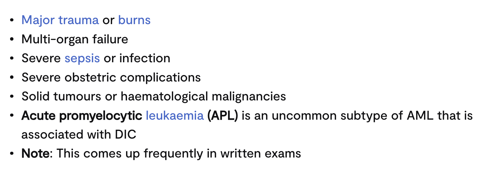
CIVD
Thrombus formation (in microvasculature) and depletion of clotting factors and platelets, which then results in bleeding.
CIVD etiology
CIVD tests
FSC (thrombocytopénie)
Frottis: schistocytes
Bilan de coag.: ↑PT, ↑PTT, ↓fibrinogen
↑d-dimers
CIVD tx
Tx cause ss jacente
Transfusions of platelets or clotting factors
Thrombocytose essentielle
F 50-70 A
Myeloproliferative disorder typified by dysregulated megakaryocyte proliferation.
Mutation JAK2 V617F (++)
TE key sx
50% asx
Erythromelalgie (red/blue discolouration of the extremities + burning sensation)
Livedo reticularis (net like purple rash)
TE tests
FSC: plaquettes ↑
JAK2 mutation
TE tx
aspirine ± hydroxycarbamide (si haut risque)
Déficience G6PD
X-link
Jews/Filipinos/Greeks/Sardinians/Kurds/black
Triggering factors: medications (AINS, aspirin), infections, or foods (fava beans)
Acute haemolytic episodes characterised by jaundice, anaemia, and dark urine.
Déficience G6PD tests
FSC: anaemia, reticulocytosis
Frottis: Heinz bodies
Elevated bilirubin levels
Déficience G6PD tx
support
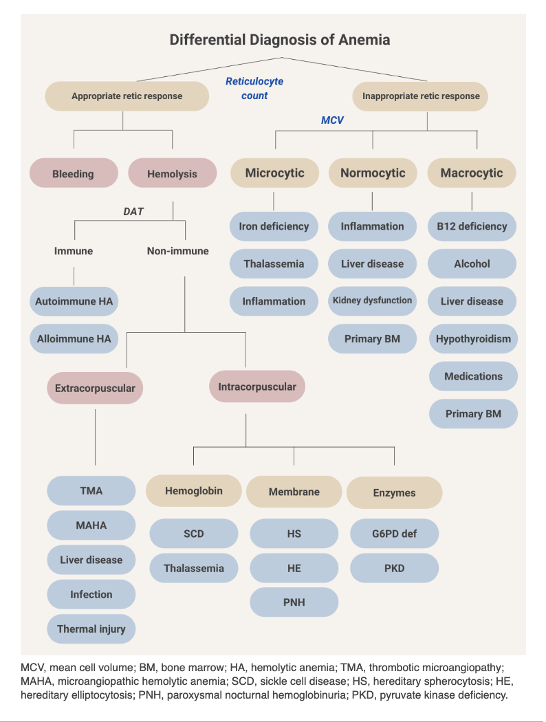
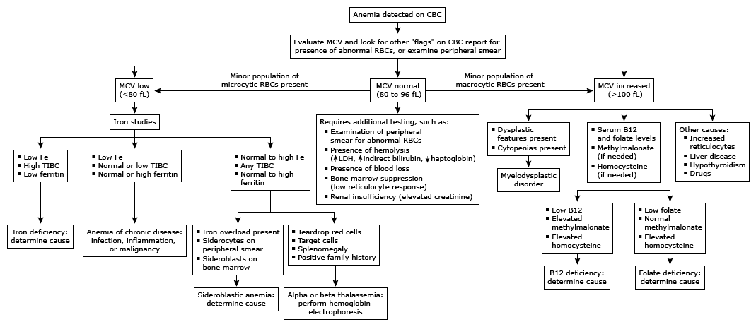
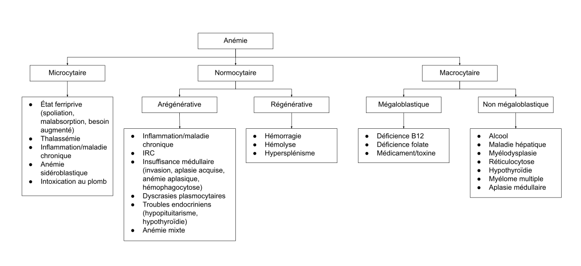
Anemia classification
Hb <135 for men <120 for women
Microcytic: TAILS (thalassemia, anemia chronic disease, IDA, lead, sideroblastic)
Normocytic: ABCD (acute blood loss, bone marrow failure, chronic disease, destruction)
Macrocytic: FATRBC (folate, alcohol, thyroid, reticulocytosis, B12 def, cytotoxic drugs)
When to consider transfusion
symptomatic, unstable, ongoing losses
Tests anemia
FSC, reticulocytes, iron profile, HBEP, frottis, LDH, haptoglobin, indirect bili, Coombs test (direct and indirect), TSH, B12, folate, INR, DIC panel (fibrinogen, d-dimer)
Bone marrow bx
Risk factors of anemia in children
poverty, poor weight gain, excessive milk intake + low iron-rich foods [malnutrition], malabsorption, GI blood loss, obesity
screen in all children between 9-12 months
IDA investigation
CBC + ferritin +/- TIBC
Consider reticulocytes
Consider Hb Electrophoresis, Blood Smear
Screen all for celiac (Anti-TTG, IgA)
Consider complete iron studies (serum ferritin, total iron binding capacity [TIBC]), and serum iron
Diagnostic trial of iron treatment x3 weeks in premenopausal women with history of menorrhagia or pregnant women
All men/postmenopausal women screened for GI malignancy
B12/folate should be checked in normocytic, inadequate response to iron treatment, or suspected (malnutrition, malabsorption, elderly - pernicious anemia)
Consider SPEP
Tx IDA
Diet
ferrous sulfate 300 die or q2d
± venofer IV
Re-check Hb in 2-4 weeks, then FU in 2-4 months
When normalized, continue tx x 3 months then d/c
Monitor periodically
Sx B12 deficiency
Anemia
Fatigue, palpitations, skin pallor
Glossitis
Neuropsych
Symmetric peripheral neuropathy/paresthesias
Ataxia/gait abnormalities
Memory loss
Personality changes
Dementia
Hémophilie A/B
Inherited bleeding disorders caused by deficiencies in clotting factors VIII (A) and IX (B).
X-linked
Hémophilie A/B sx
Deep and severe bleeding into soft tissues, joints, and muscles.
Excessive bleeding post tonsillectomy
Hémophilie A/B tx
Desmopressine (A), recombinant factor VIII (A) or IX (B)
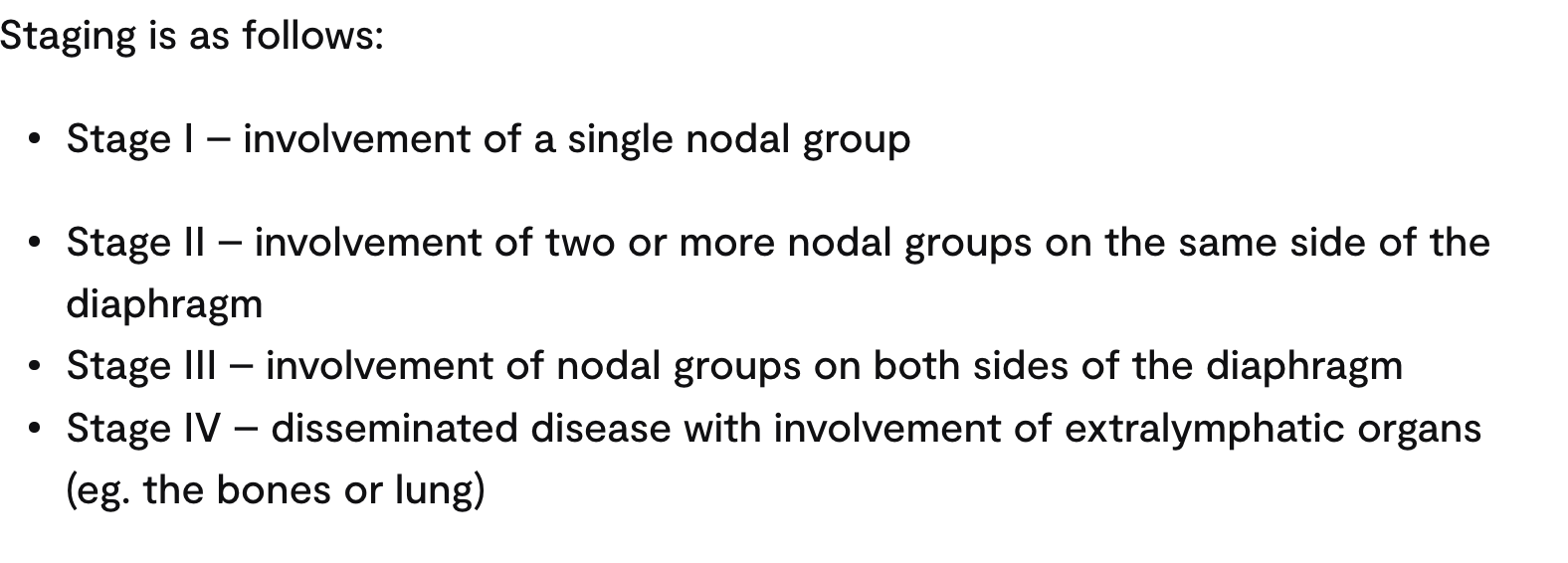
Lymphome Hodgkin présentation and dx
young adults (peak in 30s) with cervical or supraclavicular non-tender lymphadenopathy
Painful lymphadenopathy induced by ROH
Bx ganglion: Reed-Sternberg cells
Lymphome Hodgkin fdr
VIH, EBV, immunosuppression, tabagisme
PTI
Enfants
Triggered by viral infx or immunisation
Tx: observation ou CS/splenectomie si réfractaire
Anémie ferriprive tests
↓ferritine, ↓ iron, ↑ TIBC
Tx B12 deficiency
hydroxocobalamin IM
Order for tx folate and b12 deficiency
Treat b12 then add folate to avoid exacerbating neurological symptoms and precipitating subacute degeneration of the spinal cord (SACD).
SMD
Adults, > 70 ans ++
Ineffective haematopoiesis, leading to cytopaenias and dysplastic changes in blood cells.
Frottis: Howell Jolly
Bx: dysplasie ≥ 10%, < 20% blastes
Myelofibrose (aka splenomegalie myeloide)
Pancytopénie
“Dry tap” during bone marrow aspiration
frottis: tear drop poikilocytes (dacryocytes)
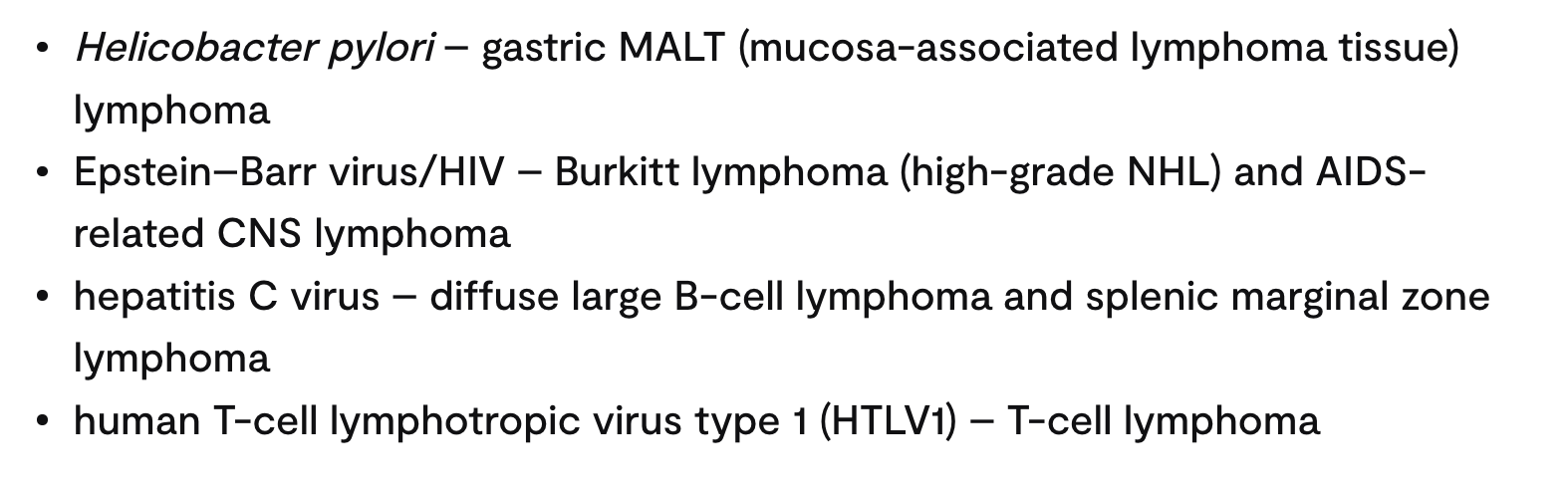
Non Hodgkin lymphoma
Commonly associated with infections and immunodeficiency.
Sx: symmetrical and painless lymphadenopathy, and is more likely to be associated with B symptoms.
Tests:
-LDH is the most important (poor prognostic marker)
-Absence of Reed-Sternberg cells
Polycythemia
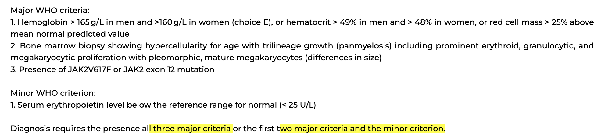
Increase in haematocrit, red cell count, and haemoglobin concentration.
Sx: myalgie, céphalée, splénomégalie
Tests: ↑ hematocrit, Hb, leucocytose, thrombocytose, JAK2 +, hypercellular bone marrow
Tx: venesection, phlebotomy, aspirin, cytoreductive therapy (high risk)
Anémie falciforme
Mutation in the beta chain on chromosome 11 leads to the production of HbS - sticky and fragile (rather than HbA).
Sx: crise vaso-occlusive, crise respiratoire
Tests:
-Frottis: drepanocytes, target cells, Howell-jolly (hyposplénisme)
Thalassémie
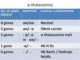
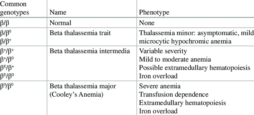
Abnormal haemoglobin production
Autosomal recessive
a-thalassemia
Tx: blood transfusions, stem cell transplantation, and splenectomy
b-thalassemia
Tx: blood transfusions and iron chelating agents
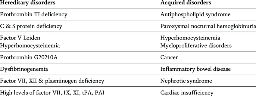
Thrombophilie
Ajout: polycythémie (acquired)
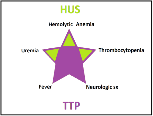
PTT
Abnormal ADAMST13 activity
Tests:
-Frottis: reticulocytes, schistocytes
-LFT, LDH, D-dimer ↑
-Haptopglobins ↓
-Dx: ↓ ADAMST13 activity
Tx: CS, plasma exchange
Maladie von Willebrand
Prolonged bleeding from minor wounds, easy bruising, menorrhagia, epistaxis, and GI bleeding.
Test: ↓ fctr VIII, PT N, ↑ APTT
Tx: desmopressin, acide tranexamique if pre-op.
Anemia classification
how to reverse dabigatran
Idarucizumab