Chapter 20 The Lymphatic System and Lymphoid Organs and Tissues
 Call Kai
 Learn
 Practice Test
 Spaced Repetition
 Match
 Flashcards
 Knowt Play
1/29

There's no tags or description
Looks like no tags are added yet.
30 Terms
Lymphatic Function
Fluid Balance: Returns leaked fluid (lymph) back to the blood to prevent swelling.
Immune Defense: Filters lymph through nodes to trap and destroy germs.
Fat Absorption: Absorbs dietary fats and fat-soluble vitamins from the digestive tract.

Three Parts of the Lymphatic System
Network of lymphatic vessels
Lymph: fluid in vessels
Lymph Nodes: cleanse lymph
Lymphoid Organ and Tissues
*Provide the structural basis of the immune system with PHAGOCYTIC CELLS AND LYMPHOCYTES.
Spleen
Thymus
Tonsils
Lymph Nodes
Lymphoid Tissues

Phagocytic Cells and Lymphocytes
Phagocytic Cells: The clean-up crew that non-specifically engulfs and digests any germs or debris on sight (innate immunity).
Lymphocytes (T & B cells): The specialized fighters that recognize specific invaders, remember them, and launch targeted attacks (adaptive immunity).

Lymphatic System
Returns interstitial fluid and leaked plasma proteins back to the blood
Lymphatic vessels: an elaborate network of drainage vessels.
3L interstitial fluid per day
Lymph
Distribution and Structure of Lymphatic Vessels
One-way system ensures lymph flows only toward the heart.
Lymphatic Capillaries
Blind-ended vessels that weave between tissue cells and blood capillaries. Not from bones, teeth, or bone marrow.
Takes up: Proteins, Cell Debris, Pathogens, and Cancer Cells
Increased Permeability
Endothelial cells overlap loosely to form one-way mini valves
Minivalves anchored by collagen filaments to matrix to increases ECF volume opens minivalves even more

Lacteals
Specialized lymph capillaries present in the intestinal mucosa. Absorbs digested fat and delivers fatty lymph (chyle) to blood.

Larger Lymphatic Vessels
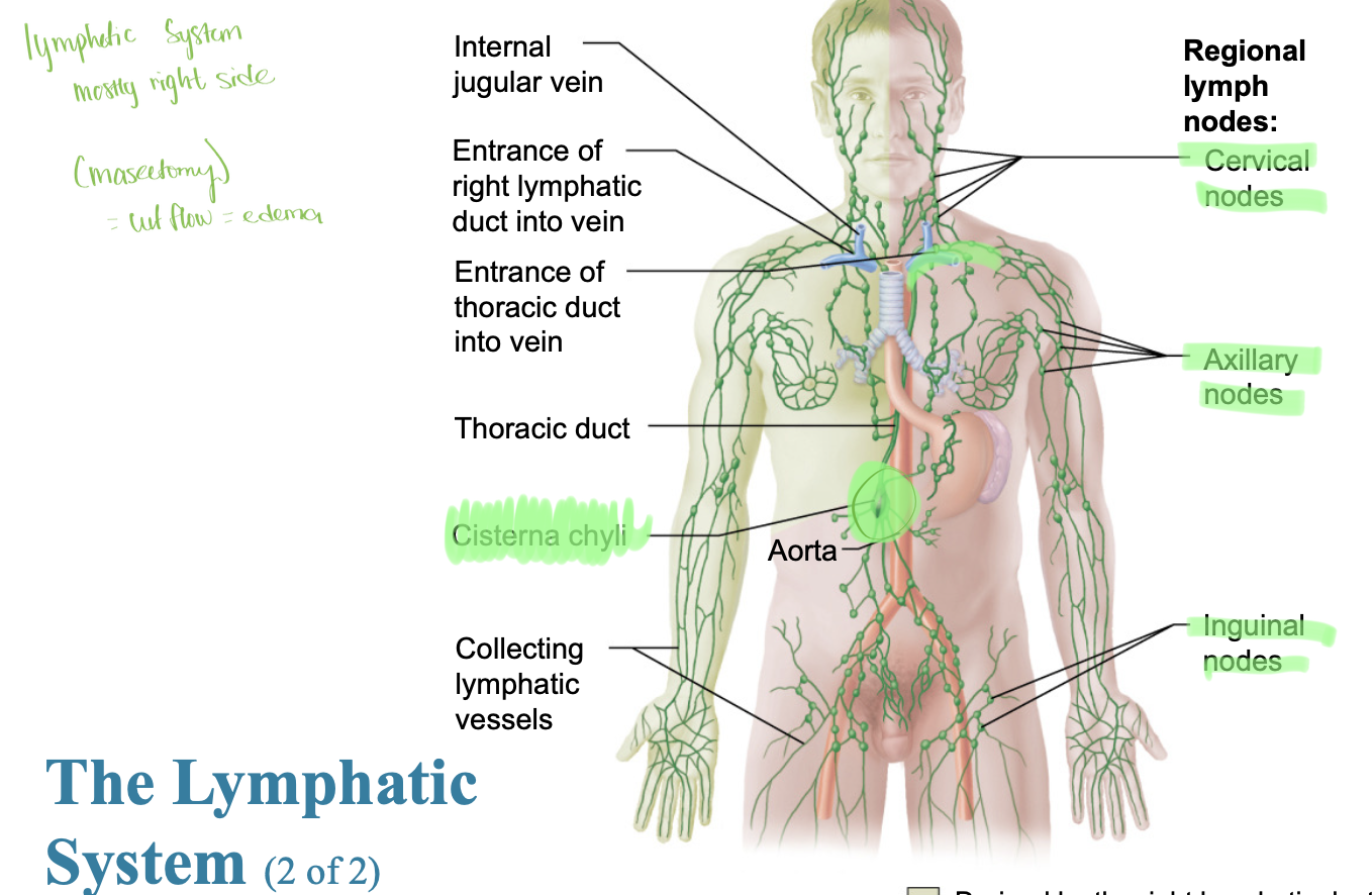
Large vessels called collecting lymphatic vessels
Vessels, trunks, and ducts.
Thinner walls, internal valves, and anastomoses
Lymphatic Trunks: largest collecting vessels that drain into
Lumbar, bronchomediastinal, subclavian, jugular trunks, and intestinal trunk.
Lymphatic Ducts
Right lymphatic duct drains the right upper arm and right side of the head and thorax.
Thoracic duct drains to the rest of the body.

Lymphatic Nodes
Cervical Nodes
Axillary Nodes
Inguinal Nodes
Cisterna Chyli

Lymphangitis
Lymphatic vessels appear as painful red lines under the skin caused by inflammation of larger lymphatic vessels that contain vaso vasora that become congested with blood.

Lymph Transport
* Low-pressure system, like the venous system
Muscle milking
Pressure changes in the thorax during breathing
Valves prevent backflow
Pulsation of nearby arteries
Contractions of smooth muscle in walls of lymphatics
**Physical activity increases flow of lymph; immobilization of area keeps needed inflammatory material in area for faster healing.
Lymphedema
Severe localized edema
Anything that prevents the normal return of lymph to the blood
Tumors blocking lymphatics or removal of lymphatics during cancer surgery.

Lymphoid Cells
Immune system cells found in lymphoid tissue and supporting cells that form lymphoid tissue structures.
Immune System Cells
Lymphocytes: Cells of the adaptive immune system; mature into one of two main types. Protect against antigens.
T cells: manage immune response and also attack and destroy infected cells.
B Cells: Produce plasma cells, which secrete antibodies.
T Cells = Thymus
They mature in the Thymus gland.
They do T-cell combat directly (cell-mediated immunity).
B Cells = Bone Marrow
They are created and mature in the Bone marrow.
They Bang out Bodies (antibodies/humoral immunity).
Macrophages: phagocytize foreign substances and help activate T cells
Dendritic Cells: capture antigens and deliver them to lymph nodes; also help activate T cells.
Reticular Cells: Produce reticular fibers called stroma in lymphoid organs. What they do: They act like a structural "skele-web" or sponge that holds these organs together and traps wandering cells (like lymphocytes and macrophages) so they can do their jobs.

Lymphoid Tissue
Houses and provides proliferation sites for lymphocytes
Offers surveillance vantage points for lymphocytes and macrophages as they filter through lymph.
Reticular Connective Tissue: Macrophages line on reticular fibers, spaces between fibers offer a place for lymphocytes to occupy when they retin from patrolling body.
What they are: Areas packed with white blood cells (mostly lymphocytes and macrophages) embedded in a supportive reticular fiber network.
What they do: They act as defense stations that monitor body fluids, trap germs, and launch immune responses.
Main Types:
Diffuse Lymphoid Tissue: Loosely organized clusters found in the mucous membranes of the respiratory, digestive, and urinary tracts (often called MALT - Mucosa-Associated Lymphoid Tissue).
Lymphoid Follicles (Nodules): Denser, solid spherical clusters of cells. Examples include the tonsils and Peyer's patches in the small intestine.
Lymphoid Organs
What they are: Distinct, organized structures enclosed by a connective tissue capsule that contain high concentrations of immune cells.
Primary Lymphoid Organs (Where cells form and mature):
Red Bone Marrow: Produces all blood cells, including B cells.
Thymus: Where T cells mature.
Secondary Lymphoid Organs (Where immune responses happen):
Lymph Nodes: Filter lymph and trap germs.
Spleen: Filters the blood, removes old red blood cells, and fights blood-borne pathogens.
MALT/Tonsils: Protect mucosal surfaces (like the throat and intestines) from incoming invaders.
Primary Lymphoid Organs
Thymus
Red Bone Marrow

Secondary Lymphoid Organs
Lymph nodes
tonsils
spleen
peyer’s patches
appendix

Lymph Nodes
Principal secondary lymphoid organs of the body.
What they are: Small, bean-shaped organs scattered along lymphatic vessels (concentrated in the neck, armpits, and groin).
What they do: They act as filtration stations for lymph, trapping bacteria, viruses, and abnormal cells before the fluid goes back into the blood.
How they work:
Lymph enters through afferent vessels.
Inside, white blood cells (macrophages and lymphocytes) destroy foreign invaders.
Cleaned lymph exits through efferent vessels.
Where they are found: cortex and medulla
cervical nodes, axillary nodes, inguinal nodes, and cisterna chyli
Structure of a Lymph Node
Cortex (Outer Region):
Outer Cortex: Contains dense clusters of B cells forming lymphoid follicles (nodules). If fighting an infection, these develop germinal centers where B cells multiply.
Deep Cortex (Paracortex): Mostly houses T cells moving between the blood and lymph, actively monitoring the fluid for threats.
Medulla (Inner Region):
Contains inward extensions called medullary cords, which are packed with antibody-producing plasma cells and macrophages.
Features medullary sinuses (open channels) where the filtered lymph collects before exiting the node.
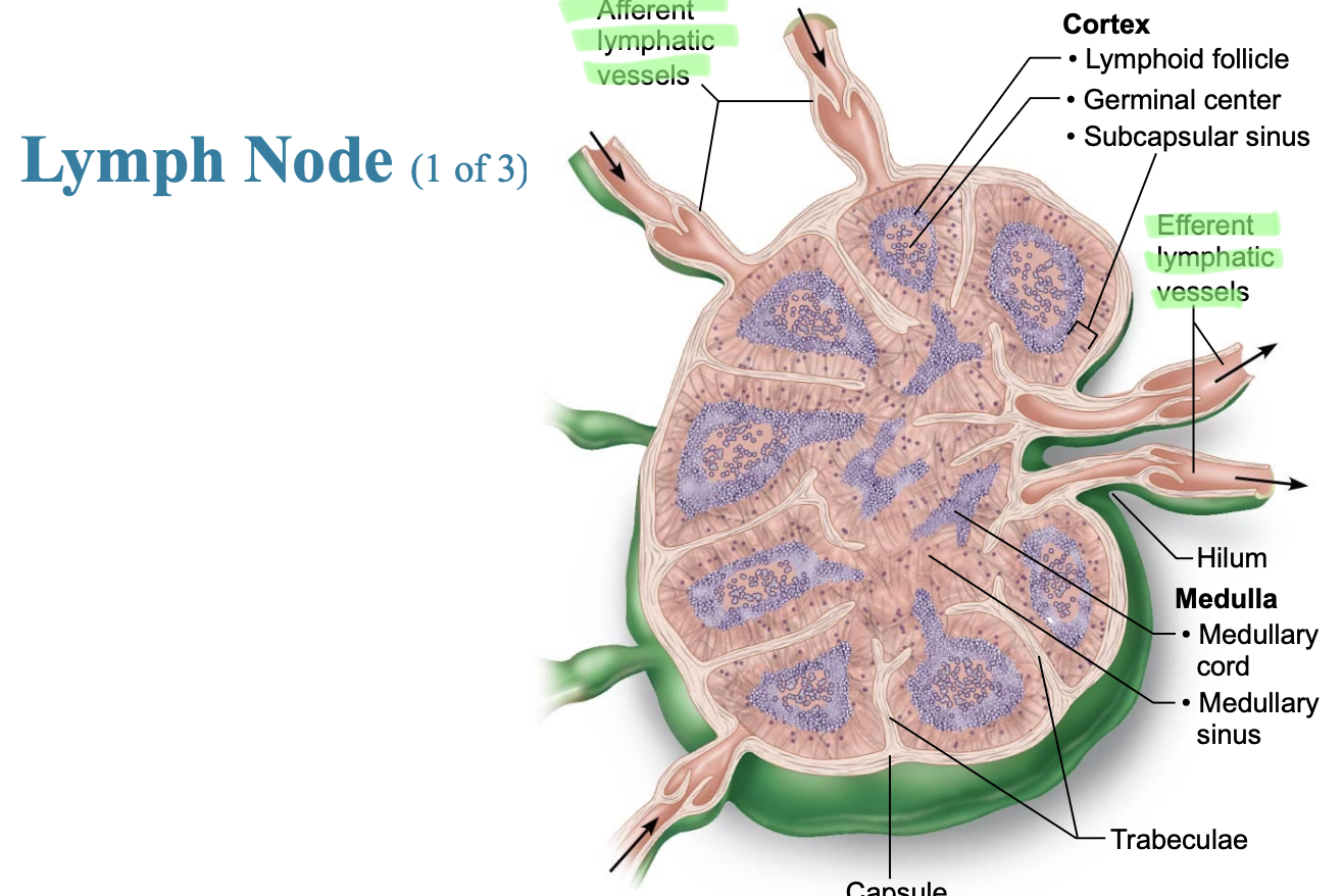
Circulation in the Lymph Nodes
Enters: Through multiple afferent vessels into the outer cortex.
Filters: Slows down as it flows via the subcapsular sinus through the cortex and medulla, where white blood cells trap and destroy germs. lymph enter medullary sinuses.
Exits: Through a single efferent vessel out the hilum.

Buboes
Inflamed, swollen, tender lymph nodes that result when nodes are overwhelmed by what they are trying to destroy. *Bubonic plague.

Spleen
Blood rich organ, located on the left side of the abdomin. Largest lymphoid organ.
Functions: immune surveillance and response, cleanses blood of aged blood cells and platelets; macrophages remove debris
What it is: The largest lymphoid organ in the body, located in the upper left side of the abdomen.
What it does: Instead of filtering lymph, it filters the blood to clean it and manage blood cells.
Two Main Areas (Pulps):
White Pulp: Packed with white blood cells (lymphocytes) that mount immune responses against blood-borne pathogens.
Red Pulp: Acts as a blood filter that destroys worn-out red blood cells and platelets, recycles iron, and stores blood platelets.

Splenectomy
Removal of a ruptured spleen to prevent hemorrhage and shock, but it can repair itself. The liver and bone marrow take over most of its functions.
MALT Mucosa-associated lymphoid tissue
What it is: Mucosa-Associated Lymphoid Tissue — a collection of small, unencapsulated lymphoid tissues scattered throughout the mucosal membranes of the body.
Where it's found: Guarding passageways that open to the outside world, primarily the digestive tract (like Peyer's patches in the small intestine), respiratory tract (tonsils), urinary, and reproductive tracts.
What it does: Acts as the body's first line of defense, intercepting and destroying pathogens (bacteria, viruses) right at the mucosal surfaces before they can enter deeper tissues.
FOUND IN
Tonsils, peyers patches, and appendix
tonsils
What they are: Masses of lymphoid tissue located in the ring around the throat (part of MALT).
Main Types:
Palatine: The ones on each side of the back of the throat (usually what people mean by "tonsils").
Pharyngeal (Adenoids): Located in the back of the nasal cavity.
Lingual: Located at the base of the tongue.
Tubual: surround opening of auditory tubes into pharynx
What they do: They trap and destroy inhaled or ingested pathogens (bacteria and viruses) before they can cause deep infections in the respiratory or digestive tracts, featuring deep pits called crypts that catch germs.

Peyer’s Patches
What they are: Large clusters of lymphoid follicles (MALT) located in the wall of the ileum (the lower end of the small intestine).
What they do: They monitor intestinal bacterial populations and prevent the growth of harmful bacteria in the gut, acting as immune sentinels for the digestive tract. also generate memory lymphocytes.

Appendix
offshoot of first part of large intestine.
Location aids in functions (like Peyer’s patches)
1. Destroy bacteria, preventing them from breaching intestinal wall
2. Generate “memory” lymphocytes
Thymus
What it is: A bi-lobed lymphoid organ located in the upper chest (mediastinum) right behind the sternum.
What it does: It is the primary site where T cells mature and learn to distinguish between the body's own cells and foreign invaders.
Key Feature: It is most active and largest during childhood; as you age, it gradually shrinks (involution) and is largely replaced by fat tissue in adults.
Developmental aspects of the lymphatic system
Embryonic Origin: Lymphatic vessels and organs (except the thymus) develop from mesoderm (specifically from lymph sacs budding off from veins).
The Thymus Exception: The thymus develops from the endodermal lining of the pharyngeal pouches.
Timeline:
The lymphatic vessels begin forming late in the embryonic period.
Lymph nodes develop later, mostly appearing just before and after birth (except for the spleen and tonsils, which form near the end of embryonic development).
Immature at Birth: A newborn's immune system is largely dependent on antibodies passed down from the mother (via the placenta and breast milk) because lymphatic organs and secondary immune responses are still immature and unexposed to antigens.