VTMD 206 - Neuro 1
 Call Kai
 Learn
 Practice Test
 Spaced Repetition
 Match
 Flashcards
 Knowt Play
1/101

There's no tags or description
Looks like no tags are added yet.
102 Terms
Astrocyte, Microglia, Oligodendrocyte, Ependymal cells
Name the 4 glial cell types of the CNS
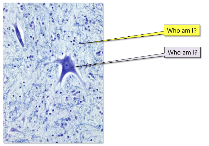
Neuropil
fibrous connections between glial cells and neurons
The space between “dots” is a dense network of neuronal and glial cell processes

Neuronal cell body
Dendrites
Nissl substance (ribosomes)
Nucleus
Nucleolus
Axon hillock
Axon
Nucleus of Schwann Cell
Myelin sheath
Node of Ranvier
Synapse
Label the Parts of the Neuron

Nissl Substance
Ribosomes that cover the neuronal cell body
For protein production
Absent from axon hillock
Astrocyte
Glial Cell of the CNS
Support and modulate the activity of neurons and maintain the blood-brain barrier
The largest and most abundant glial cells. They have numerous branching cytoplasmic processes that provide a scaffold for neurons
They have a role in regulating the movement of metabolites and wastes to and from neurons, and in maintaining ionic concentration of the extracellular space
Additionally, they modulate capillary permeability and thus, the blood-brain barrier

Microglia
Glial Cell of the CNS
Garbage collector of the central nervous system, gobble up debris by phagocytosis
The phagocytic cells of the CNS and function to remove dead neurons.
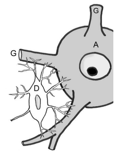
Oligodendrocyte
Glial Cell of the CNS
Produce and maintain the myelin sheath in the CNS
The multiple processes of a single ________ may myelinated several nearby axons.

Ependymal cells
Glial Cell of the CNS
Line the fluid-filled cavities of the brain and spinal cord
Form an epithelium-like lining of the canals and ventricles of the CNS, producing a fairly permeable barrier between the cerebrospinal fluid (CSF) in these cavities and the solid tissue of the brain and cord
Vary from low cuboidal to columnar, and many are ciliated or have microvilli. The beating of ependymal cilia circulates CSF

Astrocyte
Name the CNS glial cell type that is crucial for maintenance of the blood-brain barrier
Grey = Neuron
Yellow = Astrocyte, Microglia, Oligodendrocyte
Grey =
Yellow =
Pia Mater
Meninge
The innermost layer of the meninges, adhering closely to the surface of the brain and spinal cord. It is a delicate and vascular membrane that provides a protective barrier and helps nourish the underlying nervous tissue
Arachnoid
Meninge
Middle layer
Relatively thick
Has tendrils that attach it to the surface of the brain
Space between arachnoid and pia mater = subarachnoid space
Filled with cerebrospinal fluid (CSF)
All blood vessels on surface of brain also lie in this space
Mostly made of collagen
The middle layer of the meninges, located between the dura mater and the innermost layer, the pia mater. It is a thin, web-like membrane that also helps protect the brain and spinal cord
Subarachnoid space
The space between the arachnoid and the pia mater. This space contains cerebrospinal fluid (CSF), which serves as a cushion for the brain and spinal cord. It also contains blood vessels
Dura mater
Meninge
Most superficial layer
Tough collagen sheet
Fused to the periosteum in the skull (not fused in the spinal cord)
The outermost and toughest layer of the meninges, which are the protective membranes that surround the brain and spinal cord. It is a thick, fibrous membrane that helps protect the brain and spinal cord from injury
Epidural space
Space around dura mater in spine, filled with fat and some fluid for movement of spinal cord and cushioning
Not in brain, as dura is fused to the periosteum
The area between the dura mater and the vertebral column. It contains fat, blood vessels, and connective tissue. Epidural injections are sometimes used to administer medications or anesthesia in this space
Cerebrospinal Fluid
Role:
Shock absorption
Maintains intracranial pressure
Nourishes the brain
Transport
Controls ionic environment of the brain
So, not just a shock absorber, responsible for keeping the brain alive
Fills subarachnoid space,
Schwann Cell
Glial Cell of the PNS
Produce the myelin sheath of peripheral nerves
Insulate adjacent axons from one another, nurture the axon and guide its growth. Insulation is provided by axons being enclosed within recesses of the ______ cytoplasm
A. Axon
B. Nerve fibre
C. Nerve fascicle
D. Nerve
E. Endoneurium
F. Perineurium
G. Epineurium
H. Myelin Sheath
I. Adipose tissue
J. Blood vessel

Nerve fibre
Endoneurium
Fascicle
Perineurium
Epineurium

Pia mater, Arachnoid, Dura mater
Name the three meningeal layers of the CNS from innermost to outermost
Orange = Cerebrum
Green = Diencephalon
Purple = Brainstem
Blue = Cerebellum
Orange =
Green =
Purple =
Blue =

Cerebrum (Cerebral cortex)
Part of the Vertebrate Brain
Processes sensory information, supports conscious perception, learning, memory, and voluntary behavior
Majority of the brain
Neocortex
Makes up ~90% of the cerebral cortex in humans
An evolutionarily newer part of the cerebral cortex, particularly prominent in mammals
Organized into six cellular layers
Each layer has a different combinations of cell types
Supports higher-order sensory processing, learning, memory, decision-making, and voluntary behavior
Cortical folding (gyri - raised ridges and sulci - grooves) increases cortical surface area, allowing more neurons to fit within the skull
More neurons ≠ necessarily “smarter” - cognitive abilities depend on neural organization, connectivity, and specialized circuits, not neuron number alone
Birds do not have a neocortex, but their pallium contains specialized circuits that support complex cognition and behavior
Diencephalon
Part of the Vertebrate Brain
Relays sensory information and regulates homeostasis, autonomic functions, and endocrine activity
Cerebellum
Part of the Vertebrate Brain
Coordinates movement, balance, posture, and motor learning
Brainstem
Part of the Vertebrate Brain
Connects the brain with the spinal cord and controls essential functions such as breathing and cardiovascular activity
Frontal
Lobe of the Neocortex
Motor cortex, planning, long-term memories, higher functions
Temporal
Lobe of the Neocortex
Auditory cortex, memory formation (hippocampus), communication, facial recognition
Occipital
Lobe of the Neocortex
Visual cortex
Parietal
Lobe of the Neocortex
Sensory integration from multiple modalities (somatosensory cortex)
Grey matter
Mostly neuronal cell bodies
Inside of spinal cord
Outside of brain
White matter
Mostly axons (myelinated)
Outside of spinal cord
Inside of brain
Cervical, thoracic, lumbar, sacral, coccygeal
Spinal cord segments
Spinal Nerves
Emerge from the spinal cord
Contain both afferent (sensory) and efferent (motor) fibers
One pair arises from each spinal cord segment (one nerve on each side of the body)
Mixed nerves, containing both:
Afferent (sensory) fibers: carry sensory information TO the CNS
Efferent (motor) fibers: carry motor commands AWAY FROM the CNS

Dorsal root
Dorsal root ganglion
Dorsal ramus
Ventral ramus
Ventral root
Blue = Sensory axon and cell body
Red = Axon of motor neuron
c
c
c
c
c
Blue =
Red =

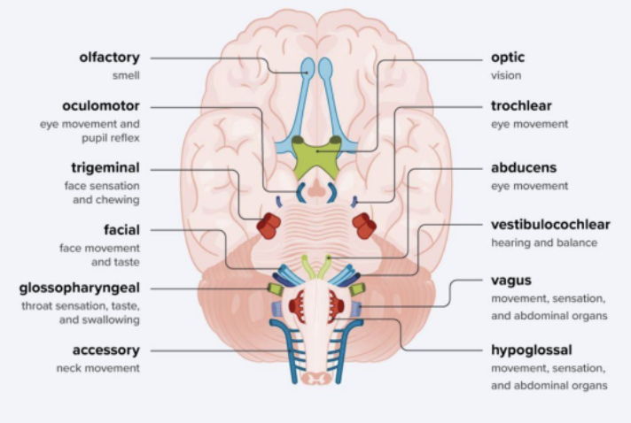
Cranial Nerves
Emerge directly from the brain
Clinically important
12 pairs in humans
May be:
Sensory (afferent)
Motor (efferent)
Mixed — both sensory and motor
Carry sensory information from the head and special senses → vision, hearing, smell, taste, and balance
Sympathetic Nervous System
Part of the PNS
“Fight-or-Flight” response
Activated during stress, danger, or physical activity
Reduces digestive activity
Prepares the body for action
Efferent neurons release epinephrine and norepinephrine
Parasympathetic Nervous System
Part of the PNS
“Rest-and-Digest” response
Promotes energy conservation and maintenance
Supports:
Digestion
Urination and defecation
Lower heart rate
Pupil constriction
Salivation
Sexual arousal
Efferent neurons release acetylcholine
Major difference is what postganglionic neuron releases at the target organ
Enteric Nervous System
Part of the PNS
Controls functions of the gastrointestinal (GI) system
Mostly works independently from CNS
Embedded within the walls of the GI tract (100 million neurons)
Controls motility and secretion
Influenced by both the sympathetic and parasympathetic systems
Can also operate independently
“The gut has its own mind”
Dendrites
Part of the Neuron
Branched projections from the cell body
Receive signals from other cells
Convey signals toward the cell body
Cell body (soma)
Part of the Neuron
Integrates incoming signals
Contains the nucleus and other organelles
Main metabolic and maintenance center of the neuron
Axon hillock
Part of the Neuron
Specialized region connecting the soma to the axon
Integrates incoming signals
If the membrane potential reaches threshold → an action potential is triggered
Action potential travels down the axon
No nissl substance
Axon
Part of the Neuron
Long projection extending from a neuron
Conducts action potentials away from the cell body
May be myelinated or unmyelinated
Synaptic terminal
Part of the Neuron
End of the axon
Stores and releases neurotransmitters
Communicates with the next cell
Synaptic cleft/junction
Part of the Neuron
Small gap between the presynaptic and postsynaptic cells
Neurotransmitters are released into the synaptic cleft
Neurotransmitters diffuse across the cleft to bind receptors on the postsynaptic cell
A stimulus brings the membrane potential to threshold, triggering an action potential (−55 mV)
Voltage-gated Na⁺ channels open → Na⁺ rapidly enters the neuron → the membrane becomes more positive (+30 mV) — depolarization
Na⁺ channels inactivate and voltage-gated K⁺ channels open → K⁺ leaves the neuron → the membrane becomes more negative — repolarization
K⁺ continues to leave the neuron briefly → the membrane becomes more negative than the resting potential (−90 mV) — hyperpolarization
K⁺ channels close and the membrane potential returns toward the resting membrane potential (−70 mV)
Steps of an Action Potential
Refractory Period
Time from beginning of action potential until return to resting state
During this period membrane will not respond normally to additional stimulus
Causes action potential to be unidirectional
Axon diameter and myelination
2 Factors that Affects the Speed of Action Potential Propogation
Chemical Synapse
Type of Synapse
vesicles that contains neurotransmitters, takes time for neurotransmitter release to occur, majority of synapses
Signal Transmission: neurotransmitter release across cleft
Speed: slower
Directionality: typically unidirectional
Effect: excitatory or inhibitory
Prevalence: most common in human brain
Examples: most synapses in CNS

Electrical Synapse
Type of Synapse
less common, 2 neurons directly connected by gap junctions, electrons can flow directly from one cell to another
Signal Transmission: Direct ion flow via gap junctions
Speed: Very fast
Directionality: Bidirectional
Effect: Synchronizes neuron groups
Prevalence: Less common
Examples: Retina, hippocampus, inferior olive, escape circuits

Glutamate
Neurotransmitter
Main excitatory NT
Amino acid NT
The primary excitatory neurotransmitter in brain
Most abundant excitatory neurotransmitter
Synthesized from glutamine by glutamine-glutamate cycle
Stored in vesicles (VGLUT transporters)
Removed by excitatory amino acid transporters (EAATs)
Amino acid neurotransmitter
Cannot cross the BBB, so must be synthesized in the brain from a-ketoglutarate (intermediate product in the CAC)
Generally has excitatory actions
Interacts with many types of receptors
3 identified ionotropic glutamate receptors
NMDA
AMPA
Kainate receptors
All allow positively charged neurons to flow across the cell membrane depolarizing the neuron and making it more likely to fire an action potential
NMDA is often involved in synaptic plasticity – important for learning and memory
Metabotropic glutamate receptors
Can have excitatory or inhibitory effects depending on the receptor
Glutamate is removed from the synaptic cleft by excitatory amino acid transporters (EAATS)
Carry glutamate into neurons and glial cells
Glutamate in glial cells is converted to the AA Glutamine, which is then transported back into neurons where it is converted back to glutamate – Glutamate-Glutamine Cycle
GABA
Neurotransmitter
Main inhibitory NT
Amino acid NT
Main inhibitory neurotransmitter
Synthesized from glutamate via GAD (glutamic acid decarboxylase)
Many general anesthetics increase _____ activity
2 classes of receptors: _____A (ionotropic) and ____B (metabotropic)
Amino acid neurotransmitter
Synthesised from glutamate
Function changes over neurodevelopment
In a mature brain, primarily inhibitory
2 types of GABA receptors
GABA A – Ionotropic
Binding causes Cl- ion channel to open and Cl- enters the cell causing hyperpolarization, making an AP less likely to occur
GABA B – Metabotropic
Binding causes the opening of K+ channels that allow K+ ions to flow out of the neuron causing hyperpolarization, making an AP less likely to occur
GABA transporters are responsible for the reuptake of GABA into presynaptic neurons or glial cells where it is degraded by mitochondrial enzymes
Increased GABA activity can have sedative effects
Alcohol and benzodiazepines increase the activity of GABA receptors
Dopamine
Neurotransmitter
Movement and reward
Monoamine NT
Particularly important in the brain, including movement, motivation, and reward
Key functions
Motor control
Mood, motivation and reward
Cardiovascular and respiratory regulation
Endocrine regulation — e.g., inhibits prolactin release
Monoamine neurotransmitter
Derived from an amino acid
Catecholamine neurotransmitter
______ pathways carry ______ from ______ rich areas to other parts of the brain
Receptors — G-coupled protein receptors
At least 5 subtypes
Removed from synaptic cleft by ______ transporter
Functions (too many to list)
Movement
Reward
Memory
Lactation
Attention
Sleep Regulation
Nausea
Motivation
Arousal
Many, many more
Dopamine-rich areas of the brain include:
Ventral tegmental area
Substansia nigra
Hypothalamus
Olfactory bulb
Retina
Dopamine pathways carry dopamine from dopamine rich areas to other parts of the brain
Nigrostriatal Pathway – Substantia Nigra to Striatum
Mesolimbic Pathway – Ventral Tegmental Area to Nucleus Accumbens and other limbic structures
Mesocortical Pathway – Ventral Tegmental Area to Throughout the Cerebral Cortex
Norepinephrine
Neurotransmitter
Important for alertness, attention, and the sympathetic nervous system
Principal neurotransmitter of most postganglionic sympathetic neurons
Monoamine neurotransmitter
Catecholamine neurotransmitter
Hormone
Synthesized from dopamine
Primarily produced in the pons and medulla of the brainstem
Locus coeruleus is the main site of norepinephrine production for the CNS
Primary neurotransmitter used by the sympathetic nervous system and is found in sympathetic ganglion cells
Released from the adrenal glands as a hormone
Acts on adrenergic receptors (G-protein coupled receptors)
At least 3 main types: Alpha-1, Alpha-2, and Beta
Each has multiple subtypes
Removed from the synaptic cleft via reuptake by the _______ transporter
Functions depend on the type of receptor that it activates and where the receptor is located
Associated with arousal, alertness, and attention in the CNS, but more complex than that
Release in sympathetic nervous system is associated with increased activity/fight or flight responses
Epinephrine
Neurotransmitter
Best known as a hormone released from the adrenal medulla during the fight-or-flight response
Principal hormone released from the adrenal medulla during sympathetic activation
Serotonin
Neurotransmitter involved in mood, gastrointestinal function, and many other physiological processes in animals
Functions and Clinical Significance:
5-hydroxytryptamine
Regulates mood, aggression, thermoregulation, sleep, and pain perception
Plays a role in GI motility and platelet aggregation
Actions primarily terminated by reuptake into the presynaptic neuron and enzymatic degradation
Dysfunction can lead to behavioural changes, GI disturbances, serious conditions such as serotonin syndrome
Increased ______ Function:
Overstimulates ______ receptors leading to serotonin syndrome
Agitation, hyperactivity, tremors, GI upset, seizures, altered mental status
Timely intervention is crucial, can be life threatening
_______ neurons are primarily found in the brainstem in clusters called raphe nuclei
Project throughout the brainstem and brain and provide serotonin to the rest of the CNS
Serotonin receptors
7 different families
6 families are G-protein coupled receptors
1 family is ligand gated ion channel
14 different receptor subtypes have been identified
Serotonin is removed from the synaptic cleft via reuptake by serotonin transporter (SERT)
Often linked to mood and many other functions
Neurotransmitter
Mood and sleep
Monoamine NT
Serotonin Neurotransmission
After being released from the presynaptic neuron, ____ acts on postsynaptic receptors and is then transported back into the presynaptic terminal by the _____ transporter (SERT)
Once inside the neuron, _____ is degraded by the enzyme monoamine oxidase, ensuring that its effects are appropriately terminated
Serotonin Transporter (SERT)
Key Terms in Serotonin Regulation
A protein that reuptakes serotonin from the synaptic cleft back into the presynaptic neuron, regulating neuron, regulating it’s levels
Monoamine Oxidase
Key Terms in Serotonin Regulation
The enzyme responsible for breaking down serotonin within the presynaptic neuron, terminating its action
Serotonergic receptor
Key Terms in Serotonin Regulation
A receptor on the postsynaptic cell that binds serotonin and mediates its effects
Acetylcholine
Neurotransmitter central to neuromuscular transmission and voluntary movement
Functions and Clinical Significance:
Primary excitatory neurotransmitter at the neuromuscular junction
Trigger skeletal muscle contraction
Essential for voluntary movement
Also supports attention, learning, memory, cognitive functions in CNS
Involved in both somatic and parasympathetic nervous systems
Mediates parasympathetic responses, including pupil constriction, digestion, urination
Dysfunction leads to muscle weakness, impaired movement, disturbances in autonomic functions
Decreased _______ Function:
Diminished excitatory signaling at the neuromuscular junction
Leads to reduced muscle contraction and clinical signs such as muscle weakness, exercise intolerance, and respiratory compromise
Disorders include myasthenia gravis, where animals may present with fatigue after minimal activity or difficulty swallowing due to muscle weakness
Neurotransmitter
Muscle and autonomic signalling
Works in many completely different areas of the nervous system
CNS: learning, memory and attention
Neuromuscular junction: skeletal muscle contraction
Parasympathetic nervous system : rest and digest
Autonomic ganglia: neurotransmitter released by all preganglionic neurons
Other CNS functions: arousal, sleep–wake regulation and pain modulation
Main neurotransmitter at neuromuscular junctions, responsible for muscle contraction
Widely used in autonomic nervous system
In all preganglionic neurons and postganglionic parasympathetic neurons)
Important in memory, arousal, and attention
Acetylcholine Neurotransmission
___ is synthesized in nerve terminals, stored in vesicles, and released into the synaptic cleft when a nerve impulse arrives
When released, binds to specific receptors on the muscle cell membrane, triggering muscle contraction
Action of ___ is terminated by acetylcholinesterase enzymes, which breaks it down, so the cycle can repeat efficiently
Glycine
Neurotransmitter
Inhibitory neurotransmitter
Key transmitter for inhibitory neurotransmission in the CNS (spinal cord and brain stem)
_____ receptor (GlyR)
Receptor on post-synaptic neuron
Effect of neurotransmitter depends on…
Diffusion out of synaptic cleft
Reuptake into presynaptic neuron
Enzymatic degredation
Fates of Neurotransmitter After Release
Postsynaptic potential
A change in the membrane potential of the postsynaptic cell caused by neurotransmitter binding to its receptor
Can be excitatory or inhibitory
Excitatory Postsynaptic Potential
Makes the postsynaptic membrane more likely to reach threshold
Usually depolarizes the membrane
Inhibitory Postsynaptic Potential
Makes the postsynaptic membrane less likely to reach threshold
Usually hyperpolarizes the membrane
Graded potential
Occur at synapses when neurotransmitters activate receptors and can lead to an action potential
Can have an additive or opposing effect
Do not travel far from the area of stimulation → local potentials
Depolarizations add to each other until threshold is reached
Opening or closing of ion channels changes the membrane potential
Na⁺ or Ca²⁺ influx → excitatory
Cl⁻ influx → inhibitory
K⁺ efflux → inhibitory
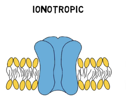
Ionotropic receptor/Ligand-gated ion channel
When the receptor is activated, the ion channel opens
Different neurotransmitters activate specific ion channels
Ions flow across the membrane → excitatory or inhibitory potential
Fast response → milliseconds
But, voltage-gated ion channels are faster
When a neurotransmitter binds, causes the opening of an ion channel that allows ions to flow across the membrane of the neuron, potentially changing the activity of the cell

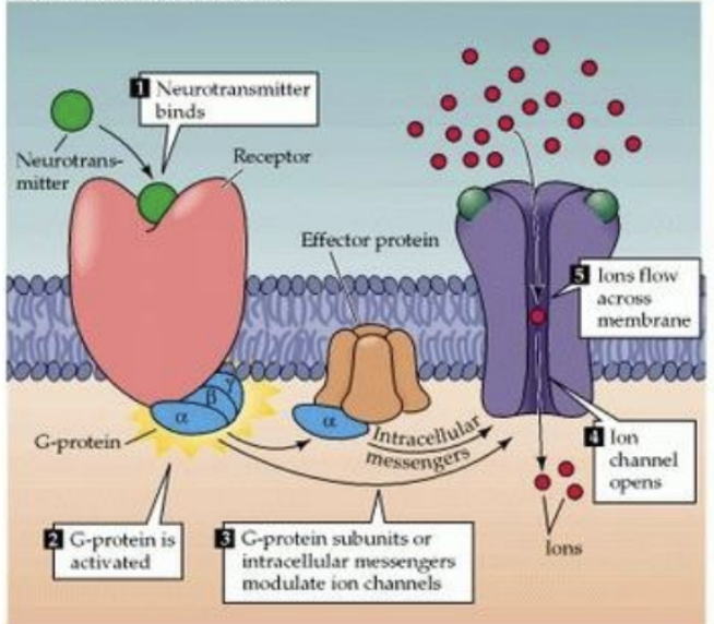
Metabotropic receptors/G-protein coupled receptors
Receptor activation triggers a signaling cascade involving secondary messengers
The cascade can eventually affect ion channels and cell excitability
Different neurotransmitters activate specific receptors
Slower response → seconds to minutes
Effects last longer and can have a more widespread effect
Neurotransmitters bind to the receptor and cause the activation of a ______
The _____ can cause the opening of ion channels, or other effects such as activating enzymes or initiating signalling cascades
Nicotinic receptor
Type of Cholinergic Receptor
Ionotropic
Cause excitation
Ionotropic
Found at neuromuscular junctions, autonomic ganglia, and adrenal medulla
Neurotransmitter binding directly opens an ion channel
Na⁺ enters → rapid depolarization
Fast response → e.g., skeletal muscle contraction
Muscarinic Receptor
Type of Cholinergic Receptor
Metabotropic
Effects depend on the subtype of the receptor
Metabotropic
Found in the CNS, heart, smooth muscle, and glands
Neurotransmitter binding activates a G protein
Triggers intracellular signaling → changes ion channels or cell activity
Slower, longer-lasting response
5 subtypes: M1–M5

Basal forebrain
Nucleus basalis
Medial septal nucleus and nucleus of the diagonal band
Brainstem
Pedunculopontine nucleus
Laterodorsal tegmental nucleus
Locations of Cholinergic Neurons
Efferent
Describes nerves that carry signals from the brain to the peripheral nervous system
CNS to PNS
Afferent
Describes neurons that carry signals from the periphery to the central nervous system
PNS to CNS
Somatic Nervous System
Under voluntary control
Descending input from the brain and the spinal cord (upper motor neuron (UMN)) to the skeletal muscle (via) lower motor neurons (LMN)
The will of the central nervous system is manifested through lower motor neurons
Somatic efferents specifically control skeletal muscle
Somatic Efferent Pathways
Motor impulses exit the motor cortex (brain)
Signals travel via upper motor neurons (spinal cord)
Signals are transferred to lower motors neurons (exit at ventral horn)
Every muscle cell receives axonal input
Somatic Afferent Pathways
Sensory receptors in peripheral tissues transmit signals to afferent neurons
Afferent neurons have cell bodies in the dorsal root ganglion and then enter the spinal cord via the dorsal horn
Afferent neurons transport info to the appropriate brain center for processing *Assuming no reflex arc has occured
Reflex Arc
Somatic afferent pathway
Response to stimulus can circumvent to brain to deliver an effect before an interpretation
Allows you to respond to a stimulus without perceiving a stimulus at the level of the brain
Works similarly as normal afferent pathways, except, it will branch and one branch attaches to an interneuron (short circuit that allows ventral horn and other part of spinal cord to communicate)
Interneuron allows cross talk between afferent and efferent
Sensory receptors in peripheral tissues transmit signals to afferent neurons
Afferent neurons have cell bodies in the dorsal root ganglion and then enter the spinal cord via the dorsal horn
Signal is transferred to interneurons to immediately perform action
Signals are interpreted by the appropriate centers in the brain
Proprioception
The ability to know where your body parts are in space without thinking about it
Constantly relayed to the brain (cerebellum) through somatic afferent pathways
_______ neurons are constantly sending feedback about muscle position and movement
Proprioceptive Pathways
These pathways are virtually always activated
Constant feedback to the cerebellum
You’ll notice there is also a reflex arc here
Involuntary control of body position/posture
Ie. Clinically, think of the “knuckling” test
Plugged into 2 spaces
Cerebellum – responsible for fine tuning movement, provides descending input to do so
A lot of reflex arcs in the pathway that allow to bypass the cerebellum and correct posture, etc. without you realizing it, spine does it for you
Via interneuron transmission
Visceral Nervous System
Sometimes termed the autonomic nervous system, responsible for involuntary control
______ efferents provide innervation to the following:
Smooth muscle, visceral organs, glands and cardiac muscle
The hypothalamus is the primary center for the integration of the _______ nervous system
Hypothalamus is the regulatory body for the entire _____ NS
Unlike the somatic nervous system, there are two neurons (instead of one) involved in each efferent ______ pathway
Referred to as preganglionic and postganglionic neurons
The _____ nervous system can be further subdivided into the parasympathetic and sympathetic nervous systems
Sympathetic: fight or flight
Parasympathetic: rest and digest
Efferent tracts are divided into preganglionic and postganglionic neurons in the periphery which interact at a peripheral ganglion
Afferent tracts contain one neuron in the periphery (first order) and synapse with second order neurons in the spinal cord
Many of the functions of the visceral nervous system are controlled by spinal reflexes (ie. blood pressure and micturition)
The hypothalamus can override and fine tune these reflexes
Visceral Efferent Pathways
There are two neurons (instead of one) involved in each ______ ______ pathway
Referred to as preganglionic and postganglionic neurons
Bring into from brain to target organs for autonomic control
Divided into sympathetic and parasympathetic pathways

Parasympathetic pathways
Type of Visceral Efferent Pathway
Originate at the brainstem
Preganglionic neuron → Ganglion (group of cell bodies) → Postganglionic neuron
Ganglion provides an additional center for consideration of response/control
Additional regulation center
More throughout than somatic pathways
Target organ
Sympathetic Pathways
Type of Visceral Efferent Pathway
Originates at the spinal cord
Preganglionic neuron → Ganglion (group of cell bodies) → Postganglionic neuron
Target organ
The majority of sympathetic pathways go through 2 neurons except for adrenal glands, only one neuron
Bcs fight or flight and need an immediate response
Hypothalamus
_________ is the regulatory body for the entire visceral NS
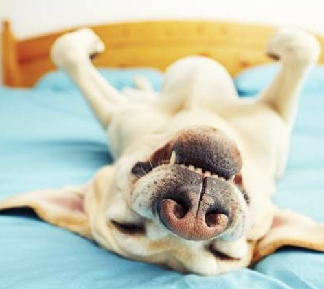
Parasympathetic tone
The level of activity of the parasympathetic nervous system
Image = High __________ _________
Sympathetic tone
The level of activity of the sympathetic nervous system
Image = High ____________ ________

Visceral afferent pathways
Located throughout viscera (ie. Gastrointestinal tract, urinary bladder etc) and provide feedback to the central nervous system
Take stimulus from viscera and relay to the brain
Common stimuli:
Stretch, distention, pressure, temperature, chemical gas concentrations etc
Ie. Bladder distension causing the urge to urinate
Viscera are largely insensitive to stimuli that the surface of the body responds to
Ie. Touch and cutting
These pathways are largely insensitive to pain and don’t respond to those pathways the same way other pathways do
The afferent neurons originate at the target tissue, and then “plug into” sympathetic and parasympathetic centers
A lot of it is done under reflex control
This is a one neuron system in the periphery
Neurotransmission
The process by which neurons communicate with each other throughout the body
Primarily occurs through chemical synaptic transmission, where one neuron releases chemicals that influence the activity of another
The precise coordination of neurotransmitter release, receptor activity, and signal termination allows the nervous system to regulate everything from movement to mood
Disruptions in these processes can lead to significant clinical consequences
Neuron
Key Term in Neurotransmission
a specialized cell that transmits nerve impulses and forms the basic unit of the nervous system
Synapse
Key Term in Neurotransmission
the junction between two neurons where communication occurs via neurotransmitter release
Neurotransmitter
Key Term in Neurotransmission
a chemical messenger released by neurons to transmit signals across a synapse to another cell
Chemicals that neurons use to communicate with one another across synapses
Synapses consist of a neuron sending a signal (presynaptic neuron) and one that is receiving a signal (postsynaptic neuron) and a small space between them known as the synaptic cleft
Stored in vesicles in the presynaptic neuron before being released into the synaptic cleft when an electrical impulse/action potential arrives at the presynaptic neuron
Bbind to receptors on the postsynaptic neuron
Presynaptic cell
Key Term in Neurotransmission
the neuron that releases neurotransmitters into the synaptic cleft during communication
Synaptic cleft
Key Term in Neurotransmission
The small gap between the presynaptic and postsynaptic cells where neurotransmitters are released
Excitatory
Neurotransmitter Effects on Postsynaptic Neurons Can…
Increase the likelihood that the postsynaptic neuron will fire an action potential